A novel label-free microscopy technique to image red blood cells and oxygenation
Researchers from the Laboratory for Optics and Biosciences (LOB, CNRS / École Polytechnique / INSERM), a member of the Ile-de-France Sud node of France-BioImaging, and from the Developmental Biology and Stem Cells Department (UMR3738, CNRS / Institut Pasteur), developed a new form of multiphoton microscopy providing label-free imaging of red blood cells and oxygenation. This technique is called color third-order sum-frequency generation microscopy, and is described in an article recently published in Light: Science & Applications.
Keeping resolution, saving time
Current methods used to map microcirculation and blood oxygenation at high resolution typically require the injection of fluorescent or phosphorescent markers. In addition, they usually require relatively long pixel times and thus are limited in spatio-temporal resolution. The novel approach is based on label-free third-order sum-frequency generation (TSFG) and third-harmonic generation (THG) contrasts. In practice, this microscopy is based on the illumination of samples by two pulsed infrared lasers and the simultaneous detection of several TSFG and THG signals emitted at different colors. This method has the advantage of providing simultaneous measurements at several wavelengths spanning the hemoglobin absorption spectrum. This simultaneity makes TSFG microscopy appropriate for studying dynamic samples…
To better understand brain and tissue physiology
…such as biological tissues! Biological tissues are supplied with oxygen by red blood cells, responsible for circulating hemoglobin through the body. Scientists have shown that the intensity of the different signals detected by TSFG microscopy depends on their spectral proximity to the absorption wavelength of hemoglobin. As the color of hemoglobin depends on its oxygenation state, the TSFG makes it possible to image red blood cells circulating in live zebrafish larvae – and even to probe their oxygenation state. Researchers also demonstrated that this contrast modality is also compatible with deep-tissue microscopy and can be used to observe the brain of a live adult zebrafish. An example that confirms the broad range of application of this novel imaging technique.
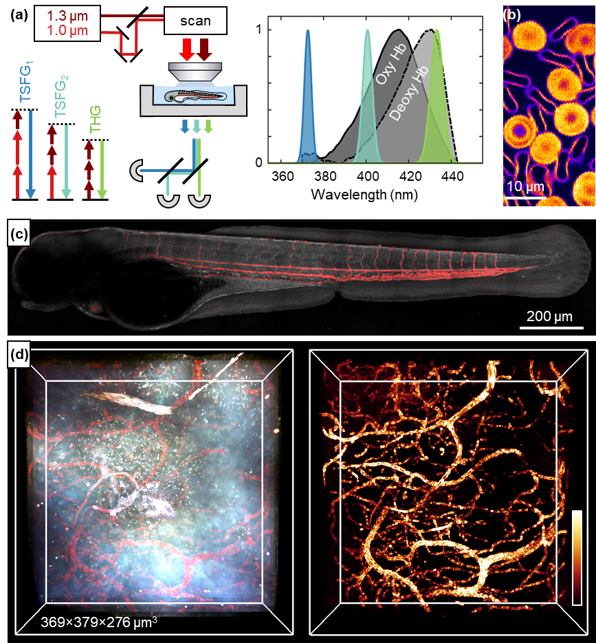
(c) Imaging of a zebrafish embryo ; (d) 3D in vivo imaging in an adult zebrafish brain.
© Laboratoire d’optique et biosciences (LOB, CNRS / École Polytechnique / INSERM)
Reference: Ferrer Ortas, J., Mahou, P., Escot, S. et al. Label-free imaging of red blood cells and oxygenation with color third-order sum-frequency generation microscopy. Light Sci Appl 12, 29 (2023). https://doi.org/10.1038/s41377-022-01064-4