Service
IdFsud gathers an important task force in biology, physics, applied mathematics and complex systems modelling. These disciplines synergize to develop innovative methodologies and tools in microscopy imaging and image analysis for applications in developmental biology, cell biology, plant biology, physiopathology and neurosciences. IdF Sud gathers three core partners with internationally acknowledged expertise: BioEmergences (CNRS-IBiSA), Imagerie-Gif and Polytechnique LOB (Lab for optics & biosciences, CNRS – INSERM).
The imaging facilities of IdFsud are developed and coordinated by: Nadine Peyrieras (INAF, CNRS, Gif-sur-Yvette), Sandrine Lecart (I2BC, CNRS, Gif sur Yvette), Emmanuel Beaurepaire (Ecole Polytechnique, CNRS, Palaiseau).
The activity of the Ile-de-France Sud node (IdFsud) consists in developing novel optical biological imaging methods, in giving access to a broad range of imaging approaches to a large community, and in providing adequate training in light microscopy and CLEM.
The cell biology pole Imagerie-Gif is localized on the CNRS campus of Gif sur Yvette, in a new building dedicated to platform activity. This IBiSA platform provides efficient access to high quality services and state of the art technologies. It is open to the whole academic scientific community and to industrial partners. The management and development of this pole is under the responsibility of the team “Dynamics of cell compartmentation” (group leader S. Lecart, Institut des Sciences du Végétal, Gif sur Yvette). It uses cell biology approaches and multiscale imaging (cytometry, bio-imaging and electron microscopy) to explore the cell. The development of new protocols and the mastering of update imaging approaches are part of the R & D objectives of the platform. Those are then transferred to platform services and disseminated through numerous training and teaching events, and opened to the whole scientific community. The cell biology pole of Imagerie-Gif activities contributes to the working groups “super resolution”, “Probe development” and “CLEM” within the France-BioImaging consortium.
The Imaging and Cytometry Platform (PFIC) is one of the 9 scientific Platforms of the UMS AMMICa of Gustave Roussy, one of the first European comprehensive cancer center, located in the south of Paris.
Supporting basic and clinical research programs on cancer, the PFIC is a service, training and R&D center at the interface of basic, translational and clinical research.
The PFIC provides research and industry with an open center of expertise in multi-scale photonic imaging from molecular to tissue, and from animal models to the human. Run by 9 engineers from which 5 dedicated to imaging, the PFIC is organized into specialized units to offer expertise:
The PFIC is also strongly involved, with industrial partners, in innovative developments in new optical devices, new fluorescent probes and specific requests for clinical transfer of photon imaging.

The Morphoscope / Polytechnique BioImaging Facility received the IBiSA labelled in 2019. It is hosted and operated by the Laboratory for Optics & Biosciences (LOB) at Ecole Polytechnique, one of the founding members of the France BioImaging infrastructure. The Morphoscope facility results from a major equipment investment grant initiated in 2013 (Equipex Morphoscope2 project). It is based on the expertise of the LOB Advanced Microscopies group (9 tenured scientists, 1 tenured research engineer, 1 zootechnician, students, and access to LOB electronics & mechanics workshops). One important specificity of this platform is its emphasis on technology development and methodological innovation. In particular, the host team is a world leader in multiphoton microscopy of tissues, and the platform hosts both state-of-the- art commercial systems and lab-built systems based on cutting-edge technologies.
Imaging equipment is organized along three complementary needs:
In addition, we have an expertise in large-volume bioimage informatics (e.g. managing & processing 3D images with >10^10 voxels)
We can provide the following:
Application-oriented setups (access through collaborations or reservation):
Methodology-oriented setups (collaborative access):
Zebrafish facility. Capacity 6000 fish.
Secured image server with an OMERO installation (70 TB).
One GPU compute server with NVidia v100x3
Several image processing/analysis workstations, including 3 Imaris licences.
Microscopy systems available @Morphoscope
The imaging and Cytometry facility is part of Généthon, which is a pharmaceutical organization specialized in understanding rare diseases and designing new approaches of therapy, mostly gene transfer. The imaging and Cytometry facility offers services in imaging and cytometry, acquisition and analysis. The facility organizes its activity around 3 main missions: services, training, Research and Development.
The facility interacts with different partners:
The facility is open to any user outside Genopole, and is currently working with the CEA, the institute of myology, the “Institut de Recherche Biomédial des armées (IRBA)” etc.
Scope of activity: The facility concentrates on quantitative imaging of muscles and neuromuscular disease.
Ecole Polytechnique is an internationally attractive institution combining research, teaching and innovation. Laboratory for Optics and Biosciences (LOB) is affiliated with the French Research institutions Inserm and CNRS, and Ecole Polytechnique. The Laboratory employs researchers with expertise in optics, molecular and cellular biology with the aim to explore new concepts and methods. Two LOB research teams are involved in optical imaging developments co-funded by France BioImaging. Access to instruments is currently provided on a collaborative basis and will be extended within the timeframe of the project.
* “Advanced Microscopies and Tissue Physiology” (E. Beaurepaire, M.-C. Schanne-Klein, W. Supatto, G. Gallot, M. Joffre et al). LOB “advanced microscopies” pole is a leading group in nonlinear microscopy of live tissues and small organisms, and develops pioneering approaches based on multimodal multiphoton imaging (multicolor 2PEF, SHG, THG, FWM, CARS), polarimetry, light-sheet illumination (SPIM), photomanipulation, wavefront control, pulse shaping.
* “Nanoemitters and Single Molecule Tracking” (A. Alexandrou, C. Bouzigues, et al). LOB “nanoemitters” team develops innovative assays based on non-blinking lanthanide-ion doped nanoprobes and microfluidics devices to study signaling processes in live cells.
The Imaging group within the Nanobio team is composed of 8 people, with 1 CNRS researcher, 1 MCF, 4 PhD students and 1 engineer on temporary contract. The team develop new fluorescence imaging microscopy modalities for live cell imaging, FLIM and super-resolution.
For most biological questions, it is essential to monitor in vivo and in real time the complex cellular machinery with extreme sensitivity and ultimate resolution. The fluorescence thus remains the tool of choice for specific monitoring in biology. Beyond imaging intensity to a specific location, fluorescence and its various spectroscopic properties can be used to locally probe its environment or reveal interactions. The photophysical properties of fluorophores are the basis of recent developments, which have enabled microscopy to turn into nanoscopy, exceeding the resolution limit imposed by diffraction, enabling the detection of single molecules with nanometer resolution. This level of resolution allows to consider the study of biological systems at a scale unprecedented in optical microscopy. The developments of the team aim to correlate functional and structural 3D information in different biological applications. In particular we develop different strategies to take advantage of supercritical angle fluorescence (SAF) emission and also introduced a time approach to localize single molecule in structured excitation (ModLoc)
The OV-Cytology and Imaging Team (OV-CI) is part of a multi-level IBISA plant phenotyping facility named “Observatoire du Végétal (transcriptome, proteome, metabolome, but also cell and tissue organization). It is hosted at the INRAE at the Institut Jean-Pierre Bourgin (Saint-Cyr l’Ecole) near Versailles, in Ile de France-Sud, and is associated with the University Paris-Saclay.
The OV-CI team is animated by 8 permanent people (full or part-time), with 1 INRAE researcher, and 7 INRARE engineers. The OV-CI team is mainly devoted to take up challenges in plant Imaging.
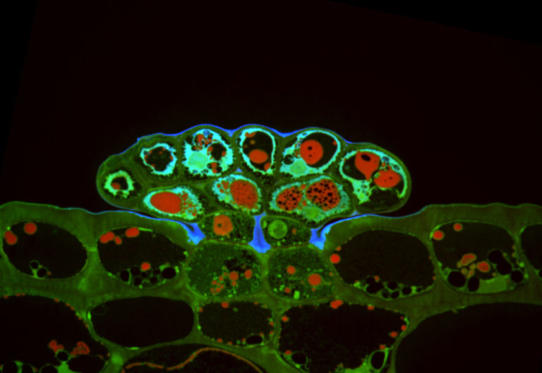
It provides new methods, protocols, or devices/equipment in BioImaging necessary to tackle the plant front science. Recent developments include a workflow for live meristem imaging, microfluidic devices for roots imaging, specific configuration for plant vertical fluorescence imaging to study tropism, sensors and growth monitoring, etc.