What’s up in multimodal imaging? The FBI CLEM day is renewed for a 2023 edition on March 13 at the Institut Pasteur in Paris.
This event is a great opportunity to discuss about multimodal imaging with expert presentations.
In addition to these talks, poster sessions will intersperse the day.
Registration is free but mandatory.
Please registrate before March 8, 2023 through the following link:
https://docs.google.com/forms/d/e/1FAIpQLScLGEUzzJmeJiZToKjak2myJwJeu3aLwLItt787doaGTKWrSA/viewform?vc=0&c=0&w=1&flr=0
Program down below
From February 6th to February 10th, France-BioImaging organised a group meeting on the project “FBI.data” in Bordeaux. For a week, participants focused on the architecture and the implementation of image data management tools. A user-friendly response to the challenges of never-ending data production.
New imaging technologies are very greedy in terms of image processing and data management. Beside the image itself, biological imaging generates a huge amount of metadata. The FBI.data project, one of the key missions of France-BioImaging, addresses the questions related to the computational analysis and handling of image data.
Speeding up the implementation of tools across the infrastructure
Although the distributed FBI.data team meets once per week, the FBI.data Sprint aims at only focusing on data management scenarii and accelerating the project. Two essential aspects have been discussed:
- First of all, the data management system architecture must be simple for them to be implemented across France-BioImaging nodes. It also has to be compatible with long-term data storage and of course, to be user-friendly – we want to keep it easy for our users!
- The second point is all about anticipating as many data management cases as possible. Running through all the needs of bioimaging experts and users, the team lists the specific features of each case and considers the perfect solutions for all of them.
Working for the FAIRisation of data
The FAIRisation of data for Open Science is an initiative fully endorsed by France-BioImaging. Meaning that data are Findable, Accessible, Interoperable and Reusable, the benefits for the bioimaging community are numerous. It improves transparency and reproducibility, enhances quality of results, accelerates scientific progress and method development and finally boosts collaboration within the scientific community.
OMERO, developed by the University of Dundee & Open Microscopy Environment teams, on which the FBI.data group is working, is one of the software making user data FAIR. Being a microscopy image data management decentralised platform, it helps organise, access and archive data. Besides, it combines image and metadata storage, a viewer and data analysis resources. Furthermore, OMERO is linked to the most valuable tools for bioimaging experts (ImageJ, Napari, QPath, etc.). And users can access their data from anywhere and keep them safe.
But much more has to be set up to have functional solutions: to ease user authentication and management, manage big data transfer, and have an adequate metadata scheme. Accompanying users is one of the mission of the FBI.data team, and the FBI.data Sprint is also the occasion to join efforts from the training working group led by the training mission officer Fabrice Cordelières (Bordeaux Imaging Center) to produce adequate training material on data management.
Sharing efforts and helping the community
The FBI.data working group is composed of:
- Perrine Paul-Gilloteaux, research engineer at MicroPICell core facility and FBI.data mission officer
- Emmanuel Faure, researcher at LIRMM (Laboratoire d’informatique, de robotique et de microélectronique de Montpellier) and FBI.data mission officer
- Guillaume Gay, research engineer at LIRMM, working full time for the FBI.data project
- Marc Mongy, research engineer at MicroPIcell, working full time for the FBI.data project
- Guillaume Maucort, research engineer in image analysis at Bordeaux Imaging Center (BIC)
- Jean-François Guillaume, research Engineer on the BIRD facility, Nantes and Pays de La Loire mesocenter (shared IT system engineer between FBI and Institut Français de Bioinformatique)
- Thierry Pecot, research engineer in image analysis at Rennes (Bretagne Loire Node)
- Further recruitments are on-going and will be reinforcing the team
By joining their skills and experience, they are working together on setting up tools and good practices for the management and FAIRisation of data inside France-BioImaging nodes but also for the entire bioimaging community. With this in mind, the project has collaboration with, among others, the Institut Français de Bioinformatique (IFB) and the Centre National de Ressources Biologiques Marines (EMBRC), and other infrastructure through the MUDIS4LS Equipex+ project. Moreover, the FBI.data project has an open GitLab, providing image data management codes in open source, and a blog with tutorials, recommendations and so much more!
Check the France-BioImaging OMERO web portal: https://omero-fbi.fr/
And its gitlab FBI data · GitLab (in2p3.fr)
Learn more about our missions and working packages: https://france-bioimaging.org/about/work-packages/
This is the first edition of our Summer School outside of France, going to South America in synchrony with the IEEE SPS-EMBS ISBI Conference in Colombia.
The spirit of our Summer School was established in French Brittany in 1994 (by Christian ROUX and Jean-Louis COATRIEUX). This Summer School has become a worldwide reference with international lecturers from 20 countries and accessible to young scientists from all around the world. Our Summer School is an open yet privileged place for exchanges and discussions on major on-going research and technologies. Informal and warm, we always select a location and design a program where ample time is dedicated to interactions between lecturers and students.
The Summer School is open to graduate students (MSc., PhD), post doctoral scientists, radiologists, biologists, researchers and engineers in industry.
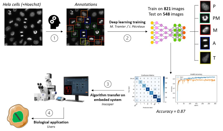
Being designed in response to imaging challenges, the Roboscope is the product of a collaboration between Marc Tramier’s team (FBI Bretagne-Loire node) with Julia Bonnet-Gélébart, research engineer, Jacques Pécréaux’s team of the Institut Génétique & Développement de Rennes (IGDR), and the Inscoper company, spin-off of the lab. This technology could become a great timesaver for fluorescence microscopy.
Allowing the automation of fluorescence microscope acquisitions, the Roboscope is an embedded technology based on a deep learning algorithm. To be precise, it is a predesigned event-driven acquisition (PEDA) based on a learning automatization of any cellular changes tracked by fluorescence. Catching rare and fast cellular events then becomes possible!
The use of the Roboscope would also save precious time of research, providing users with results without the need to stand by the microscope during acquisition. This technology goes beyond as they will be able to recover the data already classified and with only the specific points of illumination that they have previously triggered.
A broad range of applications
The teams have almost finished to develop an entire algorithm monitoring the cell cycle progression in mitosis. These events specific to the cellular division correspond to major challenges in the control and treatment of cancer progression (Kops, 2005). As the cell cycle study is needed to understand several biological processes helping the development of targeted drugs, the technology aims to monitor efficiently and automatically simple cell models through their division cycle.
And this is not its only benefit: this automatized fluorescence microscopy acquisition can be adapted in very diverse fields. From a cell cycle progression analysis to specific analysis, organelles, proteins and biological events can be tracked or activated within cells. A noteworthy advantage of the integrated device that – we hope – will be deployed widely in the future.
- Deadline: February 28th, 2023
- For users from a unit attached to the INSB who do not usually use Research Infrastructures’ services for their projects
- For access to technologies and related expertise
- For access to expertise in data analysis
- The scientific project must be a project in the process of being finalized
The field of Life Sciences has undergone major developments over the past two decades. The change of scales, both in spatial and temporal resolution, and the integration of data from a wide variety of sources, as induced by the development of technologies, have revolutionized the exploration of life. These technologies call for expensive investments and specific knowhow, carried out by highly qualified personnel, having led to the creation of common infrastructures such as research infrastructures (IRs) open to the entire scientific community.
The Institut des Sciences Biologiques (INSB) from Centre National de la Recherche Scientifique (CNRS) is launching the second edition of a call for projects to fund full access to the national infrastructures in health and biology “INBS Access to national research infrastructures”. This call aims to encourage teams to get a first access to the services offered by Research Infrastructures to help their research project. The objective is to demonstrate the significant impact of these services to improve the quality of their results (resolution, reproducibility, change of scale, etc.) or to help remove technological barriers. We also aim to augment the awareness of our teams to the cutting-edge technologies, methods and expertise offered by these national infrastructures.
Program description
Access to Research Infrastructures is open to the entire French and international scientific community, with contribution to the running costs of equipment being charged to the users on quote, after project feasibility confirmation by the infrastructure. The purpose of this program is to facilitate and finance access to these infrastructures. Target audience are INSB teams new to a technology or method offered by these infrastructures, seeking to validate the contribution that the infrastructures could make to their research topic by removing barriers that impair the completion or finalization of an ongoing project of the team. A new project is not eligible to this call. The list of eligible Research Infrastructures, to which the research team could apply, is available here: https://www.insb.cnrs.fr/fr/infrastructures-nationales. Other infrastructures may be considered depending on the proven needs of the project. Several types of access can be supported by this call for projects:
- Access to technologies and related expertise, upon quote from the infrastructures and confirmation of feasibility in 2023
- Access to expertise in data analysis, upon quote from the infrastructures and confirmation of feasibility in 2023
In addition to the costs of access to infrastructure, upon a quote emitted by the infrastructure at the second stage of the call, travel/mission costs, as well as consumables that are not covered by the costs of access to infrastructure, may be covered. The funding for each project will be in the range of 10 to 30 K€.
Instructions for submission
This is two-stage submission.
The first stage consists of a letter of intent, prepared by the scientific leader of the project, outlining the project in finalization in which the application fits and highlighting the barriers that could be removed by access to IRs. The novelty for the team of the usage of the technology and methods to which this application would give access has to be underlined. This letter of intent must also be signed by the director of the applicant’s unit. The identification of the infrastructures to access is possible at this stage but not mandatory and the application can focus on the technology and methods needed. An estimated timeline is however welcome.
The letter of intent (maximum of three pages including figures and references, in French or English), signed by the unit director, accompanied by a CV of the project leader (maximum of two pages) must be sent before Tuesday 28th of February 2023 to the following address: insb.ain@cnrs.fr. These proposals will be screened for eligibility criteria and selected projects will go to the second phase on 8th of March: The second phase consists of a consultation between the INSB and the national Research Infrastructures. This will lead to putting the applicant in contact with the infrastructures that are able to meet the expressed needs, in order to evaluate the feasibility and to establish a quote from their official invoicing cost models.
This stage will lead to the final submission of the project, including the requested budget, signed by the heads of the involved IR(s), no later than Wednesday 22th of march 2023. Funding decision will be sent to the scientific leader beginning of April.
Project eligibility and selection criteria
Access to the infrastructure must be fully implemented before 31/12/2023 (deadline for the engagements of funds by the teams). A scientific and financial report will be asked to the granted team in June 2024.
The scientific project must be a project in the process of being finalized. Access to infrastructure must unlock or accelerate the project. The start of a scientific project is not eligible for this call.
The access to the infrastructure technology or expertise required should be new to the scientific leader.
A project can call to different infrastructures.
In the first stage, budget is not expected, but the project should demonstrate its feasibility from the team side before the end of 2023 if access to the required technologies or expertise is granted (for example identified human resource from the team to undertake the project) and by providing an estimated timeline. The infrastructure(s) will confirm it in the second stage.
Project will be screened for scientific quality of the scientific leader and of the project in finalization, the interest of the targeted method and technology to finalize the project, the novelty of access to the infrastructure. In the second stage, the quote produced by the infrastructure will assess the feasibility and adequacy to the budget.
Eligible expenses are:
- Billing from the infrastructure based on quote presented at the second stage
- Mission fees to access the infrastructures
- Consumables needed to prepare samples for access if not provided by the infrastructures
MicroPICell core facility offers access and services to a broad range of bioimaging technology and expertise, specialized in cell and tissue imaging. Based in Nantes, the core facility is now certified ISO 9001 and NFX 50-900, demonstrating their investment in providing quality services to its users.
The ISO 9001 :2015 and NFX 50-900 :2016 standards ensures good practices in terms of organization and management of a life science core facility. These standards are focusing, among other criteria, on the ability to fulfill its scientific and technological research missions, to consistently provide products and services that meet customer and legal requirements and finally, aims to increase the satisfaction of its customers through the effective application of its management system.
A supported process
To implement the first standard, experts from MicroPICell core facility have been trained in the management of the required quality system. The core facility staff have then been supported, in close collaboration with the GIS Biogenouest, by the head of the IBISA quality mission in order to build and implement the quality system according to both standards. This “Groupement d’intérêt scientifique” also has a quality network, Iquare, to which the core facility participates in, to exchange, share and be advised in the implementation of the quality system. The NFX 50-900 :2016 standard has been applied on R&D projects such as the establishment of a digital histology center delivering deep learning data processing, smart microdissection or the imaging of thick samples in collaboration with the company Kaer Labs.
Guaranteeing quality to users
This double certification is a recognition of the core facility’s quality approach, and allows MicroPICell to guarantee to their industrial and academic users that the implemented tools and procedures meet the requirements of the standards. This quality approach facilitates day-to-day management and internal communication at the platform and with the various parties concerned. Above all, it is a way of continuously improving bioimaging core facilities and ensuring that the missions are carried out efficiently.
In coordination with the African BioImaging Consortium and Imaging Africa, two recently created initiatives, France-BioImaging wishes to extend its partnership with colleagues in Africa that have interest in using advanced microscopy approaches for their own research programs and projects. With this in mind and in the framework of the Horizon Europe Programme, France-BioImaging designed two calls to strengthen collaboration between African and French researchers in biology.
The call description and eligibility criteria are available here.
- Call 1: External Access
The application form to be completed by the applicant is available here and must be submitted through the submission form below.
- Call 2: Twinning program African BioImaging-France-BioImaging communities
The application form to be completed by the applicant is available here and must be submitted through the submission form below.
Deadline for submission of proposals: May 31st at 23h59 CET.
En coordination avec l’African BioImaging Consortium et Imaging Africa, deux initiatives récemment créées, France-BioImaging (FBI) souhaite étendre son partenariat avec des collègues en Afrique qui ont un intérêt à utiliser des approches avancées de microscopie pour leurs propres programmes et projets de recherche. Dans cette optique et dans le cadre du programme Horizon Europe, France-BioImaging a conçu deux appels pour renforcer la collaboration entre les chercheurs africains et français en biologie.
La description des appels et les critères d’éligibilité sont disponibles ici.
- Appel à projet 1 : Accès externe
Le formulaire à remplir est disponible ici et doit être soumis via le formulaire ci-dessous.
- Appel à projet 2 : Programme de jumelage/échanges des Communautés en BioImagerie Africaines et Françaises
Le formulaire à remplir est disponible ici et doit être soumis via le formulaire ci-dessous.
Date limite de soumission des projets: 31 mai à 23h59 CET.
Proposal submission
Call 1: External Access/Accès externeThis form is currently closed for submissions.
Call 2: Twinning program African BioImaging-France-BioImaging communities/Programme de jumelage/échanges des Communautés en BioImagerie Africaines et FrançaisesThis form is currently closed for submissions.
The next Euro-BioImaging User Forum will be taking place on 21.03.2023 from 2-5 pm CEST, focusing on the topic of “Cardiovascular Research”.
Euro-BioImaging is looking forward to featuring some of the excellent science supported by the work of EuBI nodes via presentations from your users. The presentations will be 15 min long and will include the opportunity to briefly introduce your Node. In addition the event will feature two keynote presentations.
Abstracts can be submitted here – https://forms.gle/XriAc5HTMiLAhACG6
The deadline for abstract submission is on February 6th.
All users who are working in the area of cardiovascular research are welcome ! The topic is broad as it includes vascular and cardiac development and/or regeneration, development of cardiovascular disease, inflammation in response to cardiovascular injury, etc. The users also do not have to be Euro-BioImaging users.
Euro-Bioimaging is looking forward to receiving your abstracts!
France BioImaging and all the French community aims to develop and promote innovative imaging technologies and methods. But microscopy images can also take an artistic, creative look and make the invisible world beautiful, allowing people to see the visual appeal of the life sciences.
We enjoyed the diversity of the images submitted with many different microscopy techniques, models and applications represented. A big thank you to all the participants!
The National Coordination Team and the Executive Board are proud to announce the winners of the FBI Image Contest 2022:
- 1st Place: Carole SIRET, Van de Pavert Team, Centre d’Immunologie de Marseille-Luminy
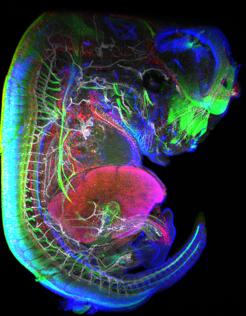
“Little Monster”
The embryonic formation of lymph nodes, small organs essential for the immune response, is now known. Using light sheet microscopy, scientists were able to determine the dynamics at work in this 13.5-day-old mouse embryo. In blue, the lymphoid cells (LTi), derived from the haematogenous endothelium, a specific tissue of the embryo. They pass into the liver where they proliferate before migrating through the body to give rise to lymph nodes. The 3D information obtained thus makes it possible to follow the interactions of lymph nodes with their environment, in particular with nerve cells, in green, and blood vessels, in white. The lymphatic endothelial cells and some macrophages are visible in red.
Lightsheet Microscopy
- 2nd Place: Magalie BENARD, Plateforme de Recherche en IMAgerie CEllulaire de Normandie (PRIMACEN), Research infrastructure HeRacLeS, Inserm US 51, CNRS UAR 2026,
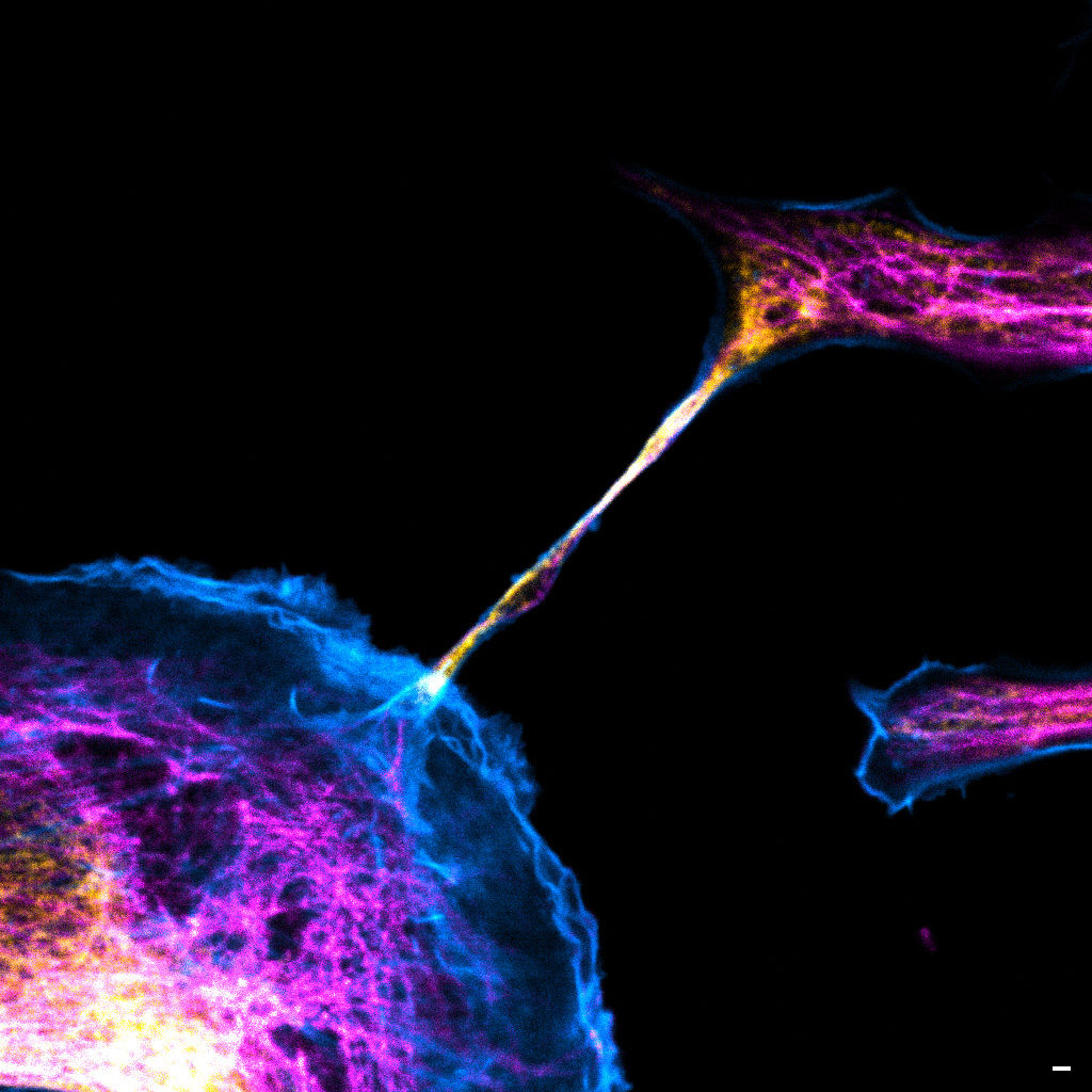
“The communication link with others”
Image of a cellular interconnection between two human tumor cells whose cytoskeleton has been labeled with anti-tubulin (ATTO-647N), anti-vimentin (AlexaFluor594) antibodies and with Phalloidin probe (AlexaFluor488). Scale bar 1µm.
Confocal microscopy
- 3rd Place: Frédéric FERCOQ, Parasites et Protistes Libres (PPL), Museum National d’Histoire Naturelle
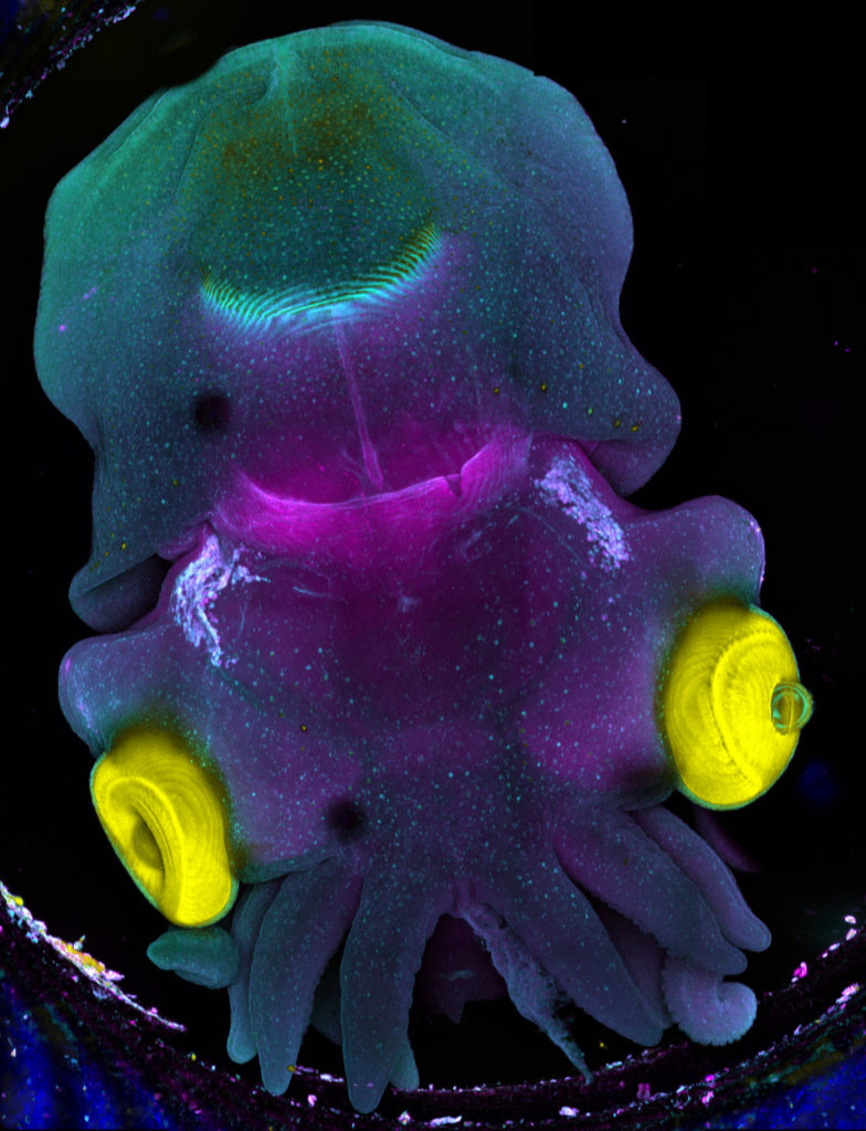
“Sepia”
Stage 25 cuttlefish embryo (Sepia officinalis) observed under a confocal microscope.
The cuttlefish was cleared and the tissue autofluorescence was captured.
This image was produced in collaboration with Laure BONNAUD-PONTICELLI and Luis MOLINA from the BOREA laboratory.
Confocal microscopy
Congratulations to the winners!
Explore all the images submitted here:


As stated in the Terms & Conditions of the contest, foreign participants non-affiliated to a French institution are featured in the gallery, but were not evaluated as part of the contest.
Since 2019, the “Cristal collectif” medal rewards teams supporting research with their technical expertise, the collective dimension, their innovation and outreach. Both nationally and internationally recognised, the Bordeaux Imaging Center (BIC) from the France-BioImaging node of Bordeaux received this award for providing access to innovating technologies and for the quality of its training. The BIC was commended for its investment in training, especially for its partnership with the International School of Neurosciences, a unique partnership in Europe. The CNRS also has particularly highlighted the core facility’s activities of research and development in implementing new techniques and image analysis. Among its achievements, the BIC has succeeded to optimize a homemade Lattice Light Sheet, which has the benefit of being a good compromise between resolution, acquisition speed, imaging depth and low phototoxicity.
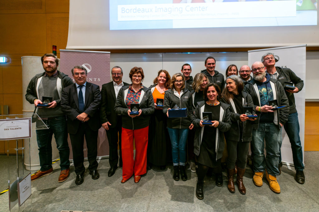
Laureates :
- Lysiane Brocard, Plant Unit manager
- Fabrice Cordelières, Image analysis manager
- Mathieu Ducros, R&D Lattice Light Sheet Microscopy manager
- Mónica Fernández Monreal, R&D CLEM manager
- Étienne Gontier, Electronic Unit manager
- Sabrina Lacomme, Transmission Electron Microscopy manager
- Florian Levet, R&D software manager
- Sébastien Marais, Confocal and Two-photon Microscopy manager
- Magali Mondin, Super-resolution Microscopy manager
- Melina Petrel, Cryo-preparation and immunomarking manager
- Christel Poujol, Photonic Unit manager
- Isabelle Svahn, Scanning Electron Microscopy manager
- Jérémie Teillon, Clarification and Light-Sheet Microscopy manager
More information: www.cnrs.fr/fr/talent/index
The Thematic Institute of molecular and structural biology of the French Alliance for life science (Aviesan) is programming a 2 days symposium dedicated to RNA biology and applications, that is a very active field with emerging topics.
Coding and non-coding RNAs are today central players in biology and medicine. RNA binding proteins act as modulators of RNAs functions, dysregulated RNA-protein interactions being linked to human pathologies, leading for example to misfolding or aggregations in membraneless organelles. In addition to human RNAs, bacterial RNAs and RNA viruses, are regarded as drug targets.
Epitranscriptomics, with modifications found in all RNAs and associated proteins have also come to the focus of intense research revealing their roles in many aspects of RNA biology and diseases. Finally, RNA medicines are actively developed for different therapeutic applications. RNA-based drugs are coming of age as a consequence of the improved knowledge of RNA biology combined with the development of sophisticated technologies.
This symposium will take place in Paris, on February 2-3 2023 end in Amphi Buffon, 15 Rue Hélène Brion, 75013 Paris. The meeting is built around different scientific sessions, namely RNA therapeutics, RNA/RNP biogenesis and structure, RNA modifications and new technological developments. The meeting is for structural, molecular and cellular biologists to have an overview of the latest results and concepts that are currently under development in the field.
A poster session will be organized and short talks will be selected.
Deadline for abstracts submission: January 12th, 2023.
Dowload the preliminary program
The organizing committee:
Edouard Bertrand, Christiane Branlant, Carine Giovannangeli, Chantal Pichon, Carine Tisne.