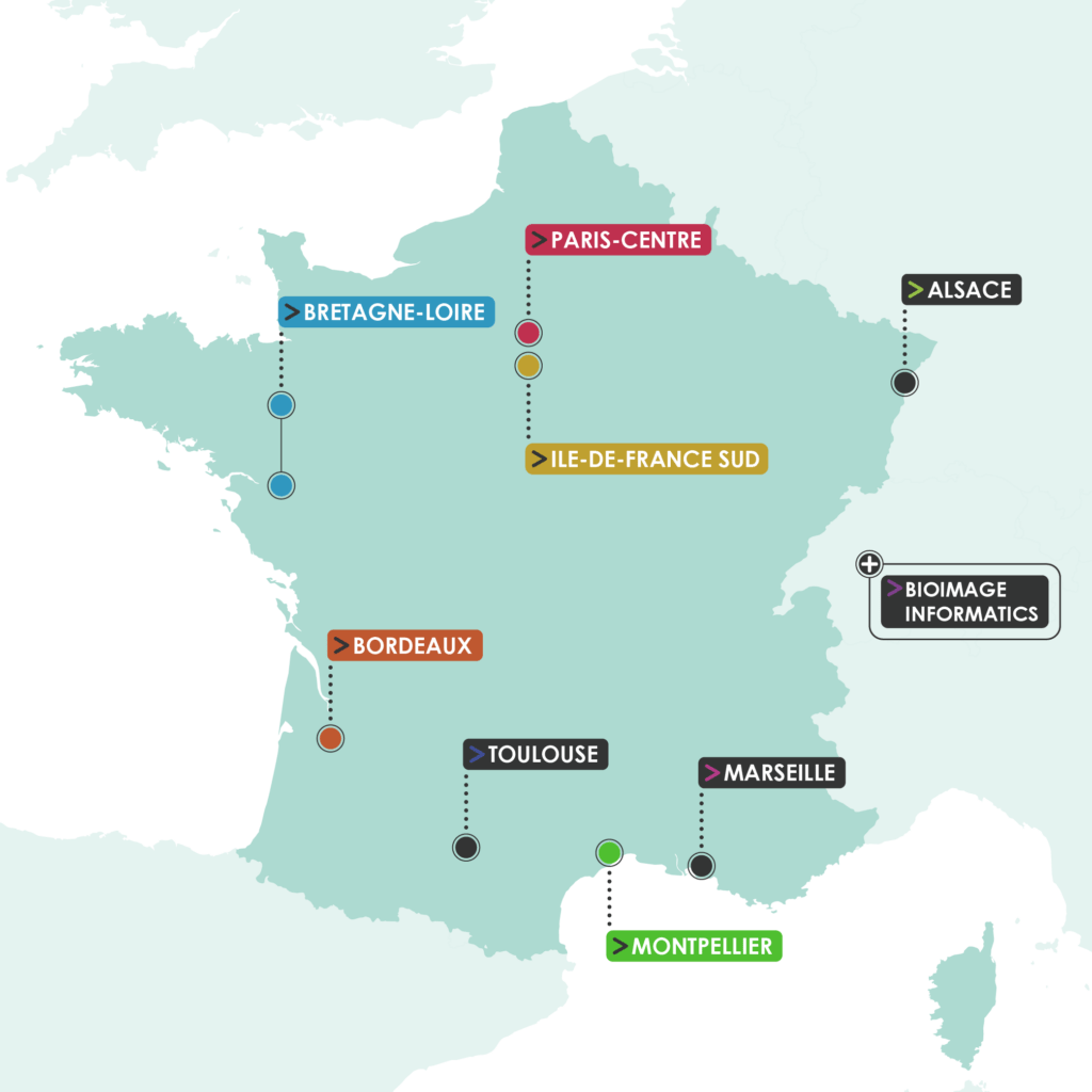
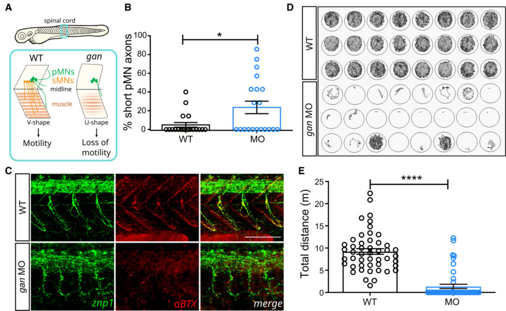
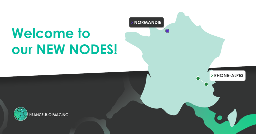
Euro-BioImaging is organizing a sixth online User Forum on Thursday, October 12, 2023, from 14:00-17:00 CET. The topic is “Understanding Plant Biology.” This event will highlight the importance of cutting-edge imaging technologies in support of plant biology and agroecology. We will showcase the specific expertise available at our Nodes across Europe through case studies presented in tandem with the research community.

Have a look on the program ! france-bioimaging.org/wp-content/uploads/2023/09/User_Forum_Plant-Imaging_October-12.pdf

The France-BioImaging Image Contest is back for its 5th edition!

This image contest is open to all within the imaging community: core facility staff and users, R&D labs teams and co-workers, students… Submit your best microscopy images for a chance to showcase your skills, research and creativity to the French bioimaging community and beyond, allowing people to see the visual appeal of the life sciences. Images from the contest will be featured on France-BioImaging communication tools, online and in print.

France-BioImaging and all the French community aims to develop and promote innovative imaging technologies and methods. But microscopy images can also take an artistic, creative look and make the invisible world beautiful.

We are all eager to see your work !

Prizes

1 to 3 images will be awarded depending on the quantity and quality of the entries submitted. France-BioImaging will cover the registration fees for one 2024 microscopy related event of the winners’ choice (FOM, ELMI, EMC, COMULIS conference, etc.).

Important: Only French or foreign participants affiliated to a French institution can enter the contest. Foreign participants non-affiliated to a French institution can submit images and will be featured in the gallery, but will not be evaluated as part of the contest.

Submission deadline: Friday, November 10th, 2023, 23h59 UTC+2. 

Click here to consult the terms and conditions of the contest. When you are ready, submit your entry by filling the form below. You can check out last edition’s entries for inspiration. One participant can submit several entries (up to 3).

(If you have any issues when submitting your image, please contact communication@france-bioimaging.org)

FBI – Image Contest 2023 Submission
First LAST
Will be used as caption in the FBI gallery. Please detail: the object observed, the microscopy technology used, the purpose of the research for which this image was produced (if applicable), and any other information you deem interesting or important.

Maximum file size: 104.86MB

Images in high resolution are preferred (1000×1000 pixels and above). Max file size: 300 MB. File format: JPG, PNG.
Precise credits and copyrights if required (if this field is left clear, the credits will be: Author name – Institution & Lab)
Remember that FBI will provide the registration fees if you win the contest, but will not cover the travel or accommodation costs to the event. FBI is not responsible for the pre-registration or the acceptance of your participation to the event (organizers’ prerogatives). As per the Terms & Conditions of the contest, foreign participants not affiliated to a French institution can submit images but will not be entered in the contest.
Sending

Discover last year’s submitted images on this following link: https://france-bioimaging.org/announcement/winning-images-fbi-image-contest-2022/

Our National Coordination visited 2 of our 9 nodes in one week: the Montpellier node and the Ile-de-France Sud node!

This visit was the perfect occasion to have a look on the broad range of technologies and expertise both nodes are providing in open access to their users. They cover a multitude of imaging techniques in electron microscopy, light microscopy and cytometry and working on various models going from the single cell to small animals. Besides, they both provide excellent services in – of course – imaging expertise, but also in sample preparation and bioimage analysis.

Furthermore, we discussed about the future of the nodes and their promising perspectives. The core facilities and R&D teams are working towards novel innovative imaging technologies that, we hope, will be in open access very soon!

Thank you to all of the following core facilities and R&D teams for the warm welcome:

We were happy to meet everyone! Until next time!

Photos from our visit at the Montpellier node:

Photos from our visit at the Ile-de-France Sud node:

We are happy to announce that our 1st Africa-France Joint Initiative for Biological Imaging calls, in coordination with the African BioImaging Consortium and Imaging Africa, is funding 11 projects!

As a reminder, these two calls (“External access” and “Twinning exchange”) have been primarily designed to strengthen collaboration between African and French researchers and engineers in all fields in biology, health and agro-ecology, where the contribution of the rapidly expanding technologies of digital imaging has become essential. The ambition of these calls is in line with the anticipation of bilateral research funding programs between Europe and Africa in the framework of the Horizon Europe Program.

Multiple scientists from the entire African continent have responded to the calls, covering 8 countries: Senegal, Togo, Nigeria, Ivory Coast, Burkina Faso, Morocco, Uganda and South Africa. Besides, we are very glad that 5 of our nodes (Paris-Centre, Montpellier, Bordeaux, Ile-de-France Sud and Bretagne-Loire) are going to work in the framework of the Africa-France Joint Initiative for Biological Imaging.

Finally, the projects have been selected for the need to access imaging resources in response to key challenges that African researchers are facing. Among them, scientists tackle issues in several topics going from infectious diseases to marine biology, but also cancer research, plant biology and climate change

Here are the selected projects

Call 1 “External Access”:

  • Production of digitised teaching tools (anatomical and histological sections) for bachelor’s and master’s studentsAliou NDIAYE, Département de Biologie végétale, Université Cheikh Anta DIOP, Dakar, Sénégal – with Montpellier node

  • VCAPE – Effects of climatic variability on the anatomical properties of Pterocarpus erinaceus Poir. wood in TogoKossi Novinyo SEGLA, Laboratoire de recherche forestière, Université de Lomé, Togo – with Montpellier node

  • TRITUICAFH – The role of imaging techniques to understand the initial cellular aspects on functionalized hydrogels David Olubiyi OBADA, Multifunctional Materials Laboratory, Department of Mechanical Engineering, Ahmadu Bello University, Nigeria – with Paris-Centre node

  • AfluBio – Botanical quality of plant raw materials used in African pharmacopoeia products: histological study and localisation of active biomolecules by autofluorescenceAkoua Clémentine YAO, Plateforme Microscopie électronique et Bioproductions, Centre Suisse de recherches Scientifiques en Côte d’Ivoire (CSRS) / Université NANGUI ABROGOUA, Côte d’Ivoire – with Bordeaux node

  • Ultrastructural Evaluation of Tramadol-induced Testicular Toxicity in Wistar RatsAdebanji AKINGBABE, Department of Anatomy EKSU, Ekiti State University, Nigeria – with Ile-de-France Sud node

  • MeCap – Histological analysis of Carica papaya roots infested by Meloidogyne javanicaLaëtitia COULIBALY, Laboratoire Mixte International (LMI) crée entre l’Institut de l‘Environnement et de Recherches Agricoles (INERA) et l’Institut de Recherche pour le Développement (IRD) à Bobo Dioulasso, Université Joseph KI-ZERBO, Ouagadougou, Burkina Faso – with Montpellier node

  • OTOSHAPE – Study of otolith shape as a tool for determining the structure of sole (Cynoglossus senegalensis) stocks using ImageJ: towards sustainable management of fishery resourcesKhady DIOUF, Laboratoire de Biologie marine, Institut fondamental d’Afrique noire cheikh Anta Diop / Université Cheikh Anta Diop de Dakar, Senegal – with Bretagne-Loire node

  • Study of the interface of native endomycorrhizae using high-resolution microanalysis methodsMalik NDIAYE, Laboratoire de biotechnologies végétales, UCAD, Senegal – with Montpellier node

Call 2 “Twinning Exchange”:

  • Twinning between the electron microscopy core facility of the Centre National de la Recherche Scientifique et Technique (CNRST) of Rabat (Morocco) and the Plateforme d’imagerie cellulaire et tissulaire (PICT) from the Institut Curie ParisMohamed EL BOUJI, Centre National pour la Recherche Scientifique et Technique (CNRST), Laboratoire de microscopie électronique en transmission – division UATRS, Rabat, Morocco – with Paris-Centre node

  • Africa-France Open BioImaging Initiative (AFOBII)William WASSWA, Mbarara University of Science and Technology (MUST), Medical Imaging and Artificial Intelligence Lab (MIAL), Mbarara, Uganda – with Paris-Centre node

  • Exchange of expertise in light sheet microscopy (lattice and others) and serial block face electron microscopy. Comparison of strategies to prepare challenging samples and EDX in biological samplesMadelaine FRAZENBURG, Microscopy Unit, University of Stellenbosch, Cape Town, South Africa – with Paris-Centre node

Congratulations to all the laureates! We are eager to welcome you on our core facilities!

Stay tuned to know more about these project’s unfolding!

This school aims at training students and researchers to master fluorescent markers used in advanced fluorescence bioimaging: their diversity, how they actually work and what are their current development.

It will be organized at the Ecole de Physique des Houches, in the french Alps, from March 17th to 22th, 2024.

Objectives of the school :

Fluorescence microscopy techniques play an essential role in biology and medicine. A very rapidly developing field concerns nanoscale imaging (“nanoscopy”), which today ties together different fields of biology such as structural and cell biology. Similarly, “single molecule” imaging and spectroscopy techniques are in rapid development and play a central role in life science research. All fluorescence microscopy methods are based on labelling samples with appropriate markers: fluorescent proteins, organic fluorophores or nanoparticles. All these type of markers are characterized by very complex photophysical behaviors, leading to blinking, chromatic conversion, photoreactivity, etc… However, nanoscopy and single molecule spectroscopy or imaging, specifically rely on the ability to understand and control these properties.

The school we offer is motivated by the fact that more and more laboratories around the world are committed to implementing advanced microscopy techniques, especially super-resolution, with an increasing number of dedicated platforms. However, there is a lack of overall knowledge about the selection of fluorescent markers, their properties and mechanisms, and the type of artifacts they can create. The school’s objective, through interdisciplinary teaching between physics, chemistry and biology, is to overcome this gap by transferring knowledge to the participants’ laboratories. Our school also aims to make participants aware of new developments in the field in order to prepare them to become major players in the breakthroughs that may result from them.

Invited Teachers

Confirmed teachers for the 2024 edition

Dominique Bourgeois, Institut de Biologie Structurale, CNRS, Grenoble, France

Claire Deo, EMBL Heidelberg, Germany

Ulrike Endesfelder, Bonn University, Germany

Viktorija Glembockyte, LMU Munchen Germany

Mike Heilemann, Goethe Universitat Frankfurt, Germany

Ludovic Jullien, Sorbonne University and ENS Paris, France

Doory Kim, Hanyang University, Korea

Andrey Klymchenko, CNRS, Université de Strasbourg, France

Emmanuel Margeat, Centre de Biologie Structurale, CNRS, Montpellier, France

Ivana Nikic-Spiegel, U. Tuebingen, Germany

Franck Riquet, Lille University, France

Markus Sauer, U. Würzburg, Germany

Nathan Shaner, UCSD, USA

Alison Tebo, HHMI Janelia Research campus, USA

Organizers

Dominique Bourgeois, Institut de Biologie Structurale, Grenoble, France

Claire Deo, EMBL Heidelberg, Germany

Ulrike Endesfelder, Bonn University, Germany

Emmanuel Margeat, Centre de Biologie Structurale, Montpellier, France

More information and registration on: https://fluorescenceleshouches.wordpress.com/

We are happy to announce our 8th France-BioImaging Annual Meeting! Happening on December 13th and 14th 2023, this year’s edition will be hosted by our new Toulouse node at the Centre de Biologie Intégrative (Toulouse, France).

The Annual meeting will highlight France-BioImaging’s development and perspectives. Imaging scientists and users from the infrastructure’s nodes will present their key projects and demonstrate how they have benefited from France-BioImaging and its community.

More information about the program and the registration coming soon!

The “Imaging life: the future” symposium will take place October 17-19 at the Corum congress centre of Montpellier (France).

Over the last 20 years, biological imaging has undergone a revolution in the spatial and temporal resolution of images, in the diversity and power of synthetic or genetically-encoded fluorescent probes and in image analysis. These advances have been transformative for many fields of biological research.

What does the next decade hold for this field?

This 2.5-day international symposium aims to foresee how future developments in light/electron/soft X-ray/multimodal microscopy and image analysis will further transform research in the biology of prokaryotes and eukaryotes. 

Seven major fields will be covered (image analysis, cancer biology, plant biology, 3D genome architecture and transcription, neuroscience, cell and development biology, infection and immunity). Two plenary lectures will complete the programme, one on imaging and image analysis at the other end of the scale spectrum, in astrophysics, the other will open up to the numerical modelling of biological processes.

Very attractive early bird registration prices are proposed until July 31st, 2023. Abstracts should be submitted by July 31st if you would like to give an oral presentation. By August 31st if you are only applying for a poster. 

More information and the registration pages can be found on the meeting’s website: https://www.atoutcom.com/futureimaging/

High-content screening (HCS) is a technology used in drug discovery and research to analyze cell phenotype. HCS has created new opportunities for studying biological phenomena as it combines high-throughput screening methods with automated microscopy on microplate format. Using automated cell manipulations and microscopy platforms, it is possible to easily screen the effects of selected drugs on the model’s phenotype. This technology, available at France-BioImaging at Montpellier Ressources Imaging (MRI), has been used in a recent study identifying therapeutic drugs for a fatal neurodegenerative disorder, the Giant Axonal Neuropathy (GAN). Benoît Bordignon and Cédric Hassen-Khodja, screening experts at the MRI and co-authors in this recent publication, tell you more about the reasons why the HCS technology was essential!

Get routinely quick acquisition of numerous samples with HCS

As mentioned before, High-Content Screening uses automated cell manipulations and automated microscopy platforms to easily visualize the modification of the phenotype. In other words, it allows image acquisitions and analysis of a large number of samples, and collects data of biological parameters of interest directly for statistical analysis. Image acquisition and analysis are done in batches and rapidly, which limit potential bias as the process will be identical from one well to another. A huge advantage that prevents issues and saves time!

Imaging can help therapeutic research

Giant axonal neuropathy (GAN) is a fatal neurodegenerative disorder for which there is currently no treatment. Affecting the nervous system, GAN starts in infancy with motor deficits that rapidly evolve toward total loss of ambulation. Using the gan zebrafish model that reproduces the loss of motility as seen in patients, scientists conducted the first pharmacological screening for the GAN pathology. They combined behavioral, in silico, and high-content imaging analyses to identify drugs restoring locomotion, axonal outgrowth, and stabilizing neuromuscular junctions in the gan zebrafish. 

This is when imaging becomes essential! To validate at cellular level the last 103 selected hits, they have developed a custom-made High-Content Screening method to check if the phenotype was also restored at the cellular level in gan zebrafish, in particular on neuromuscular junction (NMJ) and axonal function. The postsynaptic nature of the drug’s cellular targets provides direct evidence for the pivotal role the neuromuscular junction holds in the restoration of motility. 

The results of this study identify the first drug candidates that can now be integrated in a repositioning approach to fasten therapy for the GAN disease.

Scientists working on this study therefore created an automated image analysis protocol using HCS to quantify neuromuscular junctions’ number/size and measure axon length in treated gan zebrafish. Thus, image data treatment had a central place in this study. And this is just an example! This novel high-content imaging methodology represents a useful automated procedure that can be transferred to other neuromuscular conditions for mechanistic studies and drug screening.

Thanks to Benoît Bordignon and Cédric Hassen-Khodja for providing very helpful insights of the study!

The gan zebrafish mimics the loss of motility described in GAN patients
A – Schematic of the behavioral and cellular defects described in the gan zebrafish model. Underlying the loss of motility in the gan zebrafish, the architecture of the spinal cord is remodeled with shortening and/or absence of MN axons (in green, primary MNs; in red, secondary MNs) and loss of neuromuscular junctions (NMJ). The neuromuscular phenotype is accompanied by a change in the shape of myofibers, which adapt a “U-shape” instead of a normal “V-shape.”
B – The percentage of shorter pMN axons (< 70 μm) per fish is significantly higher in gan morphants (n = 20) than in noninjected WT (n = 20) at 48 hpf.
C – Representative images for the neuromuscular junctions (znp1: green; αBTX: α-bungarotoxin: red) in WT and gan morphants at 48 hpf. Note the shorter pMN axons and sparse AChR clusters in gan morphants.
D – Representation of the cumulative tracking of the spontaneous locomotion of 5-day-old larvae for 1 h, in noninjected and MO-injected animals.
E – Quantitative measures of the traveled distance (m: meter) show total loss of locomotion in 79.2% of gan morphant; n = 48 (WT), n = 48 (MO).

Get access to one of our services!

You need HCS or another imaging technology or expertise that France-BioImaging provides? To get open access, please login via Euro-BioImaging website! You just have to choose the technology you want to use, then submit your proposal. All applications will be processed by the Euro-BioImaging Hub in close relation with France-BioImaging. And of course, all scientists regardless of their affiliation, area of expertise or field of activity can benefit from open access services! Users whose projects will be validated by Euro-BioImaging will benefit from a waiver for the access cost on France-BioImaging core facilities (france-bioimaging.org/access)

Lescouzères, L., Hassen‐Khodja, C., Baudot, A., Bordignon, B., & Bomont, P. (2023). A multilevel screening pipeline in zebrafish identifies therapeutic drugs for GAN. EMBO Molecular Medicine, e16267. https://doi.org/10.15252/emmm.202216267

COMULISglobe aims at consolidating and extending a collaborative and innovative network that promotes MultiModal Imaging and analysis across scales (MMI) from biological research to clinical diagnostics. To this aim, they will help bridge the gap between biological and clinical imaging, identify, fund, and showcase novel multimodal pipelines, and develop, evaluate, and publish correlation software through dedicated networking activities. COMULIS have two calls and support schemes open:

1. Global Showcases: This call can fund up to 3 showcase projects (with up to 12k Euros each) that tackle an outstanding biomedical research question using two or more imaging modalities across countries. Eligible expenses are travel costs for lab exchanges, consumables, instrumentation access, supplies, or sample mailing fees. Please send your 2-page applications to andreas.walter@hs-aalen.de by July 23rd, 2023. Projects need to take place between September 1st, 2023 and April, 30th,2025.
Find more info here: https://www.comulis.eu/comulisglobeshowcase-projects

2. Lab Exchanges/Mobility Grants: These lab exchange fellowships are aimed at supporting individual mobility, strengthening the existing networks and fostering collaborations by allowing PhD students, Early Career Investigators (ECIs), facility staff and experienced researchers in the field of MultiModal Imaging to visit an institution, laboratory or industry in another country. The lab exchange will be offered on regular competitive calls throughout the duration of the CZI grant.
Find more info here: https://www.comulis.eu/comulisglobe-lab-exchange


Expansion microscopy (ExM) is a relatively new super-resolution method based in the isotropic dilatation of the biological sample in order to overcome the diffraction limit of conventional microscopy. Since its development in 2014, many laboratories have been implementing and adapting the technique to their needs, and the biological applications of ExM grow exponentially.

The ExM working group from the French fluorescence microscopy network (RTmfm), with the support of France-BioImaging, is organizing a workshop on ExM at Bordeaux on October 2, 2023. During the workshop, we will cover different aspects of ExM protocols with experts in biochemistry and biology, and we will present examples of biological applications.

Invited speakers:

Marine Laporte (Institut NeuroMyoGène, Université Claude Bernard, Lyon).

Sven Truckenbrodt (E11 Bio, Alameda, California, US).

Maxence Wisztorski (University of Lille, Inserm, CHU Lille, U1192).

You can register here to enjoy the ExM community during a day, and to show you work with a poster or a short talk. We have limited seats!!! To send abstracts please contact monica.fernandez-monreal@u-bordeaux.fr and jim.dompierre@ibgc.cnrs.fr.

Deadline for applications is July 15th 2023.

Preliminary program: download here.

Atomic Force Microscopy (AFM) is a scanning probe microscopy technique that relies on measuring the interaction forces between a sharp tip and the surface of a sample to generate high-resolution images of its surface features and mechanical properties. A very broad range of sample types can be imaged with this technique at a very high resolution – at sub-nanometer level for some of them! Discover the AFM at the Montpellier node of France-BioImaging with Christine Doucet from Integrative Biophysics of Membranes team of the Centre de Biochimie Structurale.

Quickly visualize dynamic biological processes with High-Speed AFM

AFM provides images in physiological conditions, in liquid, over a length-scale ranging from few nanometers (single biomolecules) to tens of micrometers (living cells). In fact, the resolution depends on the tip radius and sample properties. For some of them, you can routinely obtain a nanometer lateral resolution and Angstrom axial resolution!

You want a video-rate version of the biological samples you are imaging? The High-Speed AFM, permits the acquisition of movies at approximately 10 images per second, enabling the visualization at nanoscale of dynamic biological processes involving biomolecular interactions, diffusion or conformational changes. It delivers nanometric resolved images typically at the same speed as conventional fluorescence microscopes!

Unravel the chemical information of your sample by combining AFM with…

AFM in ambient conditions and in liquids has a key limitation in that it does not directly provide chemical information about the sample being imaged. However, this limitation can be overcome by combining AFM with other techniques to obtain additional information about the sample’s composition. 

One commonly used technique in correlation with AFM is fluorescence microscopy. This combined approach of fluorescence labeling and AFM provides valuable insights into the chemical and biological properties of the sample. It was recently used on the Montpellier custom-made correlative AFM / fluorescence setup to observe the sublocalization of proteins in HIV-1 budding sites 1. They also used it to unambiguously attribute some unexpected configurations of the nucleoplasmic sides of Nuclear Pore Complexes 2. In these two cases, fluorescently-labeled proteins were imaged by dSTORM (direct STochastic Optical Reconstruction Microscopy). Of note, the lateral resolution of dSTORM and AFM are both in the 20 nm range with such samples, which makes their combination ideal!

In addition to fluorescence microscopy, AFM can also be correlated with other complementary techniques to obtain chemical information about the sample, such as Raman spectroscopy, Infrared Spectroscopy, X-Ray spectroscopy, microscopy and scattering.

Learn more about AFM applications

Here are 2 studies where Atomic Force Microscopy were essential: 

  • Structure and mechanics of the human nuclear pore complex basket using correlative AFM-fluorescence superresolution microscopy

Combining mechanical and superresolution measurements to reveal the plasticity of the Nuclear Pore Complexes

Nuclear pore complexes (NPCs) are the only gateways between the nucleus and cytoplasm in eukaryotic cells, facilitating the transport of selected cargoes of size from a few up to hundred nanometers. This versatility implies an important pore plasticity. Here, by combining atomic force microscopy (AFM) and single molecule localization microscopy (SMLM), a group led by France-BioImaging R&D team members Christine Doucet and Pierre Emmanuel Milhiet revealed that the NPC basket is very soft and explores a large conformational landscape: apart from its canonical basket shape, it dives into the central pore channel or opens, highlighting  how this structure can adapt, and let morphologically diverse cargoes shuttle through NPCs.

Vial et al., Nanoscale, 15, 5756-5770 (2023) 

  • The structure of pathogenic huntingtin exon 1 defines the bases of its aggregation propensity

Structural Biology meets Correlative Imaging

Huntington’s disease is a neurodegenerative disorder caused by an extended polyglutamine (poly-Q) tract in huntingtin. Here, using NMR, the team of Pau Bernado (CBS Montpellier) demonstrated that this poly-Q tract adopts long α-helical conformations. By adding correlative Atomic Force Microscopy and Fluorescence Microscopy data obtained in the FranceBioImaging facility PIBBS in Montpellier, they could demonstrate that the stability of this α-helix is a stronger signature than the number of glutamines, in defining the aggregation kinetics and the structure of the resulting fibrils, potentially linked to their pathogenicity.

Elena Real et al., Nature Structural & Molecular Biology, 30, 309–320 (2023)

How to use Atomic Force Microscopy at France-BioImaging?

Atomic Force Microscopy is open to collaborations under Proof-of-concept studies via Euro-BioImaging webportal (www.eurobioimaging.eu/service)! At the Montpellier node of France-BioImaging, you will be in contact with Dr Luca Costa (costa@cbs.cnrs.fr) with whom you will talk about the feasibility and the inherent experimental constraints linked to the technique. The collaboration procedure is discussed on a case-by-case basis, depending on the duration and technicity of the required experiments. Feel free to submit your project!

Thanks to Christine Doucet and Emmanuel Margeat for providing helpful information!

1. Dahmane, S., Doucet, C., Le Gall, A., Chamontin, C., Dosset, P., Murcy, F., Fernandez, L., Salas, D., Rubinstein, E., Mougel, M., et al. (2019). Nanoscale organization of tetraspanins during HIV-1 budding by correlative dSTORM/AFM. Nanoscale 11, 6036–6044.

2. Vial, A., Costa, L., Dosset, P., Rosso, P., Boutières, G., Faklaris, O., Haschke, H., Milhiet, P.-E., and Doucet, C.M. (2023). Structure and mechanics of the human nuclear pore complex basket using correlative AFM-fluorescence superresolution microscopy. Nanoscale 15, 5756–5770.

  FBI opens a call for the recruitment of its next Deputy Director for International Affairs

Deadline: June 1st, 2023

Mission

The Deputy Director for International Affairs will oversee a key strategic mission: develop France-BioImaging’s international activities. This includes enabling France-BioImaging to expand and enhance its engagement in the international community and within targeted communities, with a focus on Europe and Africa. The three primary goals for this position are to increase FBI’s participation in Horizon Europe programme; aid in developing FBI’s approach to international outreach; and build a long-term engagement with FBI’s international partners. The Deputy Director will work closely with the Manager of External Affairs to tailor specific cooperation activities, prepare strategic decision-making and prioritize needs for future international initiatives.

Organisation

France-BioImaging is a national research infrastructure distributed throughout France that provides researchers with access to the latest innovations in biological imaging and aims to accelerate the transfer of technological and methodological innovations in biological imaging to the 22 platforms that constitute the infrastructure. The Deputy Director for International Affairs will contribute to the development and strengthening of France-Bioimaging international relations and outreach activities.

The Deputy Director for International Affairs will work within the national coordination of the France-BioImaging infrastructure. He/she will work within a team distributed between Montpellier and Bordeaux, composed of 4 people: the Scientific Director and the Deputy Director for International Affairs, the Internal Affairs Manager, and the External Affairs Manager.

Mandate of the Deputy Director for European Affairs

  • Help to develop and implement the FBI’s international cooperation activities
  • Promote and organize a watch on the existing European and/or international programmes/funding
    devices in the fields of interest, and promote and organize the diffusion of information on international cooperation within FBI
  • Advise the management team in the area of international cooperation in relation to the infrastructure’s scientific policy, synthesize and prepare elements to assist in decision-making
  • Advise the management team in the field of research development in relation to the institution’s scientific policy
  • Represent the infrastructure and to lead the relations with the partners, in particular with the Euro-BioIamging ERIC, as CNRS representative of the infrastructure within the European Euro-BioImaging Infrastructure Board.
  • Promote the scientific and structuring activities of the infrastructure at the international level, to seek out and federate potential partners, to initiate and manage international cooperation programs, and to provide expertise and advice.
  • Organize a consultation and a prospective reflection on the development of new initiatives and on the actions to be carried out (i.e. preparation of the participation of the infrastructure in the European calls for projects of the Horizon Europe framework program)

Time commitment: 20% of his overall working time

Mandate duration: 5 years

More details and information in the pdf below 

Entry into function is planned as soon as possible, September 1st, 2023 at the latest.

If you are interested,
please send a short CV and a motivation letter (2 pages) at direction@france-bioimaging.org
BEFORE the 1st of June