3D Res/volution event – FIB-SEM and Lattice SIM Elyra 7
▽ Scroll down
Author: alban.belloir
Imagerie-Gif core facility, from our Ile-de-France Sud node, is pleased to announce the acquisition of a Scanning Ion Beam Electron Microscope (FIB-SEM) and a Lattice Structured Illumination Microscope (SIM) Elyra 7. For the occasion, the core facility is organizing “3D Res/volution“, a scientific event on high-resolution 3D imaging on December 15, 2022 from 2:00 pm to 5:00 pm at B21 amphitheatre. This event will be a great opportunity to introduce to you the possibilities of these 2 new systems available at Imagerie-Gif.
Initiated a few years ago, the Inria-IPL-NAVISCOPE (“Image guided NAvigation and Visualization data sets in live cell imaging and microscopy”) project aims at overcoming challenges of bioimaging observation. Virtual and augmented reality could become the new way to visualize and analyze microscope image renders.
Despite incredible progresses in microscopy, imaging biomolecular dynamics in cells remains a challenge. A lack of sensitivity, limited recording speed, photobleaching and phototoxicity associated have restrained, for a long time, our capacity to study biomolecules in their natural environments. As microscopy image is commonly observed on 2D screens, it can narrow human capacities to grasp volumetric, complex, and discrete biological dynamics. Following new modes of visualization including virtual reality (VR)/augmented reality (AR) approaches, the NAVISCOPE project allows more accurate analysis and exploration of large time series of volumetric images, such as those produced by the latest 3D + time fluorescence microscopy.
Why should cell biologists be interested in this project?
The project to which 4 FBI-teams from the BI-IPDM node participate, aims at engineering a technology made with and for biologists. For VR/AR approaches to be adopted by the broader bioimaging community, it is, indeed, important that they are evaluated by the biologists, on their own datasets.
The potentials of VR/AR technologies for scientists are numerous: navigating into multidimensional, large data sets with another view angle or perception, interacting with these data especially by selecting subregions, quantifying features of interests, etc. New VR/AR approaches also provide specific quantification tools to show distances, angles, counting, local density, and histogram profiler or include a selection of regions of interest for further analysis such as the 3D Timelines. Moreover, because communication with analysis software coded in Java or Python is now integrated, more post-treatment analysis is possible on selected features, providing a multifaceted and accessible tool for biologists.
A promising future ahead
In practice, immersion of the user within 3D + time microscopy data still represents an acculturation challenge for the concerned community. Thus, to promote a broader adoption of these approaches by biologists, further dialogue is needed between the bioimaging community and the VR&AR developers. Nonetheless, future innovation can already be foreseen as there are multiple way to upgrade this technology. For example, using eye-tracking (Günther et al., 2020) or haptic interfaces (Petit et al., 2020) can improve human perception by providing local sensations, which would improve the selection of responses in a 3D + time space. Besides, a better integration of multiple channels with high pixel resolution or the addition of vector representations could add information about the orientation, movement of molecules or organization of structures such as cytoskeleton elements or membrane lipids. The prospects initiated by the NAVISCOPE projects are, as mentioned above, endless and could be a technology that reshapes the way we see biology at the hearth.
Ranging in size from 20 microns to 1 milimetre, Meiofauna is a crucial link between micro- and macro- marine ecosystems. Valentin Foulon, a research engineer in marine biology, is part of the Blue Revolution program that aims to develop a taxonomic identification protocol for these tiny creatures. He approached the Bretagne-Loire node of France-BioImaging, in Nantes, where a Single Plane Illumination Microscopy (SPIM) light-sheet available in open access helped him to further reach his goal.
Valentin Foulon loves imaging and microscopy – and he loves meiofauna. He works on the Blue Revolution program, whose goal is to accelerate the taxonomic identification of meiofauna using artificial intelligence algorithms. But while the confocal-microscopy-based protocol worked well for small plankton, the researchers quickly realized that it had to be adapted to work with meiofauna.
Choosing the right microscopy approach
Identification of Meiofauna is usually operated manually on a classical microscope, which is “incredibly challenging” says Valentin. “And we only have one focal plane. To see important details, we need a 3D view of organisms.” explains Valentin. “That’s why we got curious about light-sheet imaging, because we would be able to see the organisms in 3D.” As France-BioImaging provides this type of microscopy in open-access in Nantes, “we clearly saw an opportunity there,” explains Valentin. His project proposal, which was submitted to the Euro-BioImaging pilot User Access fund, was fully funded by France-BioImaging, in a generous initiative introduced by France-BioImaging to fund all user projects to its facility received by Euro-BioImaging for the pilot User Access fund.
“I had never tried light sheet microscopy before,” says Valentin, “And in Nantes, the lab didn’t have experience with marine organisms. Together, we found a compromise between different imaging parameters such as the resolution or the acquisition time.” Once the set-up was fine-tuned, he sent his samples, and the engineer at the lab did the imaging work. “It was a very nice experience,” expresses Valentin. “I discovered a new technique, which is always enriching. I also think the lab was happy to work with a new sample type. And finally, we validated our proof-of-concept, proving that it is possible to image these organisms in 3D.
Image data management
But the project doesn’t end there. “The next step is data management. We imaged between 200-300 different organisms. It’s not enough for automatic classification, but nevertheless, we have 6-7 TB of data. Now we must process the data for this classification with machine learning. It’s an essential part of the project, to make the link between sample imaging, and data processing. We are working to close that gap.”
“The outcome of this project is to validate a proof-of-concept, from the sample collection to the image classification. With this new taxonomic identification protocol, we will be one step forward in the comprehension of meiofauna, these mysterious marine organisms, who may hold the key to understanding the impact of human activity on marine ecosystems,” concludes Valentin.
Thank you Marianna Childress, communication officer of Euro-BioImaging, for the original article.
As the 2022 edition of the France-BioImaging Image Contest admissions is coming to an end, we wanted to highlight our previous winners and their projects. Here is a quick throwback to our 2021 winners.
Before getting to the heart of the matter, we want to remind you that you still have time (before November 11th) to submit your best images and try to win your registration fees for one 2023 microscopy-related event! Please make sure you upload your images on the following link:
Last year, we enjoyed the winning images submitted for their artistic take and their quality. Thanks to Léna Meneux, Eunice HoYee Chan, Camille Boutin et Nicolas Brouilly for their beautiful images!
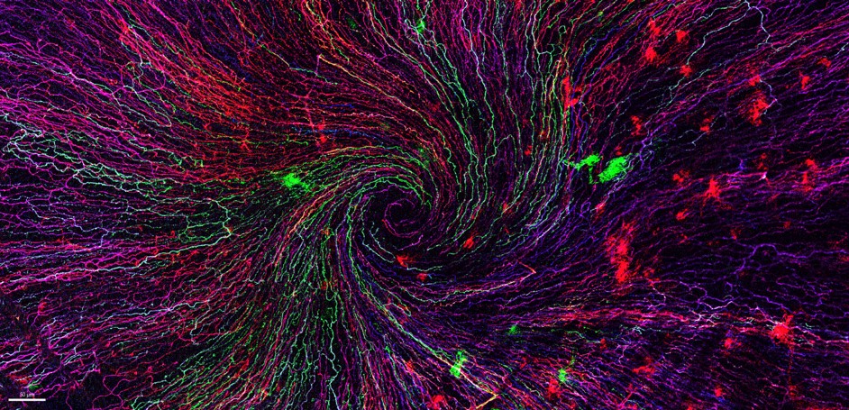
1st place:Léna Meneux, Eye Team, Institut des Neurosciences de Montpellier
"The eye of the storm"
Sensory fibers of a mouse cornea imaged with a confocal microscope. The corneal nervesconverge toward the centre forming a vortex. This particular transgenic mouse model allows stochastic expression of fluorescent proteins, unravelling the heterogeneity of the fiber origines inside the corneal epithelium. Acknowledgements to Karine Loulier for the mouse model and Laetitia Hudececk for her help during the acquisition.
In the Institut des Neurosciences de Montpellier since 2020, Léna is a PhD student working in the team Eye lead by Dr. Frédéric Michon. This team is investigating the mechanisms related to the preservation and the integrity of the anterior part of the eye, including the lacrimal gland, the tears and the cornea. Léna’s project focuses on the cellular and molecular effects of the corneal innervation on the corneal homeostasis. The project goes further as they aim at highlighting new targets able to prevent and/or repair corneal damage.
The image she submitted for the 2021 France-BioImaging Image Contest (The eye of the storm) represents the sensory fibers of a mouse cornea. This innervation follows a typical pattern where all the nerves converge toward the centre forming a vortex. This particular transgenic mouse model allows random expression of fluorescent proteins, unravelling the heterogeneity of the fibers’ origin inside the corneal epithelium. As cornea is the most innervated tissue in the whole body, this model shows the differences between fibers. In pathological context, for example wound injury, it is thus possible to follow a specific fiber during the healing process.
France-Bioimaging sponsored her participation to the FOM (Focus on Microscopy)2022 congress where she presented her project through a poster. Even though the congress was online, it gave her the opportunity to share her results with experts and as a consequence, to gather advice on her ongoing experiments.
2nd place:Eunice HoYee Chan, Muscle Dynamics Team, Developmental Biology Institute of Marseille (IBDM)
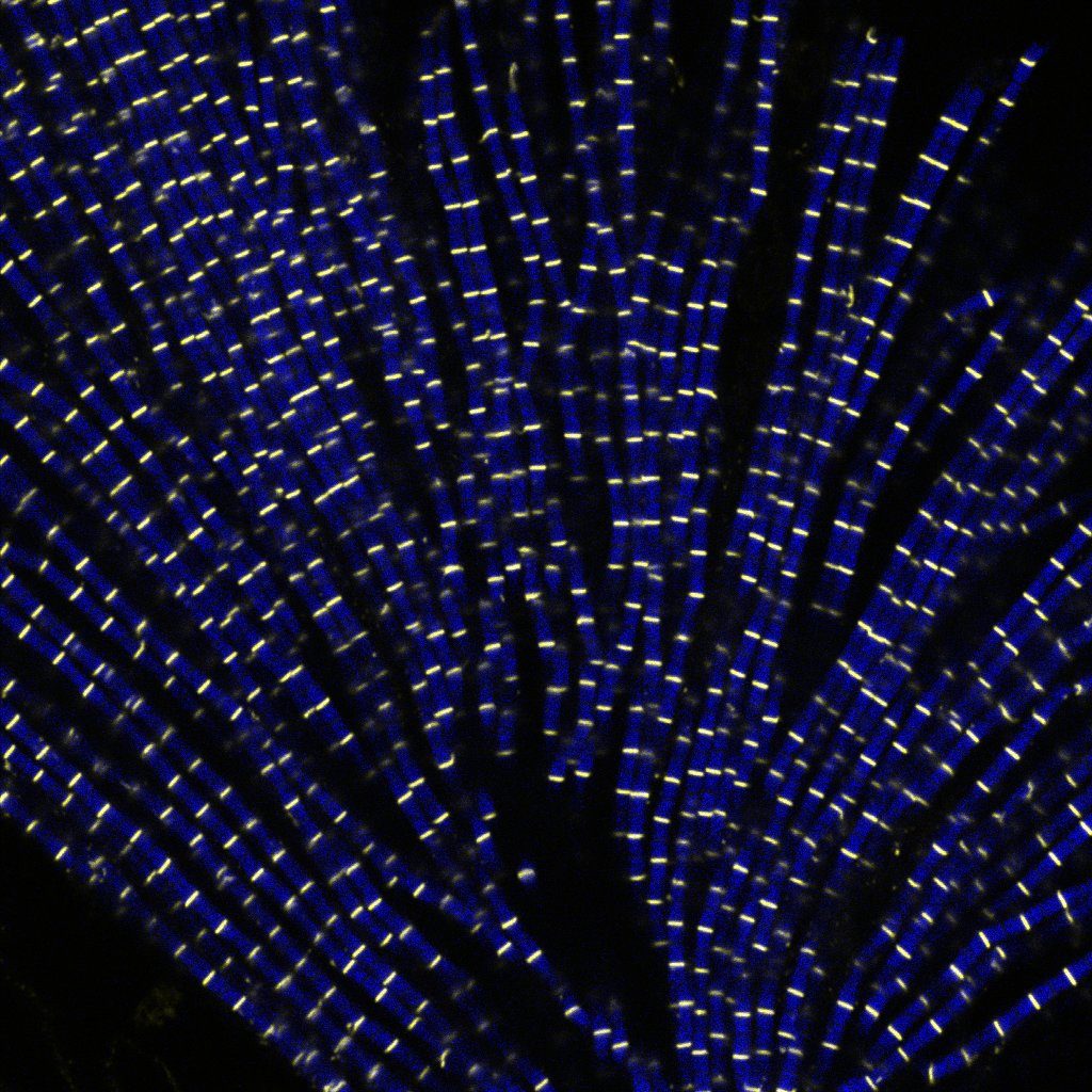
"Sarcomeric bouquet"
Myofibrils isolated from Drosophila indirect flight muscle labelled with titin (yellow) and actin (blue). Image captured from confocal microscope. We are studying the role of titin protein in muscle mechanics and organisation during development.
Research engineer in Frank Schnorrer's team at Institut de Biologie du Développement de Marseille (IBDM), Eunice focuses her research on Drosophila muscle dynamic and organisation during development using advanced biophysical and imaging techniques.
The image she submitted named “Sarcomeric bouquet" was from one of her very first muscle myofibrils isolation experiment. She dissected flight muscles from flies and labelled the individualised myofibrils with Llama nanobodies conjugated with different epitopes. Those labelled myofibrils were then subjected to various imaging methods including standard confocal microscopy, super resolution microscopy and cryo electron-tomogram. Using these novel labelling tools and imaging techniques, her team could study the dynamic and organisation of muscles during development in details.
France-BioImaging sponsored her registration to the 49th European Muscle Conference in Prague (22-26 September 2022). As she is new to the muscle field, this conference offered a great opportunity to have a broad view on different kind of state-of-the-art imaging techniques. Besides, she gave a presentation during the conference, highlighting her work and initiating discussion.
3rd Place:Camille Boutin, Biology of multiciliated cells Team, Developmental Biology Institute of Marseille (IBDM) &Nicolas Brouilly, PICsL Imaging facility, Electron Microscopy department
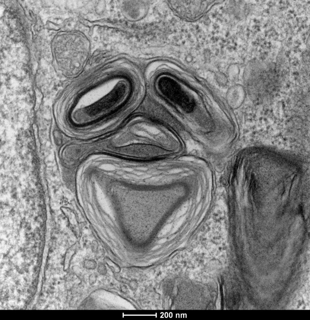
"Clown"
Lamellar structure in a differentiating multiciliated cell observed by transmission electron microscopy with a Tecnai G2 200kV FEI.
Camille is a researcher in Laurent Kodjabachian’s group at the Institut de Biologie du Développement de Marseille (IBDM). She develops projects as a principal investigator on the compartmentalization and sizing of multiciliated cells. With this in mind, she routinely uses confocal and super-resolution microscopy but also scanning and transmission electron microscopy and tomography.
Nicolas is in charge of the Electron Microscopy Unit of the Plateforme d’imagerie commune du site de Luminy (PICsL). In addition to the routine sample preparation and 2D TEM imaging, this imaging facility offers, to internal and external users, advanced sample preparation (cryo-methods, immunolabelling...) and advanced imaging (tomography, CLEM, serial blockface…).
To understand the production of multiple centrioles in multiciliate cells, they focused on the deuterosome, a membrane-less organelle that has been described 50 years ago but whose composition, organisation and function remain unknown to this day. In this context they have developed an inducible multiciliated cells line. This image was taken during the initial characterisation of this cell line by transmission electron microscopy.
Thanks to the France-Bioimaging Image Contest, Nicolas participated to the COST COMULIS Conference that was held by the Cyprus Institute in Nikosia. It was a great opportunity to exchange with the people at the cutting edge of the multi-modal imaging field. The program covered subjects such as the sample preparation for multi-modal imaging, image analysis and integrated industrial partners.
Published on August 23rd, 2022 in EMBO reports, this article questions the way that core facilities should be recognized in the scientific literature and their key contributions to data lifecycle. An initiative endorsed by France-BioImaging.
Core facilities are an integral part of the life science research landscape as providers of centralised access to technological resources and expertise. This article’s working group has estimated that between 40 and 80% of imaging, proteomics and genomics data at their institutes are generated at core facilities. The contribution of core facilities to scientific research and innovation must thus be accordingly recognised. In that respect, the most straightforward way is an acknowledgement. Unfortunately, the lack of formal rules still leaves core facilities being inadequately recognised.
This article proposes that the recognition of core facilities should be deployed via two actions and implemented in two phases: first, with the systematic acknowledgement of core facilities in all scientific publications, and second, by including core facilities and their staff in data citations (Cousijn et al, 2018).
The first step can be accomplished at the manuscript-submission stage by asking the corresponding author to confirm if any data (and associated metadata) used in the manuscript originated from a core facility, and if yes, to identify the associated core facility. EMBO Press has recently included a question in the author checklist to confirm whether the work in the publication “benefited from core facilities” and that the core facility be acknowledged accordingly.
The next step would be to make it compulsory for authors to respond to such a query and explicitly identify the core facility and relevant data (and associated metadata). The MDAR (Materials, Design, Analysis, Reporting) form (Macleod et al, 2021), wherein one needs to provide information about data availability in the Analysis section, could likewise include a question to explicitly identify core facilities involved. Eventually, the information in the author checklist could be automatically fed into the acknowledgement section.
Acknowledging will have two key positive consequences: on the sustainability of core facilities and on their staff careers. In the absence of a high number of publications, particularly as lead or corresponding authors, acknowledgements are used as a measure of a core facility and its staff’s output and impact. Second, it further motivates and incentivizes core facility staff to actively contribute to scientific research.
The acknowledgement of a core facility goes beyond professional courtesy: identifying the origin of data (and associated metadata) is essential for data traceability and reproducibility particularly since core facilities are major generators of data in life science research.
Thanks to Jean SALAMERO, our “Action inter-infrastructures” mission officer, for contributing to this article.
Katja Kivinen, Henri G A M van Luenen, Myriam Alcalay, Christoph Bock, Joanna Dodzian, Katerina Hoskova, Danielle Hoyle, Ondrej Hradil, Sofie Kjellerup Christensen, Bernhard Korn, Theodoros Kosteas, Mònica Morales, Krzysztof Skowronek, Vasiliki Theodorou, Geert Van Minnebruggen, Jean Salamero, Lavanya Premvardhan
Developed by the Serpico Inria-CNRS-Institut Curie Joint Team, member of the IPDM-BioImage Informatics node of France-BioImaging (FBI), this open-source framework could be a huge step forward in bioimaging management and analysis.
Bioimaging has a broad range of applications, addressing a variety of biological models at diverse scales of life. Thus, descriptions of novel computational approaches are often focused on target case studies. To tackle any scenario in biological imaging is a major challenge, that needs the conception and the development of a unified solution.
With this in mind, the BioImageIT project aims at providing a middleware that integrates data management with analysis using existing softwares (Omero, BioFormats, Fiji, napari, Scipy, pytorch…). The mission of BioImageIT was to design a graphical user interface (GUI) that allows any scientist without coding skills to annotate and analyze datasets using various software. By being user-centered, open-source and cross-platform (Windows, MacOS, Linux), BioImageIT created a management tool that is definitely accessible and well documented.
Started in late 2019, the project, funded by France-BioImaging, is now being deployed in 10 FBI imaging facilities. As it is a first step, the BioImageIT project have the ambition to expand the dissemination of the middleware throughout France and even further, Europe.
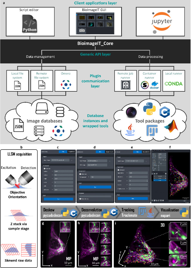
BioImageIT overview.a, Schematic view of BioImageIT architecture. The BioImageIT core is composed of data management and data processing functionalities. Users can access plugins by a script editor, Jupyter or the BioImageIT graphical interface (GUI). Data management functionalities exploit local files, remote files or databases such as OMERO. Data processing can perform computations in remote jobs, containers, or local runners. Image analysis is provided by plugins written in different languages. Developers can implement their own plugins in BioImageIT and design their own Graphical Interface. (b-i)LLSM processing workflow gathered in BioImageIT. Hela cell line expressing CD-M6PR-eGFP were stained with Tubulin TrackerTM Deep Red for Microtubules. b, Due to the geometry of LLS scanning, raw 3D images are skewed. c, g, First, realignment (deskew) of raw stacks is performed using Pycudadecon. d, h, Richardson Lucy deconvolution is performed using Pycudadecon. e, CD-M6PR-eGFP vesicles are tracked using Trackmate(FiJi). f, i, Deconvolved stacks and tracks are rendered using napari.
Prigent, S., Valades-Cruz, C.A., Leconte, L. et al. BioImageIT: Open-source framework for integration of image data management with analysis. Nat Methods (2022). https://doi.org/10.1038/s41592-022-01642-9
The France-BioImaging Image Contest is back for its 4th edition!
This image contest is open to all within the imaging community: core facility staff and users, R&D labs teams and co-workers, students… Submit your best microscopy images for a chance to showcase your skills, research and creativity to the French bioimaging community and beyond, allowing people to see the visual appeal of the life sciences. Images from the contest will be featured on France-BioImaging communication tools, online and in print.
France-BioImaging and all the French community aims to develop and promote innovative imaging technologies and methods. But microscopy images can also take an artistic, creative look and make the invisible world beautiful.
We are all eager to see your work !
Prizes
1 to 3 images will be awarded depending on the quantity and quality of the entries submitted. France-BioImaging will cover the registration fees for one 2023 microscopy related event of the winners’ choice (FOM, ELMI, EMC, COMULIS conference, etc.).
Important: Only French or foreign participants affiliated to a French institution can enter the contest. Foreign participants non-affiliated to a French institution can submit images and will be featured in the gallery, but will not be evaluated as part of the contest.
Submission deadline: Friday, November 11th, 2022, 23h59 UTC+2.
We use cookies on our website to give you the most relevant experience by remembering your preferences and repeat visits. By clicking “Accept All”, you consent to the use of ALL the cookies. However, you may visit "Cookie Settings" to provide a controlled consent.
This website uses cookies to improve your experience while you navigate through the website. Out of these, the cookies that are categorized as necessary are stored on your browser as they are essential for the working of basic functionalities of the website. We also use third-party cookies that help us analyze and understand how you use this website. These cookies will be stored in your browser only with your consent. You also have the option to opt-out of these cookies. But opting out of some of these cookies may affect your browsing experience.
Necessary cookies are absolutely essential for the website to function properly. These cookies ensure basic functionalities and security features of the website, anonymously.
Cookie
Duration
Description
cookielawinfo-checkbox-analytics
11 months
This cookie is set by GDPR Cookie Consent plugin. The cookie is used to store the user consent for the cookies in the category "Analytics".
cookielawinfo-checkbox-functional
11 months
The cookie is set by GDPR cookie consent to record the user consent for the cookies in the category "Functional".
cookielawinfo-checkbox-necessary
11 months
This cookie is set by GDPR Cookie Consent plugin. The cookies is used to store the user consent for the cookies in the category "Necessary".
cookielawinfo-checkbox-others
11 months
This cookie is set by GDPR Cookie Consent plugin. The cookie is used to store the user consent for the cookies in the category "Other.
cookielawinfo-checkbox-performance
11 months
This cookie is set by GDPR Cookie Consent plugin. The cookie is used to store the user consent for the cookies in the category "Performance".
viewed_cookie_policy
11 months
The cookie is set by the GDPR Cookie Consent plugin and is used to store whether or not user has consented to the use of cookies. It does not store any personal data.
Functional cookies help to perform certain functionalities like sharing the content of the website on social media platforms, collect feedbacks, and other third-party features.
Performance cookies are used to understand and analyze the key performance indexes of the website which helps in delivering a better user experience for the visitors.
Analytical cookies are used to understand how visitors interact with the website. These cookies help provide information on metrics the number of visitors, bounce rate, traffic source, etc.
Advertisement cookies are used to provide visitors with relevant ads and marketing campaigns. These cookies track visitors across websites and collect information to provide customized ads.