We are looking for our next Deputy Director for International Affairs!
▽ Scroll down
Author: alban.belloir
FBI opens a call for the recruitment of its next Deputy Director for International Affairs
Deadline: June 1st, 2023
Mission
The Deputy Director for International Affairs will oversee a key strategic mission: develop France-BioImaging’s international activities. This includes enabling France-BioImaging to expand and enhance its engagement in the international community and within targeted communities, with a focus on Europe and Africa. The three primary goals for this position are to increase FBI’s participation in Horizon Europe programme; aid in developing FBI’s approach to international outreach; and build a long-term engagement with FBI’s international partners. The Deputy Director will work closely with the Manager of External Affairs to tailor specific cooperation activities, prepare strategic decision-making and prioritize needs for future international initiatives.
Organisation
France-BioImaging is a national research infrastructure distributed throughout France that provides researchers with access to the latest innovations in biological imaging and aims to accelerate the transfer of technological and methodological innovations in biological imaging to the 22 platforms that constitute the infrastructure. The Deputy Director for International Affairs will contribute to the development and strengthening of France-Bioimaging international relations and outreach activities.
The Deputy Director for International Affairs will work within the national coordination of the France-BioImaging infrastructure. He/she will work within a team distributed between Montpellier and Bordeaux, composed of 4 people: the Scientific Director and the Deputy Director for International Affairs, the Internal Affairs Manager, and the External Affairs Manager.
Mandate of the Deputy Director for European Affairs
Help to develop and implement the FBI’s international cooperation activities
Promote and organize a watch on the existing European and/or international programmes/funding
devices in the fields of interest, and promote and organize the diffusion of information on international cooperation within FBI
Advise the management team in the area of international cooperation in relation to the infrastructure’s scientific policy, synthesize and prepare elements to assist in decision-making
Advise the management team in the field of research development in relation to the institution’s scientific policy
Represent the infrastructure and to lead the relations with the partners, in particular with the Euro-BioIamging ERIC, as CNRS representative of the infrastructure within the European Euro-BioImaging Infrastructure Board.
Promote the scientific and structuring activities of the infrastructure at the international level, to seek out and federate potential partners, to initiate and manage international cooperation programs, and to provide expertise and advice.
Organize a consultation and a prospective reflection on the development of new initiatives and on the actions to be carried out (i.e. preparation of the participation of the infrastructure in the European calls for projects of the Horizon Europe framework program)
Time commitment: 20% of his overall working time
Mandate duration: 5 years
More details and information in the pdf below
Entry into function is planned as soon as possible, September 1st, 2023 at the latest.
If you are interested,
please send a short CV and a motivation letter (2 pages) at direction@france-bioimaging.org BEFORE the 1st of June
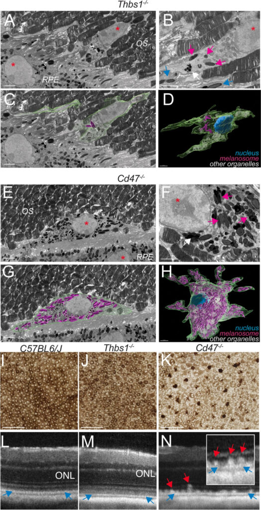
Age-related macular degeneration (AMD) affects more than 150 million people worldwide (early AMD) and 10 million of patients suffer from debilitating late stage AMD. Blurring central vision, this eye disease progresses over time, usually beginning when people are around their 50s or 60s by causing damage to the macula, in the retina. Researchers from the Institut de la Vision (Sorbonne Université, INSERM, CNRS, UMR_S 968) recently published about the AMD. Thanks to Serial Block-Face Scanning Electron Microscopy (SBF-SEM) experiments carried out at the ImagoSeine core facility (Institut Jacques Monod / FBI Paris-Centre node), they describe in this new study melanophages as a disease-progression marker.
Early or intermediate AMD is characterized by pigmentary changes and lipoproteinaceous debris accumulation between the photoreceptors and the melanosome-rich retinal pigment epithelium (RPE) or below the RPE. Later, AMD can be complicated by central choroidal neovascularization or by an expanding lesion of the photoreceptors. Even though patients with early or intermediate AMD can progress and develop late AMD, a large part of patients stay stable for years, underlining the potential usefulness of progress.
AMD is associated with the appearance of hyperreflective foci, with reflectivity comparable to melanocyte-containing RPE cells. Thbs1 and CD47 are both important for the elimination of these cells. In the absence of either of them, melanocyte-containing RPE cells would then accumulate. The goal was to determine the origin of these cells in the retina, and the main question was: are these cells RPE migrating to the wrong place, or melanosome phagocytes cells having ingested melanosomes?
SBF-SEM: the key to answer this question
The Serial Block-Face Scanning Electron Microscopy (SBF-SEM) is a 3D electron microscopy imaging technique, where an ultramicrotome is placed inside a SEM. Biological samples are beforehand stained with heavy metals and embedded in a plastic resin block. Inside the microscope, a thin-section is cut at the surface of the block and discarded. Then, an image of the surface of the block – therefore inside the sample – is made, using back-scattered electrons. The process of cutting and imaging is repeated automatically as many times as necessary to produce a 3D stack of images inside the sample, as it is progressively imaged and destroyed.
This technique allows 3D imaging of large samples for Electron Microscopy standards (up to several hundred microns in each of the X,Y,Z direction) at high resolution. This technique is often used to image whole cells, or even small pieces of tissues in 3D. The two major domains of application are to:
find a rare structure within a cell or tissue. The sample is imaged until the structure of interest is found.
understand the 3D spatial organization of organelles within cells, or of cells between them.
The benefits of bioimaging in this study
In the study, SBF-SEM was essential. As previously mentioned, AMD is associated with the appearance of hyperreflective foci, with reflectivity comparable to melanocyte-containing RPE cells. In the images produced by SBF-SEM, the retinal pigment epithelium (RPE) surrounding the melanophages in mice, where CD47 was inhibited, were markedly less pigmented and deformed compared to those where Thbs1 was blocked. This suggests that melanosomes have been transferred by phagocytosis from the RPE to nearby melanophages because they lack CD47. Finally, authors have shown that CD47 acts as a “don’t eat me” signal. The SBF-SEM was a great addition to this study where understanding the 3D spatial organization of the structure of interest was key.
Thanks to Jean-Marc Verbavatz for providing very helpful insights of the study!
Augustin, S., Lam, M., Lavalette, S. et al. Melanophages give rise to hyperreflective foci in AMD, a disease-progression marker. J Neuroinflammation 20, 28 (2023). https://doi.org/10.1186/s12974-023-02699-9
Get access to one of our services!
You need SBF-SEM or another imaging technology or expertise that France-BioImaging provides? To get open access, please login via Euro-BioImaging website! You just have to choose the technology you want to use, then submit your proposal. All applications will be processed by the Euro-BioImaging Hub in close relation with France-BioImaging. And of course, all scientists regardless of their affiliation, area of expertise or field of activity can benefit from open access services! Users whose projects will be validated by Euro-BioImaging will benefit from a waiver for the access cost on France-BioImaging core facilities (https://france-bioimaging.org/access/)
Massive intracellular accumulation of RPE-derived melanosomes in subretinal MPs of CD47−/−-mice causes subretinal melanophage formation and their clinical appearance as hyperreflective foci.
France-BioImaging was at the Euro-BioImaging‘s All Hands Nodes Meeting at EMBL in Heidelberg. It was a pleasure to share this unique moment with all the Euro-BioImaging nodes! We had great discussions from passionate people around building the future of the infrastructure and providing open access to high-end technologies and expertise.
This was the perfect time to hear about the latest news and opportunities from every European nodes. Several FBI members were there:
Caroline Thiriet, our External affairs manager, was a panelist at a discussion about “Funding for national imaging communities”, highlighting the history of France-BioImaging and how the french infrastructure works.
Perrine Paul-Gilloteaux, our Image data mission officer, gave a fantastic talk about “Linking and analyzing correlative image datasets”.
Fabrice Cordelières, our Training mission officer, and Alban Belloir, Communication officer, presented two posters on training and about our infrastructure’s structuring activities.
Melina Petrel, as an Electron #microscopy specialist, represented the FBI core facility staff.
Thanks to all Euro-BioImaging team for organizing and hosting this wonderful event! We are glad to be part of this amazing international community working together as a European Research Infrastructure!
1st Interdisciplinary Summer School on Chemical and Physical Probes for Biology
July 3rd to 7th
The fine understanding of molecular mechanisms in native biological systems is an important step in rationalizing, preventing and ultimately curing diseases. Photonic imaging plays an important role in this field. Quantitative imaging experiments designed to answer complex biological questions require the implementation of efficient probes and adapted imaging setups and data processing workflows.
This interdisciplinary school will provide an overview of these different chemical, physical and biological aspects. It will offer also interactive and interdisciplinary workshops to learn how to communicate effectively between disciplines as well as two half-days of hands-on.
The PFIC facility organizes a technological seminar on “Quantitave Phase Imaging – exploring the unseen” April 20th at 10:00 am in Espace Maurice Tubiana at Gustave Roussy, Villejuif.
Quantitative Phase Imaging (QPI) is a novel label-free microscopy technique bringing a completely new contrast into the live-cell imaging field. It allows extracting of information-rich quantitative data from unlabeled cells and monitoring their dry mass in non-invasive experiments.
This seminar will explain the principle of technology and present broad range of applications focusing on the label-free analysis of cell growth, cell death, cell migration etc. Apart from cell biology research, studies of biomolecular condensates and biomaterials will be discussed.
The seminar will be held in English by Zuzana Nováková (Telight application specialist).
Hope to see you all you interested by this technology there!
The second workshop of Holotomography microscopy will be held from April 12th to 14th on the PFIC microscopy facility.
You will test the HT-2H microscope which has more resolution than his bigger brother HT-X1.
The key features are: No label needed, High resolution with one single lens 60X NA 1.2 water immersion (120 nm XY resolution, 356 nm Z resolution, 150 fps T resolution ), quantifiable data, Low phototoxicity, fast imaging.
The microscope has both 3D Holotomograms and 3D fluorescence capabilities in one single unit.
To better prepare your samples for imaging the Tomocube will give us some mounting chambers.
Correlative X-ray imaging and electron microscopy (CXEM) is the combination of X-ray imaging and electron microscopy. It is a correlative approach that makes it possible to characterise a sample of interest and locate a structure of interest in a non-destructive way. Nicolas BROUILLY is in charge of the Electron Microscopy Unit of the PICsL imaging facility on the Marseille node of France BioImaging, where CXEM is used for developmental biology studies. As part of Euro-BioImaging’s Proof-of-Concept study, his facility is now accepting applications from external users for CXEM projects. Learn more about how this approach works and what it can be used for in the interview below.
We are today talking about CXEM imaging. Please provide a short summary of this type of imaging and tell us some applications:
Nicolas Brouilly: It is often very useful to combine 2 imaging modalities to take advantage of each while trying to lower their respective drawbacks. For example, by combining Light Microscopy and Electron Microscopy, we obtain the popular CLEM (for Correlative Light and Electron Microscopy). Visible light can then be used in combination with EM either:
To target a precise region of interest ;
To localize molecules within the ultrastructural information obtained by EM.
Using the same acronym building, CXEM corresponds to Correlative X-ray and Electron Microscopy. X-rays are photons of shorter wavelength than those from visible light, and can again be used to characterize a sample in 2 different ways:
To use their ability to easily go through tissues in order to record the 3D morphology of a sample: either by computed x-ray micro-tomography (or micro-CT) for micrometric resolution of big samples (mm to cm range) or by Soft X ray tomography for nanometric resolution of small samples (100’s of nm to um range);
To use a focused beam of high energy x-rays to analyse the localization of the elements of a sample: X-Ray Fluorescence microscopy (or XRF).
Both modalities can be used to complement the ultrastructural information obtained by electron microscopy. At the Marseille node of France BioImaging, in the Electron Microscopy Unit, we routinely use Correlative Micro-CT and Electron Microscopy to answer developmental biology questions.
What are some advantages of this technique that make it suited to addressing this type of question?
Nicolas Brouilly: The main advantage of Micro-CT (or Computed X-ray Tomography) is its ability to “see through” a sample and to reveal its overall organization in 3D without any labelling. The second advantage of Micro-CT is the fact that it is non-destructive. Thirdly, the contrast we usually give to samples for electron microscopy is compatible and even beneficial for X-ray imaging.
Altogether, this means that we can use X-ray tomography to map the microscale morphology of a sample in order to target a specific region of interest without having to go through the time-consuming and destructive collection of semi-thin sections.
We routinely use the micro-CT tool, not only to target a given organ or a given group of cells, but also to pre-orient the sample in order to cut it under a specific orientation. It is a timesaving tool within the frame of a 2D electron microscopy project, but it really is key within the frame of a 3D electron microscopy project given that Serial BlockFace and Focused Ion Beam techniques are destructive.
Tell us a bit more about a specific project that was done in your facility using this technology? What scientific questions were you addressing?
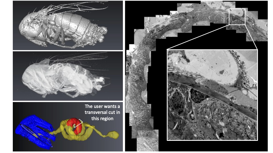
Nicolas Brouilly: Imagine that, first, you have a ball of yarn, second, you cannot untangle it, and third, you want to cut small bits of the thread at 24 cm from the end (not 22, not 26… 24 !). CXEM enabled us to do this on Drosophila gut. The micro-CT gave us the 3D map of the sample within the resin block. We could then use this map to find the best itinerary within the sample to make transverse sections of the portion of interest that was precisely indicated by the user on the micro-CT dataset. At the end of the day, the user was able to look at perfect transverse ultrathin TEM sections, at a precise position of this ball of yarn that Drosophila gut is. He could finally get precise metrics from this precise part of the gut in several samples. None of this could have been achieved without CXEM.
Like a ball of yarn… Above is an example of how CXEM can be used to find the best itinerary within a sample to make transverse sections of the portion of interest. On the left, the micro-CT provided a 3D map of the sample within the resin block. On the right, a transverse ultrathin TEM section of the drosphilia gut.Image courtesy of Nuno Luis (Schnorrer lab, IBDM) & Nicolas Brouilly (Electron Microscopy Facility, IBDM AMU/CNRS, France BioImaging).
For another example, you can have a look at the following paper where we used CXEM to map platelet aggregates within arteries in order to explore them by Serial BlockFace SEM, another example of “Find a needle in a haystack”. Have a look at movie S1, it is a wonder that we could not obtain without CXEM:
CXEM is part of the Euro-BioImagingProof-of-Concept study. The Proof-of-Concept study makes it possible to introduce exciting, new imaging technologies to our portfolio that were previously unavailable via our network. We are currently accepting applications to use these technologies at participating Nodes as part of the Proof-of-Concept study. Be part of this study – and contribute to community-wide continuous technological innovation!
All scientists, regardless of their affiliation, area of expertise or field of activity can benefit from Euro-BioImaging’s pan-European open access services. Potential users of these new technologies are encouraged to submit project proposals via our website. To do so, you can Login to access our application platform, choose the technology you want to use and the facility you wish to visit, then submit your proposal. All applications will be processed by the Euro-BioImaging Hub. As usual, users will benefit from advice and guidance by technical experts working at the Nodes, training opportunities, and data management services.
Alors que les volumes et la complexité des données augmentent de manière sans précédent, leur suivi et partage devient de plus en plus un défi pour la communauté de la recherche en science de la vie (biologie, biomédicale…). Ce manque de traçabilité a un impact négatif reconnu sur la réutilisation des données publiées. C’est pourquoi il est important de rendre les données « FAIR » – faciles à trouver, accessibles, interopérables, réutilisables – dès la conception des projets de recherche.
OMERO est la solution intégrée de référence pour gérer les données d’images pendant toute leur durée de vie, de l’acquisition à la publication.
Le RT-mfm du CNRS organise cette ANF avec la collaboration de France BioImaging et le EMBRC. Cette ANF est orientée vers les ingénieurs en plateforme d’imagerie et similaires. Elle a pour objectifs de former les stagiaires à la gestion FAIR de données au travers la base de données OMERO comme outil principal de gestion de données pour la microscopie et pendant tout le cycle de vie de la donnée : de l’acquisition à la publication.
A l’issue de la formation les stagiaires seront capables de : • Comprendre les principes FAIR et leur application dans la pratique • Comprendre les enjeux et objectifs d’un Plan de Gestion de Données. • Connaître les schémas de mise en place d’une infrastructure OMERO avec les différents scenarios de déploiement • Savoir administrer les politiques d’accès selon les différentes configurations de groupes • Importer des images dans OMERO • Être capable de décrire les images avec des métadonnées riches • Analyser les données sur OMERO et sauver les résultats sur OMERO en association avec les données source • Annoter les images en masse à partir de tableaux • Chercher des images en fonction des différentes métadonnées • Être capable de créer des figures avec OMERO-figure
• Apprendre les options d’interfaçage avec différents logiciels (ImageJ, Napari, CellProfiler, QPath,…) • Publier les données
Two projects recently received funding from the Chan-Zuckerberg Initiative (CZI), in which France-BioImaging members take part actively: COMULIS and NEUBIAS. Two community building activities breaking up frontiers to gather scientists around one goal: developing biological imaging.
COMULIS received a 2-year funding from CZI to expand their network both globally and sustainably. Being designed to harness the power of multimodal imaging (MMI) across scales, from basic to clinical diagnostics, this European initiative aims at facilitating access and training a new generation of scientists for whom multimodal imaging will be the new norm. Thanks to this grant, the project will be consolidated and it will help extend the collaborative and innovative network to establish a global multimodal imaging association (COMULISglobe) and ensure long term sustainability.
MMI integrates the best features of combined techniques and overcomes limitations faced when applying single modalities independently. MMI relies on the joint expertise from biologists, physicists, chemists, clinicians, and computer scientists, and depends on coordinated activities and knowledge transfer between technology developers and users. To achieve this inherently interdisciplinary goal, the ultimate goal is toestablish a network of scientists across continents and disciplines, from academia to industry, including transnational research facilities (e.g. synchrotrons, Euro-BioImaging ERIC), to foster and market MMI as a versatile tool in biomedical research and diagnostics.
COMULISglobe will help bridge the gap between biological and clinical imaging, identify, fund, and showcase novel multimodal pipelines, and develop, evaluate, and publish correlation software through dedicated networking activities, including conferences, training schools, open databases, and fellowships for lab exchanges, access to research infrastructures, and conference attendance. And, of course, all outputs of the project will be open access!
Please do not hesitate to join the community and help organize activities or publications – and please share the news, mobility and access grants available at: https://www.comulis.eu/comulisglobe-czi
Thanks to Perrine Paul-Gilloteaux, Bretagne-Loire node of France-BioImaging, for taking part of this amazing project!
The international Network of European BioImage Analysts (NEUBIAS), hosted by German BioImaging has also received a 2-year funding from the Chan Zuckerberg Initiative (CZI) as part of their Advancing Imaging through Collaborative Projects program. This grant will secure the sustainability of NEUBIAS, establish strong connections to similar initiatives, and share knowledge about state-of-the-art bioimage analysis tools and methods globally.
Spreading the profession of bioimage analysts and bioimage analysis knowledge internationally are the major aims of NEUBIAS. Modern life-sciences are unthinkable without advanced microscopy imaging techniques and quantitative bioimage analysis. This grant will help ensure novices and experts can access cutting edge techniques, reduce duplications of effort, and support everyone who is working to making new discoveries possible.
NEUBIAS had a tremendous impact on the community by training a powerful generation of bioimage analysts across Europe and beyond. The next step of this project will expand the network internationally and connect to related imaging and image analysis societies around the globe. With that in mind, the project includes travel grant opportunities for early-career bioimage analysts who seek to join NEUBIAS activities, explicitly including scientists outside central Europe. Besides, a dedicated team will work on collecting bioimage analysis teaching materials and make them accessible to the global imaging and life science community.
Great news for both projects that – we hope – will continue to write the great story of bioimaging!
Thanks to Florian Levet, Bordeaux node of France-BioImaging, for being a member of this fantastic project!
Brettanomyces bruxellensis is one of the most damaging spoilage yeasts in the wine industry because of its impact on the beverage’s flavor. Lysiane Brocard, research engineer specialised in plant biology at the Bordeaux Imaging Center (FBI Bordeaux node), recently co-published an article on this yeast cell surface and bioadhesion properties.
Fruits are transformed into beverages through fermentation processes carried out by microorganisms naturally present in the environment. In wine, yeasts and bacteria play this role and contribute to the development of volatile compounds. Scientists targeted Brettanomyces bruxellensis in this study, a yeast famous for the production of volatile phenols, characterized by horse sweat odors which is – usually – not very enjoyable for the consumer.
A yeast characterized by bioadhesion abilities
This specific odor comes from volatile compounds, the 4-ethylgaïacol (4EG) and 4-ethylphenol (4 EP), that winemakers try to avoid. Beside adding an unpleasable flavor to the beverage, the issue is that the spoilage yeast is persistent in cellars over several years, resulting in recurrent wine contamination. This suggests a bioadhesion process that helps the microorganism to survive in its environment. To put it simply, bioadhesion is the ability of an organism to adhere on a surface to, then, participate in the formation of a biofilm (which is defined as “a structured community of microorganisms adhered to a surface and producing an extracellular matrix”).
Here, 54 strains of B. bruxellensis were characterized for their cell surface physico-chemical and bioadhesion properties. And all of them have shown bioadhesion abilities (after only three hours on stainless steel) both on synthetic medium and wine. Enough to highlight the persistence of our favorite horse sweat flavored yeast.
How did bioimaging help in this project?
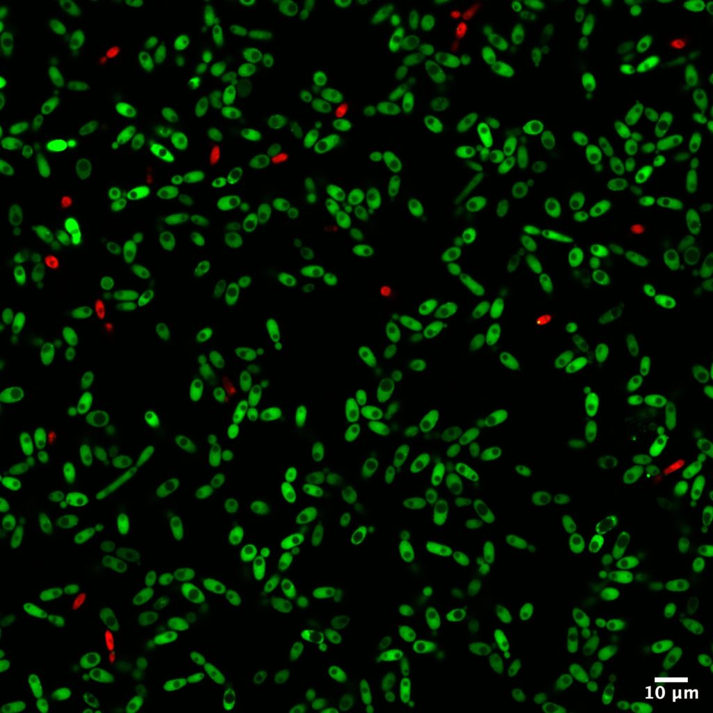
Among all the analytical methods used in this study, microscopy helped identify the structure of the biofilms formed with B. bruxellensis. Two imaging techniques were used: confocal microscopy and scanning electron microscopy. The first one offers the advantage of realizing live imaging without being too time-consuming. With fluorescent dyes, the status of cells can be easily determined at the same time as the cell repartitions and concentrations.
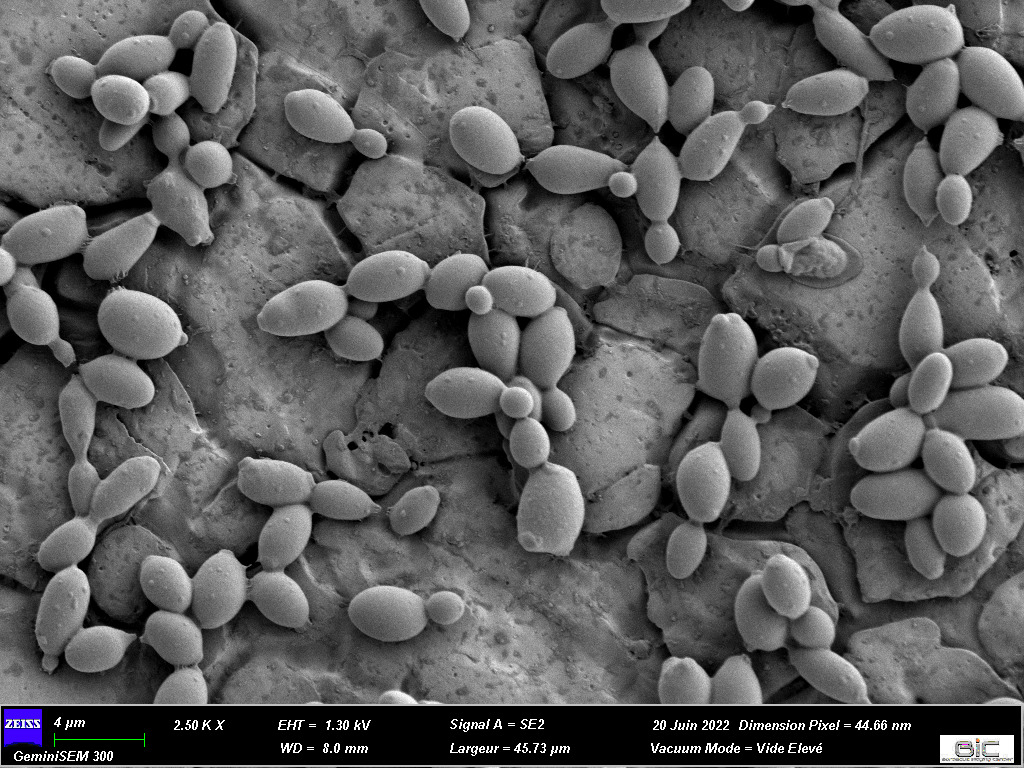
Moreover, comparisons have been made thanks to confocal microscopy to determine if some strains of B. bruxellensis could form biofilm with only one cell layer or if they proliferate in three dimensions. To complete these observations, scanning electron microscopy was performed at the Bordeaux Imaging Center (FBI Bordeaux node) with the help of Isabelle Svahn, expert of this type of microscopy. It was a great addition to this study as these observations validated the morphological variability among Brettanomyces strains.
Bioimaging helped a broader project about Brettanomyces led by Isabelle Masneuf-Pomarède who works in the Institut des Sciences de la Vigne et du Vin of Bordeaux. Isabelle studies the persistence and proliferation of Brettanomyces over the years. Isabelle’s PhD student, Paul Le Montagner, carried out most of the experiments published in this paper. Thanks to them for this amazing paper!
Confocal microscopy observations after 3h of bioadhesion of cells on stainless steel
SEM observation of 3h-aged cells adhered on stainless steel
Original article: Paul Le Montagner, Morgan Guilbaud, Cécile Miot-Sertier, Lysiane Brocard, Warren Albertin, Patricia Ballestra, Marguerite Dols-Lafargue, Vincent Renouf, Virginie Moine, Marie-Noëlle Bellon-Fontaine, Isabelle Masneuf-Pomarède, High intraspecific variation of the cell surface physico-chemical and bioadhesion properties in Brettanomyces bruxellensis, Food Microbiology, Volume 112, 2023, 104217, ISSN 0740-0020, https://doi.org/10.1016/j.fm.2023.104217
You are interested in our bioimaging services, including technologies and expertise?
An innovative technology to look at thick samples at high resolution? Marc Tramier, a group leader at the Institute of Genetics & Development of the University of Rennes/INSERM/CNRS, and scientific director of MRic (Microscopy Rennes Imaging Centre), is currently working with his team on Random Illumination Microscopy (RIM), a fast and easy to use microscopy technique with low phototoxicity. His facility, which is part of the Bretagne-Loire Node of France-BioImaging, offers RIM as a Euro-BioImaging Proof-of-Concept study, and is now accepting applications for projects. He explains the ideas behind RIM in the article below.
The idea of Random Illumination Microscopy is to use the speckle of the illumination laser in wide field to create a structured illumination pattern at the diffraction limit. By varying the pattern from image to image using a diffracting element (in our case a SLM), scientists are able to acquire a stack of images (around 100 images) on a camera which corresponds to a cumulative homogeneous illumination. By resolving the inverse problem, a super-resolved image is, then, reconstructed, at the focal plane with unprecedented optical sectioning. In comparison to conventional SIM, RIM is able to work in depth inside diffusive samples as the speckle is insensitive to diffusion.
A transfer full of advantages
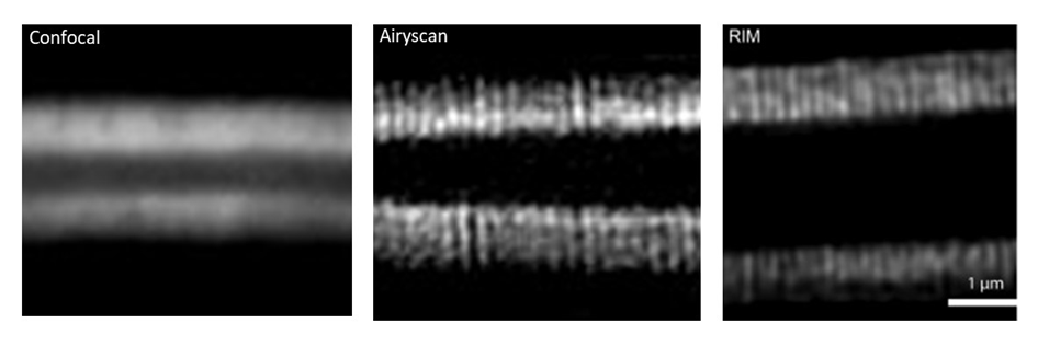
The method was first implemented by Thomas Mangeat – that we are happy to welcome in our new Toulouse node! – and collaborators in Toulouse (Mangeat et al., 2021. doi: 10.1016/j.crmeth.2021.100009). In the MRic, after the transfer of the prototype, the facility was able to image microvilli of intestine in c-elegans (depth > 50µm) having a spatial resolution of around 100 nm. This structure is impossible to be revealed by conventional confocal microscopy. Before the use of RIM, only the airyscan approach allowed us to resolve the microvilli but with higher illumination power (photobleaching of the sample) and longer acquisition time (around 10 times more). Now with RIM, we are able to follow microvilli in the living C–elegans at the second time-scale during several minutes.
RIM is one of the powerful methods to achieve super-resolved images in depth at high speed with very low phototoxicity. This makes a very nice compromise of z-sectioning and super-resolution with wide field illumination particularly adapted to thick live samples.Beside reconstruction and data analysis, MRic is offering a user-friendly system, with a complete set of microscopy methods for live sample investigation, from wide-field to light sheet including spinning disk, confocal and airyscan. And of course, this technology is available in open access through France-BioImaging and Euro-BioImaging!
How to apply to use RIM:
Random Illumination Microscopy is part of the Euro-BioImaging Proof-of-Concept study, in collaboration with our Nodes. The Proof-of-Concept study makes it possible to introduce exciting, new imaging technologies to our portfolio that were previously unavailable via our network. We are currently accepting applications to use these technologies as part of the Proof-of-Concept study. Be part of this study – and contribute to community-wide continuous technological innovation!
All scientists, regardless of their affiliation, area of expertise or field of activity can benefit from Euro-BioImaging’s pan-European open access services.Potential users of these new technologies are encouraged to submit project proposals via our website. To do so, you can Login to access our application platform, choose the technology you want to use and the facility you wish to visit, then submit your proposal. All applications will be processed by the Euro-BioImaging Hub. As usual, users will benefit from advice and guidance by technical experts working at the Nodes, training opportunities, and data management services.
Thank you Marc Tramier and Marianna Childress, communication officer of Euro-BioImaging, for the original article.
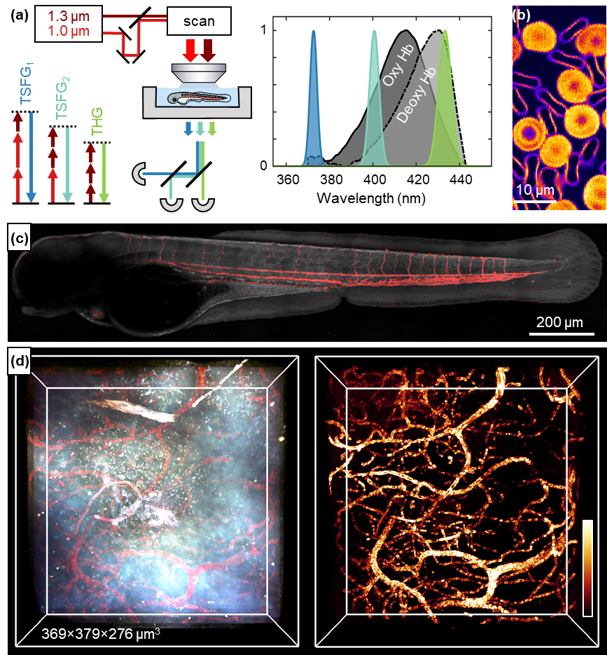
Researchers from the Laboratory for Optics and Biosciences (LOB, CNRS / École Polytechnique / INSERM), a member of the Ile-de-France Sud node of France-BioImaging, and from the Developmental Biology and Stem Cells Department (UMR3738, CNRS / Institut Pasteur), developed a new form of multiphoton microscopy providing label-free imaging of red blood cells and oxygenation. This technique is called color third-order sum-frequency generation microscopy, and is described in an article recently published in Light: Science & Applications.
Keeping resolution, saving time
Current methods used to map microcirculation and blood oxygenation at high resolution typically require the injection of fluorescent or phosphorescent markers. In addition, they usually require relatively long pixel times and thus are limited in spatio-temporal resolution. The novel approach is based on label-free third-order sum-frequency generation (TSFG) and third-harmonic generation (THG) contrasts. In practice, this microscopy is based on the illumination of samples by two pulsed infrared lasers and the simultaneous detection of several TSFG and THG signals emitted at different colors. This method has the advantage of providing simultaneous measurements at several wavelengths spanning the hemoglobin absorption spectrum. This simultaneity makes TSFG microscopy appropriate for studying dynamic samples…
To better understand brain and tissue physiology
…such as biological tissues! Biological tissues are supplied with oxygen by red blood cells, responsible for circulating hemoglobin through the body. Scientists have shown that the intensity of the different signals detected by TSFG microscopy depends on their spectral proximity to the absorption wavelength of hemoglobin. As the color of hemoglobin depends on its oxygenation state, the TSFG makes it possible to image red blood cells circulating in live zebrafish larvae – and even to probe their oxygenation state. Researchers also demonstrated that this contrast modality is also compatible with deep-tissue microscopy and can be used to observe the brain of a live adult zebrafish. An example that confirms the broad range of application of this novel imaging technique.
Reference: Ferrer Ortas, J., Mahou, P., Escot, S. et al. Label-free imaging of red blood cells and oxygenation with color third-order sum-frequency generation microscopy. Light Sci Appl12, 29 (2023). https://doi.org/10.1038/s41377-022-01064-4
We use cookies on our website to give you the most relevant experience by remembering your preferences and repeat visits. By clicking “Accept All”, you consent to the use of ALL the cookies. However, you may visit "Cookie Settings" to provide a controlled consent.
This website uses cookies to improve your experience while you navigate through the website. Out of these, the cookies that are categorized as necessary are stored on your browser as they are essential for the working of basic functionalities of the website. We also use third-party cookies that help us analyze and understand how you use this website. These cookies will be stored in your browser only with your consent. You also have the option to opt-out of these cookies. But opting out of some of these cookies may affect your browsing experience.
Necessary cookies are absolutely essential for the website to function properly. These cookies ensure basic functionalities and security features of the website, anonymously.
Cookie
Duration
Description
cookielawinfo-checkbox-analytics
11 months
This cookie is set by GDPR Cookie Consent plugin. The cookie is used to store the user consent for the cookies in the category "Analytics".
cookielawinfo-checkbox-functional
11 months
The cookie is set by GDPR cookie consent to record the user consent for the cookies in the category "Functional".
cookielawinfo-checkbox-necessary
11 months
This cookie is set by GDPR Cookie Consent plugin. The cookies is used to store the user consent for the cookies in the category "Necessary".
cookielawinfo-checkbox-others
11 months
This cookie is set by GDPR Cookie Consent plugin. The cookie is used to store the user consent for the cookies in the category "Other.
cookielawinfo-checkbox-performance
11 months
This cookie is set by GDPR Cookie Consent plugin. The cookie is used to store the user consent for the cookies in the category "Performance".
viewed_cookie_policy
11 months
The cookie is set by the GDPR Cookie Consent plugin and is used to store whether or not user has consented to the use of cookies. It does not store any personal data.
Functional cookies help to perform certain functionalities like sharing the content of the website on social media platforms, collect feedbacks, and other third-party features.
Performance cookies are used to understand and analyze the key performance indexes of the website which helps in delivering a better user experience for the visitors.
Analytical cookies are used to understand how visitors interact with the website. These cookies help provide information on metrics the number of visitors, bounce rate, traffic source, etc.
Advertisement cookies are used to provide visitors with relevant ads and marketing campaigns. These cookies track visitors across websites and collect information to provide customized ads.
 FBI opens a call for the recruitment of its next Deputy Director for International Affairs