We are happy to announce that our 1st Africa-France Joint Initiative for Biological Imaging calls, in coordination with the African BioImaging Consortium and Imaging Africa, is funding 11 projects!
As a reminder, these two calls (“External access” and “Twinning exchange”) have been primarily designed to strengthen collaboration between African and French researchers and engineers in all fields in biology, health and agro-ecology, where the contribution of the rapidly expanding technologies of digital imaging has become essential. The ambition of these calls is in line with the anticipation of bilateral research funding programs between Europe and Africa in the framework of the Horizon Europe Program.
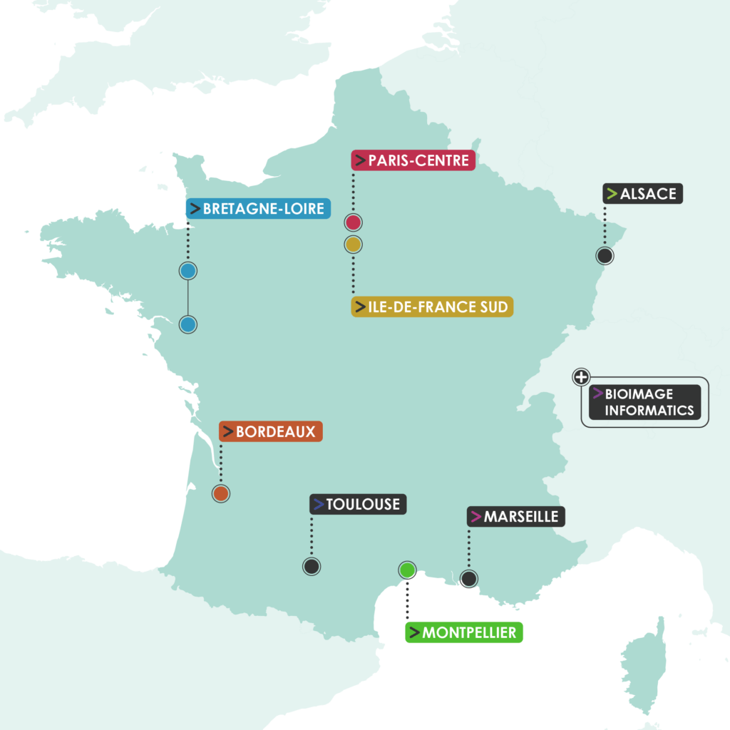
Multiple scientists from the entire African continent have responded to the calls, covering 8 countries: Senegal, Togo, Nigeria, Ivory Coast, Burkina Faso, Morocco, Uganda and South Africa. Besides, we are very glad that 5 of our nodes (Paris-Centre, Montpellier, Bordeaux, Ile-de-France Sud and Bretagne-Loire) are going to work in the framework of the Africa-France Joint Initiative for Biological Imaging.
Finally, the projects have been selected for the need to access imaging resources in response to key challenges that African researchers are facing. Among them, scientists tackle issues in several topics going from infectious diseases to marine biology, but also cancer research, plant biology and climate change.

Here are the selected projects
Call 1 “External Access”:
- Production of digitised teaching tools (anatomical and histological sections) for bachelor’s and master’s students – Aliou NDIAYE, Département de Biologie végétale, Université Cheikh Anta DIOP, Dakar, Sénégal – with Montpellier node
- VCAPE – Effects of climatic variability on the anatomical properties of Pterocarpus erinaceus Poir. wood in Togo – Kossi Novinyo SEGLA, Laboratoire de recherche forestière, Université de Lomé, Togo – with Montpellier node
- TRITUICAFH – The role of imaging techniques to understand the initial cellular aspects on functionalized hydrogels – David Olubiyi OBADA, Multifunctional Materials Laboratory, Department of Mechanical Engineering, Ahmadu Bello University, Nigeria – with Paris-Centre node
- AfluBio – Botanical quality of plant raw materials used in African pharmacopoeia products: histological study and localisation of active biomolecules by autofluorescence – Akoua Clémentine YAO, Plateforme Microscopie électronique et Bioproductions, Centre Suisse de recherches Scientifiques en Côte d’Ivoire (CSRS) / Université NANGUI ABROGOUA, Côte d’Ivoire – with Bordeaux node
- Ultrastructural Evaluation of Tramadol-induced Testicular Toxicity in Wistar Rats – Adebanji AKINGBABE, Department of Anatomy EKSU, Ekiti State University, Nigeria – with Ile-de-France Sud node
- MeCap – Histological analysis of Carica papaya roots infested by Meloidogyne javanica – Laëtitia COULIBALY, Laboratoire Mixte International (LMI) crée entre l’Institut de l‘Environnement et de Recherches Agricoles (INERA) et l’Institut de Recherche pour le Développement (IRD) à Bobo Dioulasso, Université Joseph KI-ZERBO, Ouagadougou, Burkina Faso – with Montpellier node
- OTOSHAPE – Study of otolith shape as a tool for determining the structure of sole (Cynoglossus senegalensis) stocks using ImageJ: towards sustainable management of fishery resources – Khady DIOUF, Laboratoire de Biologie marine, Institut fondamental d’Afrique noire cheikh Anta Diop / Université Cheikh Anta Diop de Dakar, Senegal – with Bretagne-Loire node
- Study of the interface of native endomycorrhizae using high-resolution microanalysis methods – Malik NDIAYE, Laboratoire de biotechnologies végétales, UCAD, Senegal – with Montpellier node
Call 2 “Twinning Exchange”:
- Twinning between the electron microscopy core facility of the Centre National de la Recherche Scientifique et Technique (CNRST) of Rabat (Morocco) and the Plateforme d’imagerie cellulaire et tissulaire (PICT) from the Institut Curie Paris – Mohamed EL BOUJI, Centre National pour la Recherche Scientifique et Technique (CNRST), Laboratoire de microscopie électronique en transmission – division UATRS, Rabat, Morocco – with Paris-Centre node
- Africa-France Open BioImaging Initiative (AFOBII) – William WASSWA, Mbarara University of Science and Technology (MUST), Medical Imaging and Artificial Intelligence Lab (MIAL), Mbarara, Uganda – with Paris-Centre node
- Exchange of expertise in light sheet microscopy (lattice and others) and serial block face electron microscopy. Comparison of strategies to prepare challenging samples and EDX in biological samples – Madelaine FRAZENBURG, Microscopy Unit, University of Stellenbosch, Cape Town, South Africa – with Paris-Centre node
Congratulations to all the laureates! We are eager to welcome you on our core facilities!
Stay tuned to know more about these project’s unfolding!
 FBI opens a call for the recruitment of its next Deputy Director for International Affairs