The Global BioImaging project entails an international job shadowing program that aims to give the opportunity to the project’s stakeholders to visit imaging facilities across the globe and learn from their peers.
The program allows both the hosting facilities and their guests to exchange experiences and ideas, while working on innovative imaging technologies and the related technical aspects. It also has the added value to support networking and prepare possible future collaborations between imaging infrastructures.
After the success of the first round of the Global BioImaging shadowing program, which took place during 2017, the call for the second round is now open!
Imaging facility staff members within the Global BioImaging (GBI) Project Network (Euro-BioImaging, Australian National Imaging Facility, Australian Microscopy & Microanalysis Research Facility, India-BioImaging) who wish to make a period of job shadowing at another GBI imaging facility can now apply to the program. Visits are foreseen to be international (from Europe to India/Australia and vice versa).
A limited number of travel grants is available for this second round of shadowing.
Applications will be scored by a panel of international external experts on the basis of applicants’ CVs and compliance between their positions and the requested job shadowing. The travel grants will be assigned to the highest scoring applications.You can find below the general guidelines for the shadowing program and a list of hosting facilities. Please read these documents carefully and if interested apply to the program by filling-in the on-line form at the following link: https://www.research.net/r/gbijobshadowing . Please be aware that you will be asked to upload a CV and a letter of approval from your supervisor/facility manager.
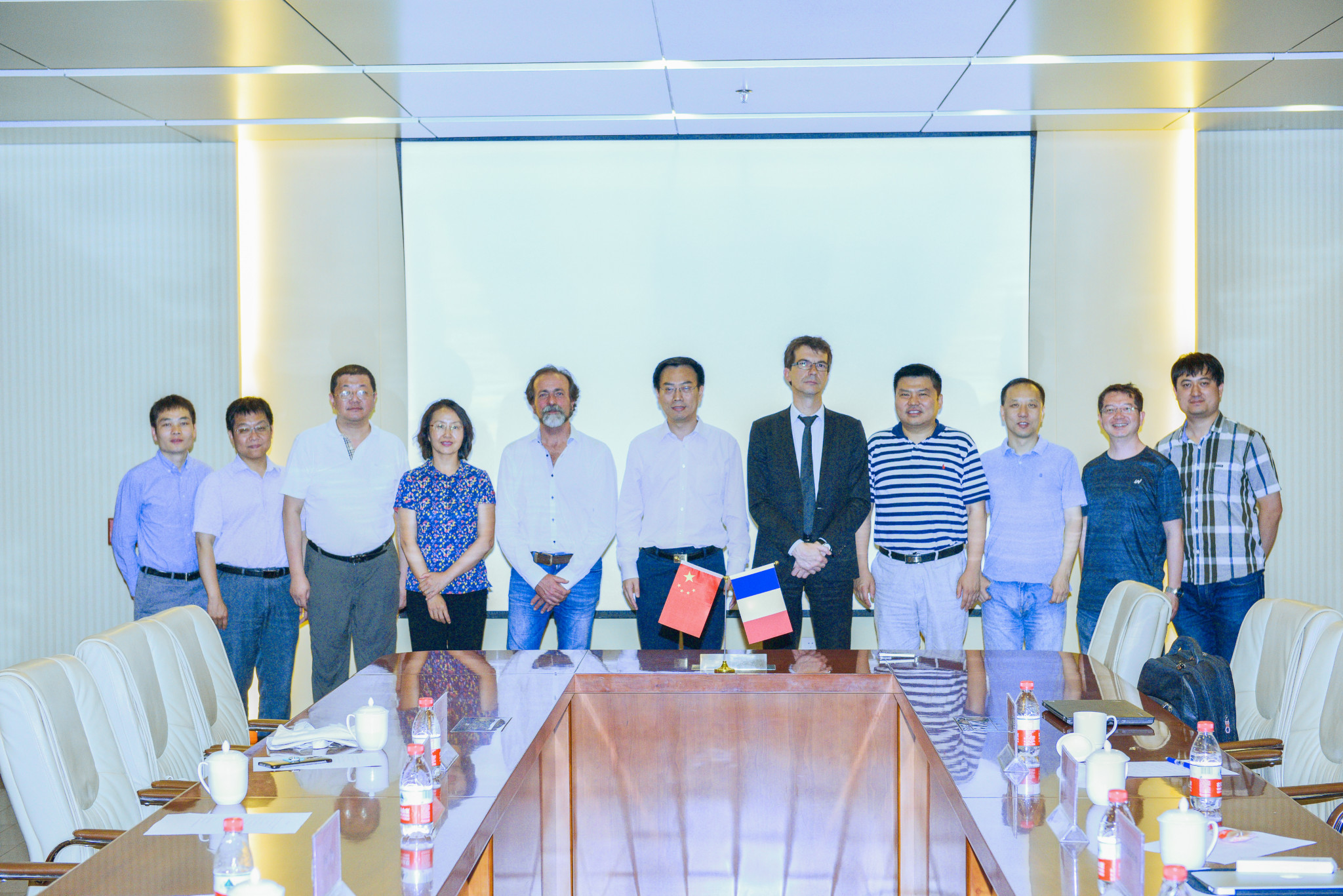
To reinforce joint research activities and publications;
To develop joint training activities for diverse categories of personnel, including imaging core facility staff;
To exchange information and materials in those fields which are of interest to both parties;
To organize joint conferences and academic programs;
To develop grant proposals for joint research, infrastructure development (center and/or consortium);
To foster technology transfer between each parties.
Several French institutions will take part in the partnership. We hope that the France BioImaging users and partners will be able to benefit from this partnership starting in 2018.
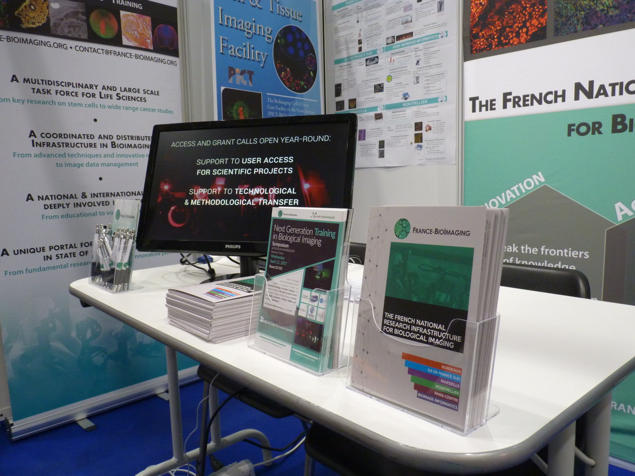
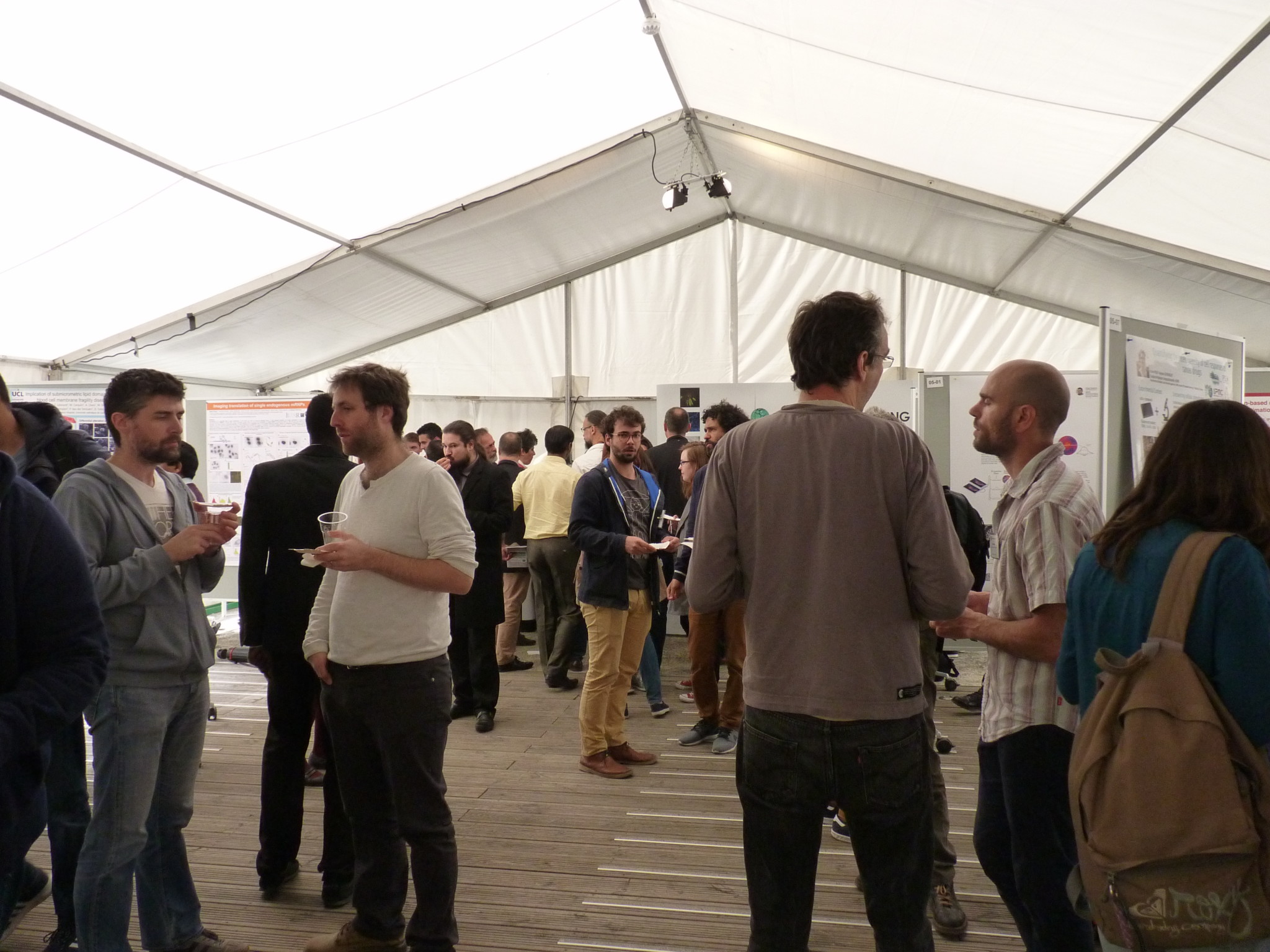
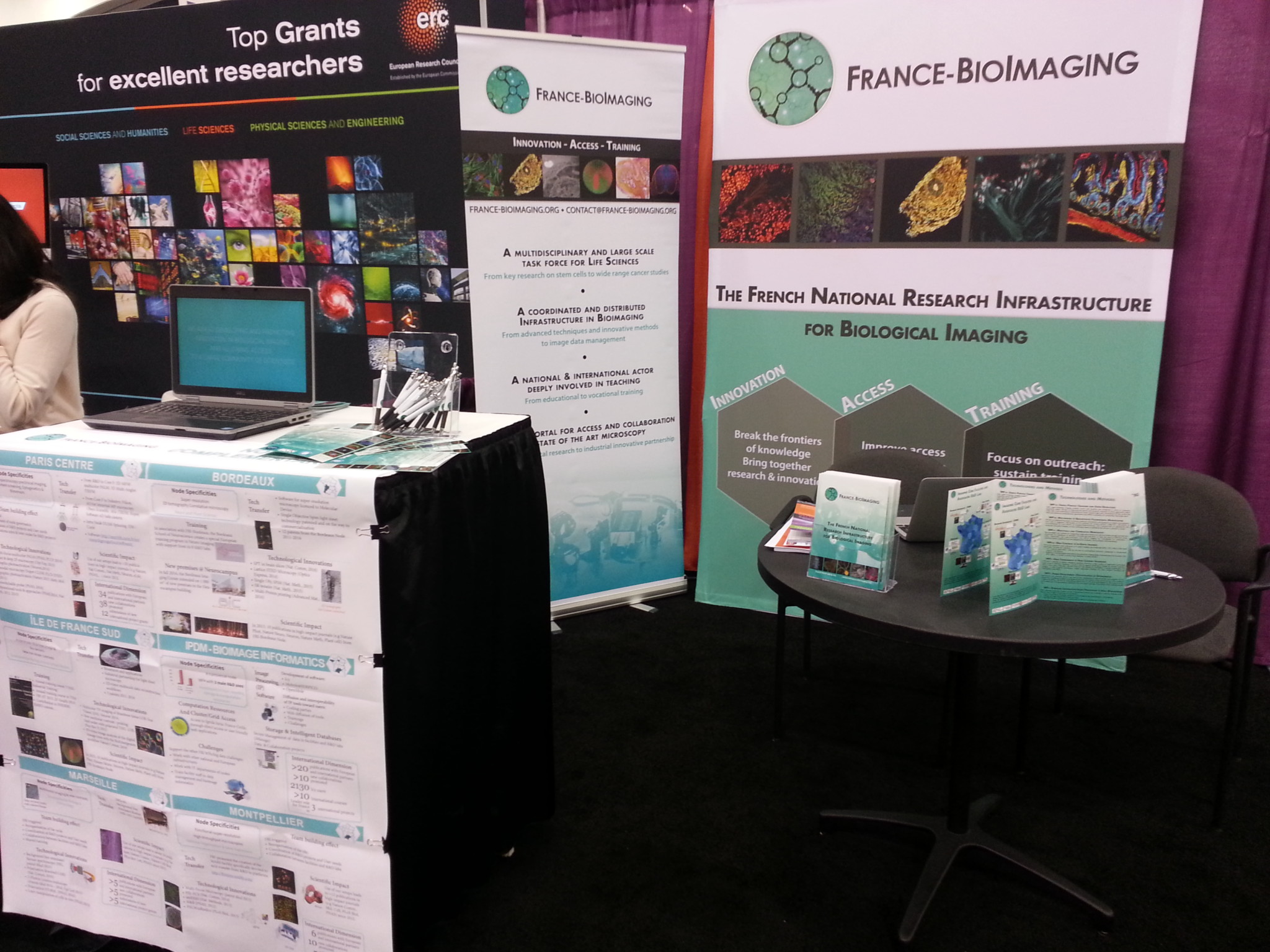
This year, the Focus on Microscopy Conference took place in sunny Bordeaux, France, from April 9th to April 12th. France BioImaging participated in two ways: with a booth in the exposition hall, and through a symposium co-organized with Euro BioImaging, held on Wednesday 12th.
Many curious attendees stopped by our booth to enquire about our activities. In particular, foreign visitors were curious to know how the infrastructure was organized, as they expressed the intention to set up similar services in their own countries. Industrials were equally intrigued by our organization, and expressed the desire to work with us towards the dissemination of their new available technologies.
Volunteers from the Bordeaux Node gracefully assisted the coordination team in setting up & animating our booth.
Symposium on Training
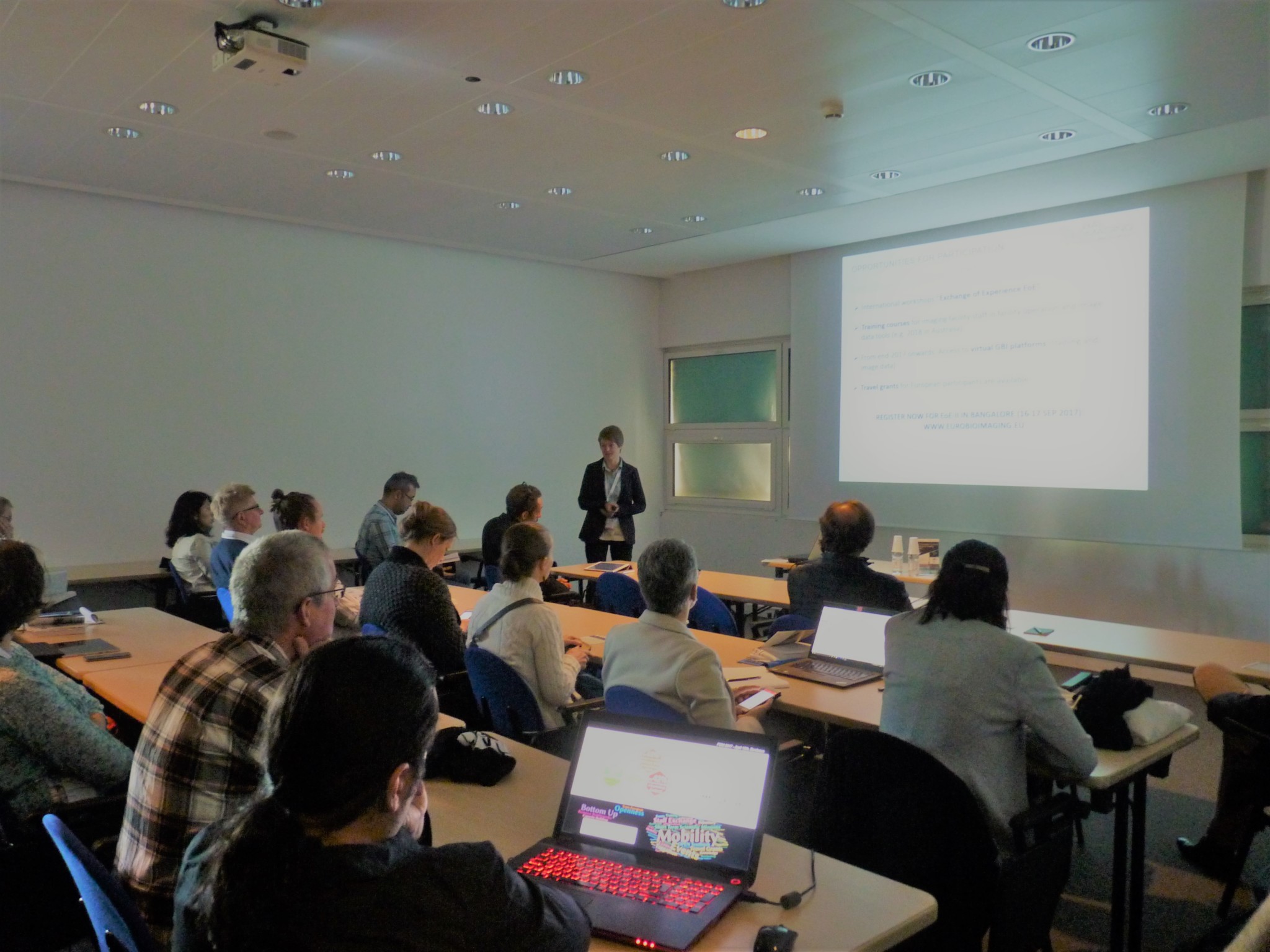
With more than 40 attendees, the Symposium on Training (Wednesday 12 April) was a great success. The public and speakers shared fruitful ideas about the past, present and future of biological imaging training at the European level.
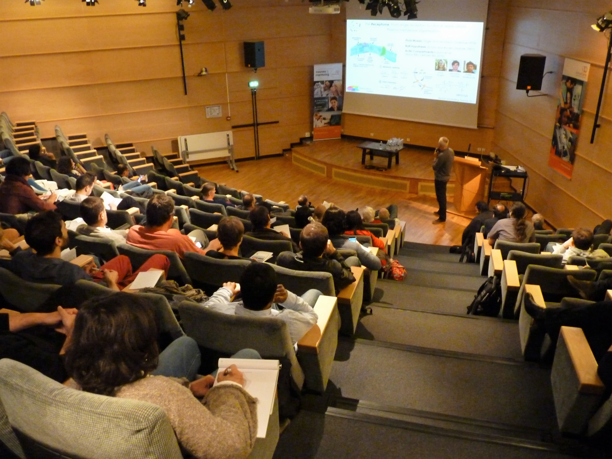
Our 4th Annual Meeting took place on Friday April 14th 2017. We had the pleasure of welcoming about 80 participants at the Curie Institute. This year, the meeting focused on the question of future challenges in biological imaging.
The four sessions of the program were centered around the following topics:
Quantification of the molecular dynamics and coordination in cells and small organisms, including at the nanometer scale
Imaging architectures and processes of life, from molecular complexes to multi cellular systems
New frontiers for imaging, sensing, and controlling biomolecules
Bioimage informatics, image processing and microscopy data management
At lunch time, the room was buzzing during the poster session.
Find all the details of the organization & program here.
The FBI Marseille node is composed of the PICsL light and electron microscopy core facility and three associated research and development (R&D) labs. The FBI-PICsL facility is located on the Marseille Luminy campus and hosted by two institutes: the Centre d’Immunologie de Marseille Luminy (CIML) and the Institut de Biologie du Développement de Marseille (IBDM). The R&D labs comprise two labs of these institutes (Lenne lab at IBDM and Marguet lab at CIML) and the biophotonics lab of the Fresnel institute (Mosaic group led by H. Rigneault). The FBI-PICsL facility via its microscopy resources is dedicated to help its users deciphering cellular mechanisms in the fields of cell biology, immunology and developmental biology.
The PICsL staff. From left to right : Fabrice RICHARD, Aïcha AOUANE, Nicolas BROUILLY, Sébastien MAILFERT, Cédric MATTHEWS, Rémi FLORES-FLORES, Elsa CASTELLANI, Mathieu FALLET, Claire CHARDES, Brice DETAILLEUR, Roxane FABRE.
In 2017, the FBI-PICsL facility comprises 40 setups. Altogether, these microscopes cover a wide range of temporal and spatial scales from the second to milliseconds and from single molecules to entire organisms. This repertoire of microscopes is composed of cutting-edge light microscopes (FCS, FCCS, STED 3D, PALM/dSTORM, Fast polarization-resolved spinning disk, Light sheet microscopes) among which 8 are homemade and 3 electron microscopes (two TEMs and one SBF-SEM). Ten staff members manage the facility under the scientific supervision of Pierre-François Lenne (IBDM) and Didier Marguet (CIML). The FBI-PICsL facility staff is also involved in teaching microscopy techniques to the facility users and within courses open to external academic and industrial users.
New set-ups on the FBI-PICsL facility
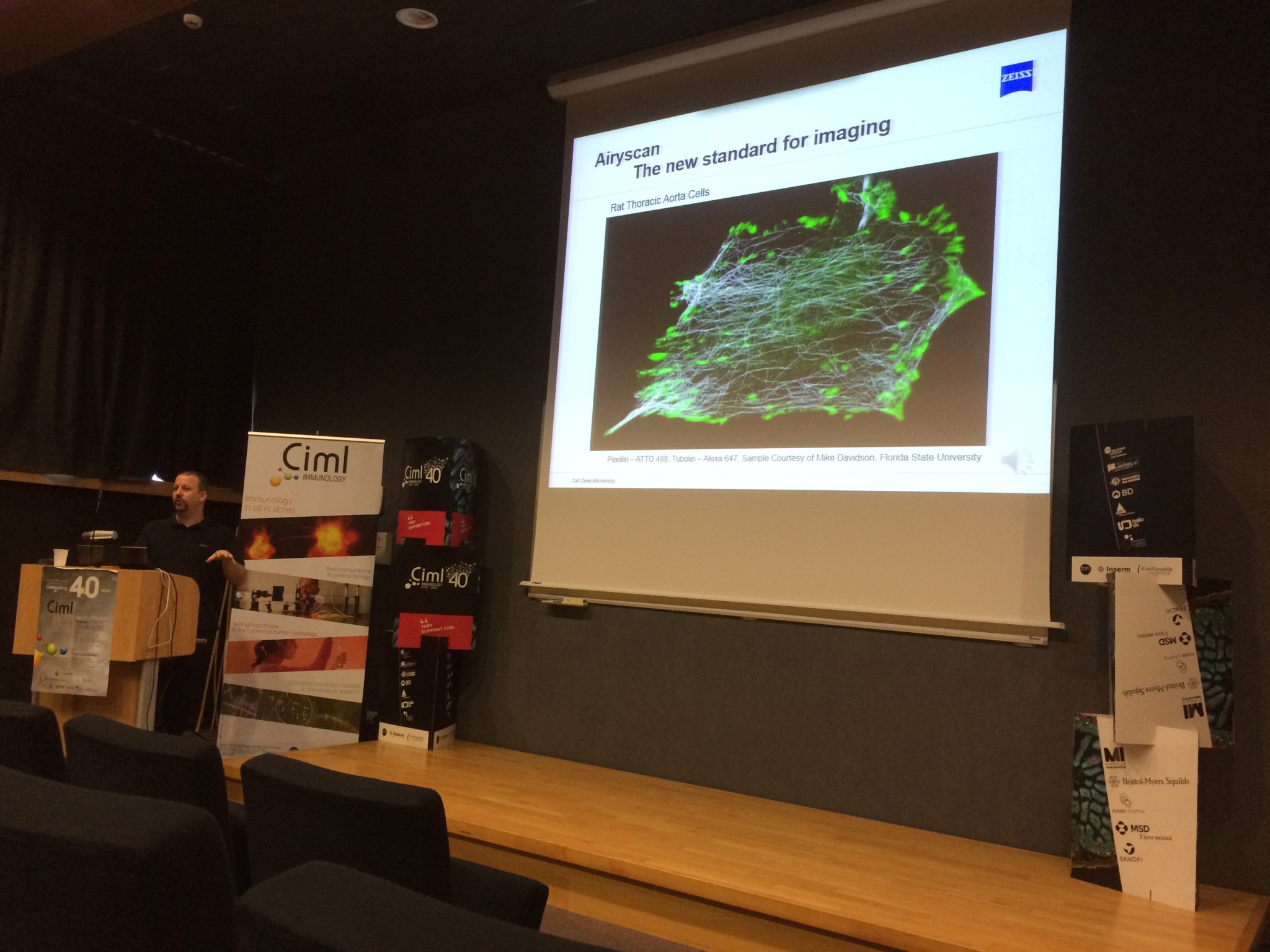
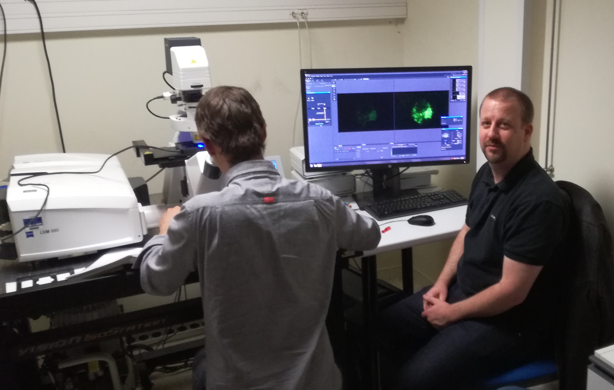
LSM 880 Fast-Airyscan
Photo credits: Carl Zeiss Microscopy GmbH
A new confocal microscope using the technology Airyscan (LSM 880 Fast-Airyscan, Zeiss) has been purchased through a co-funding FBI/Inserm/CNRS/Region PACA. The Airyscan improves spatial resolution (1.7 x) by imaging the Airy disk onto a concentrically arranged hexagonal detector array. Its detection area consists of 32 single detector elements, each of which acts like a very small pinhole. The confocal pinhole itself remains open and doesn’t block light – thus all photons of the whole Airy disk are collected. The signals from all detector elements are then reassigned to their correct position, producing an image with increased signal-to-noise ratio and resolution.
The FBI-PICsL facility also recently homed an electron microscope dedicated to automated 3D imaging.
Revealing the complex 3D architecture of cells and tissues is crucial for the structure-function correlation in biological systems. However, in most of the electron microscopy experiments, only a small portion of the sample is imaged. For instance, one 70nm-section of an average vertebrate cell only represents 0.4-1.0% of its volume. Beyond the fact that this tiny volume might not be representative to account for the entire cell ultrastructure, the sectioning also avoids a definite visualization of the cellular organization.
Until recently, the imaging of big volumes (tens to hundreds of cubic micrometres) by electron microscopy was tedious because it involved manual serial sectioning. Beyond the need of super-skilled people, manual serial sectioning has a limited z resolution of 70nm and is a time-consuming process at the sectioning, imaging and post-processing steps. Within the last 10 years, the Serial Block-Face Scanning Electron Microscopy (SBF-SEM) has emerged as the method of choice for the imaging of big volumes by electron microscopy.
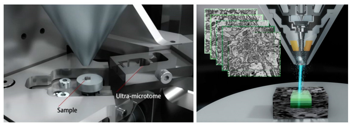
Photo credits: FEI
The concept behind SBF-SEM is to have a microtome within the chamber of a scanning electron microscope (see Figure). This set-up enables the iterative sectioning and imaging of the sample block-face for hundreds of microns in a fully automated way (Figure). When an electron beam hits a sample, it interacts with it and yields several signals that can be detected and assigned back to the scanned positions on the sample surface. Among these signals, the backscattered electrons can be collected to retrieve information on the sample electron density at a given point. Routinely, a working pixel size in SBF-SEM datasets is ±5nm and the slicing can be as thin as 20-30 nm.
In addition to be an excellent microscope in classical SEM mode, the Teneo VS microscope by FEI is currently the most advanced microscope to carry out SBF-SEM. It does not only allow you to cut and image the sample in a fully automated way but it also gives the possibility to avoid charging artefacts during imaging by putting the sample in low vacuum conditions. By limiting the charging, the microscope gives you the opportunity to retrieve more information from the sample. For instance, the sample can be scanned with different acceleration voltages in order to retrieve ultrastructural information at different depths (10nm – 20nm – 30nm – 40nm). When this multi-energy imaging is combined to a 40nm mechanical slicing, it gives rise to previously inconceivable datasets of hundreds of cubic microns of tissue with a voxel size of 5x5x10nm !
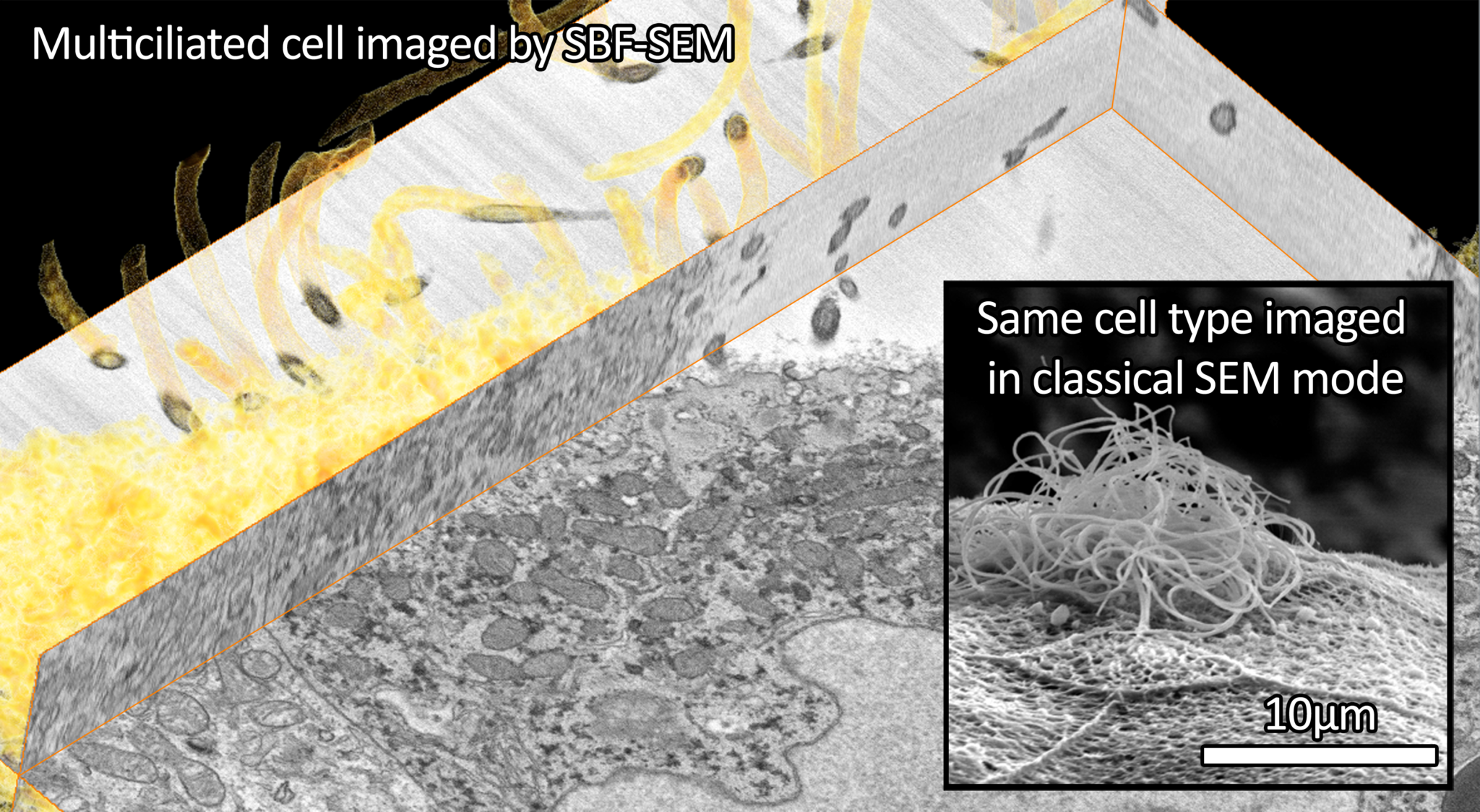
Since the delivery of the FEI Teneo VS microscope at the FBI-PICsL facility in December 2016, we tested the microscope on a broad range of biological samples in classical SEM mode (fly egg-laying apparatus, Xenope embryo, mouse face…) and in SBF-SEM mode (cell pellets, Drosophila muscle, mouse brain, marine sponges, Xenope embryo…). In particular, the group of Laurent Kodjabachian (IBDM), working on multiciliated cells, wanted to image the same cell type in both classical SEM mode and in SBF-SEM. We could 1. assess the penetrance of a mutant phenotype in classical SEM mode and 2. localize the basal bodies of these multi-ciliated cells in SBF-SEM (see Figure below).
Photo credits: Virginie Thome and Nicolas Brouilly
Major implications of the FBI-PICsL facility
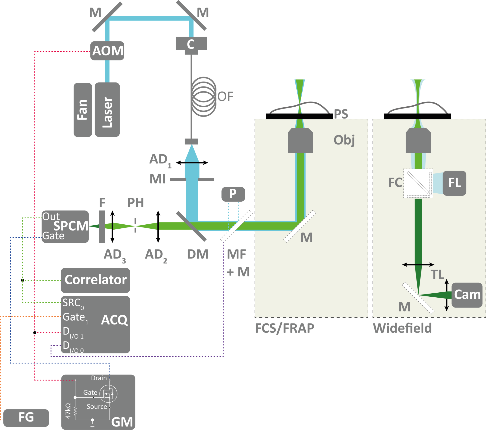
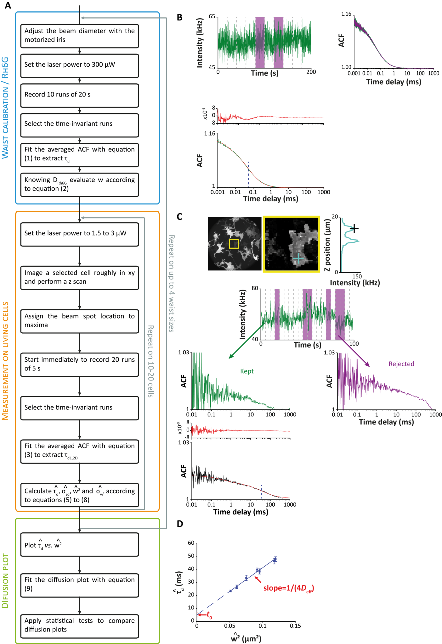
Spot variation fluorescence correlation spectroscopy (svFCS) (Hai-Tao He and Didier Marguet’s team)
In collaboration with Nicolas Bertaux (Institut Fresnel, AMU, Centrale Marseille), members of the Hai-Tao He and Didier Marguet team published a user guide for characterizing plasma membranes subdomains in living cells by sv-FCS2. This book chapter aims to serve as a guide for setting and applying the svFCS methodology to study the plasma membrane of both adherent and nonadherent cell types.
Optical setup for svFCS and fluorescence recovery after photobleaching (FRAP).svFCS recording and data analysis workflow. (Click to view the image in full size)
This technique has been used to understand a genetic disease by studying molecular diffusion at the plasma membrane. In collaboration with Christophe Lamaze (Institut Curie) and Céline Galès (Institut des Maladies Métaboliques et Cardiovasculaires de Toulouse), Hai-Tao He and Didier Marguet’s team worked on decoding the dysfunction of molecular mechanisms involving interferon-γ (IFN-γ), key protein for immune defense. This protein is not able to fulfil its natural function as a protective cytokine when its receptor is “blocked” in the wrong place on the cell membrane: a modification to the immune response causing severe infections sometimes even life-threatening for young children.
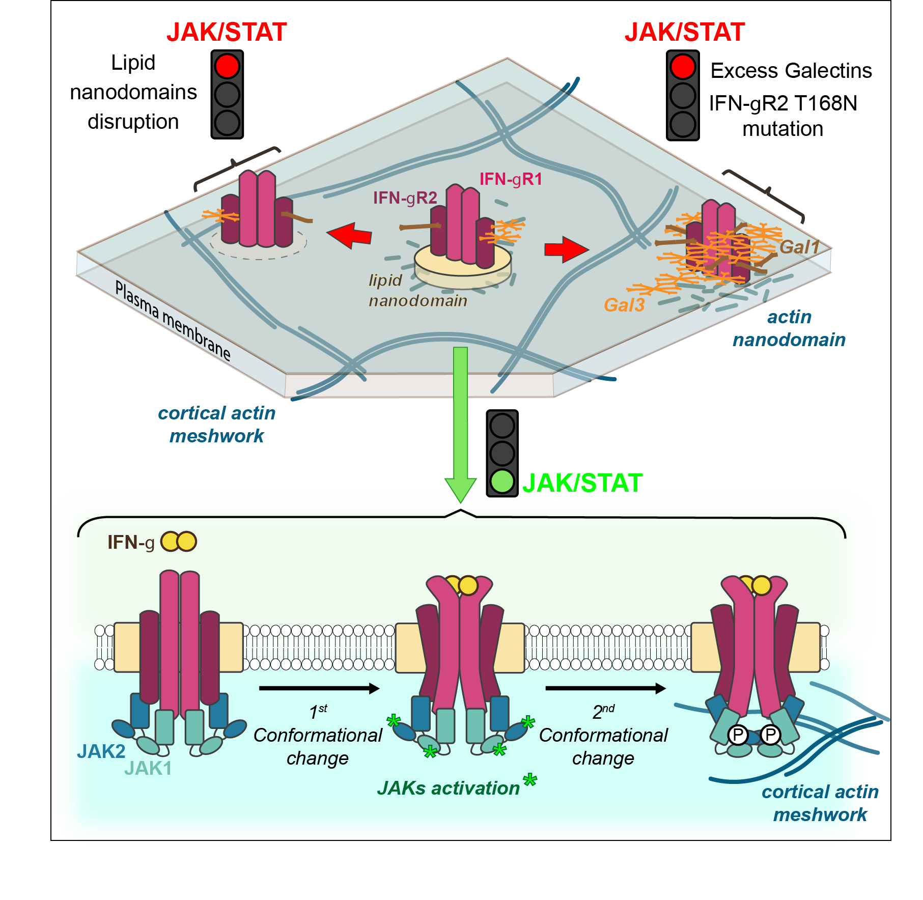
Glycosylation-Dependent IFN-γR Partitioning in Lipid and Actin Nanodomains Is Critical for JAK Activation
IFN-γ receptor is composed of two protein chains to which are generally associated six sugars. However, some patients diagnosed with Mendelian susceptibility to mycobacterial diseases syndrome (MSMD) have seven sugars. This simple gain of glycosylation affects greatly the immune response against mycobacteria.
Using their expertise in biophysics and more particularly in svFCS, this CIML team showed the existence of nanodomains and their fundamental role in signaling activity. “It was completely unexpected that only one additional sugar could modify the way the receptor is localized on the surface of the cells, affecting greatly the later signaling events”, explained Hai-Tao He. “IFN-γ receptor is then delocalized towards another membrane nanodomain populated by galectin proteins, that bind to this additional sugar” added Yannick Hamon2 (cf Figure)
As therapeutic target, galectins are an appealing perspective, particularly as turning off their expression canceled in vitro the pathologic effect linked to the glycosylation gain of the IFN-γ receptor. However, therapeutic programs towards patients requires the exploration and understanding of molecular mechanisms by uniting expertise of biologists and physicians but also mathematicians and physicists as it was the case in this discovery4.
News
In March 2017, a workshop on super-resolution is organized in Marseille by Sébastien Mailfert, Jean-Bernard Fiche and Orestis Faklaris on multicolor STORM an dSPT-pointillism data analysis, this workshop is dedicated to advanced researchers in this field. This event is supported by FBI.
The FBI-PICsL facility is also homing the 14th RTmfm convention from the 20th to the 22nd of March 2017. This convention is a great opportunity for imaging facilities staff to share their experiences and ideas on the past, present and future of their work (technical development, big data issues and solutions, link with industry).
Our facility also yearly organizes the following course: Scientific platform, instrumental sharing, how to build up and develop a service. The aim of the course is to acquire, by alternating courses, exercises and practical conditions, methods and constraints to the development of a provision of scientific equipment service. We insist on how to develop a contributory model for managing a scientific platform.
References
Mailfert, S., Hamon, Y., Bertaux, N., He, H-T. & Marguet, D. Methods in Cell Biology, 139, 1-22 (2017)
Blouin, C., Hamon, Y. et al. Cell, 166, 920-934 (2016)
Reporting on the 1st international training course on “Management and Operation of Imaging Core Facilities”, Global BioImaging H2020 Project November 16-18, EMBL, Heidelberg
20 participants attended this first course, representing European and international facilities (Argentina, Australia, India, South Africa). The course was organized around 4 sessions (Soft Skills for Core Facility Staff; Administrative Skills, Quality Management, Metrology; How to set-up an Imaging Core Facility (exchange workshop); E-learning/hands-on in virtual training platform) with a strong participation of the French Community members as speakers. All sessions were overall well received, as suggested by the feedback collected via survey. Some adjustments were however proposed that should frame the next GBI Training Course (planned in Australia, summer 2018). In the meantime, GBI will also organize the 2nd workshop “Exchange of Experience” for core facility managers, 15-16 September 2017 in Bangalore, India and will continue to develop its “shadowing” program (personal exchanges between imaging facilities worldwide; second session March – September 2017). Stay tuned! For details, read more on the Euro BioImaging website.
France BioImaging was present at the ASCB 2016 meeting in San Francisco (December 3-7). It was a great occasion to present our infrastructure on our booth and to draw future strategies in BioImaging for Cell and Development Biology.
The France BioImaging booth at ASCB 2016 (Click the image to view)
We also interacted with scientific organizations and funders, such as the Howard Hughes Medical Institute, the National Science Foundation and the Gordon and Betty Moore Foundation to whom we presented our R&D programs as well as the overall FBI organization. Most of them were amazed by what we could propose and surely other meetings and new programs could emerge from those informal although in depth discussions.
Beyond the above type of contacts, young scientists were attracted by FBI possibilities in terms of training capacity, accessibility to the most emerging technologies, software platform and lots of them were asking about FBI PhD and Post-doc programs. Certainly a prospect we could mine in the future.
As a general feedback on what were the active new fields of interest in the scientific area of the congress, let us mention the obvious developments in the Biology of Induced Pluripotent stem Cell (IPCs), CRISPR/Cas genome editing techniques, cell mechanics approaches and structure/function of macromolecular complexes in living cell and organisms. It is clear that FBI anticipation in developing super-resolution, optogenetics, multi-scale and highly sensitive imaging techniques will best serve our research teams focusing their interest in these diverse fields.
Finally, as many of our foreigner colleagues mentioned, attendance of French researchers was quite remarkable at ASCB this year. A number of them gave talks that were highly appreciated. A majority of them were coming from the direct perimeter of the FBI Nodes and clearly benefited from or even participated to the development of advanced technologies in imaging provided by France BioImaging.
The BIC is setting up in a brand new space
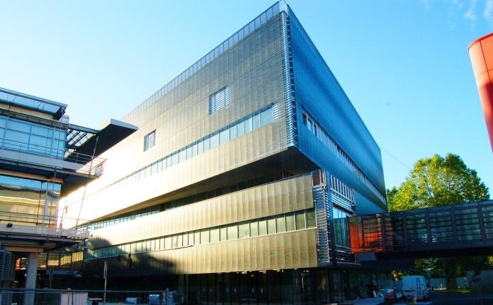
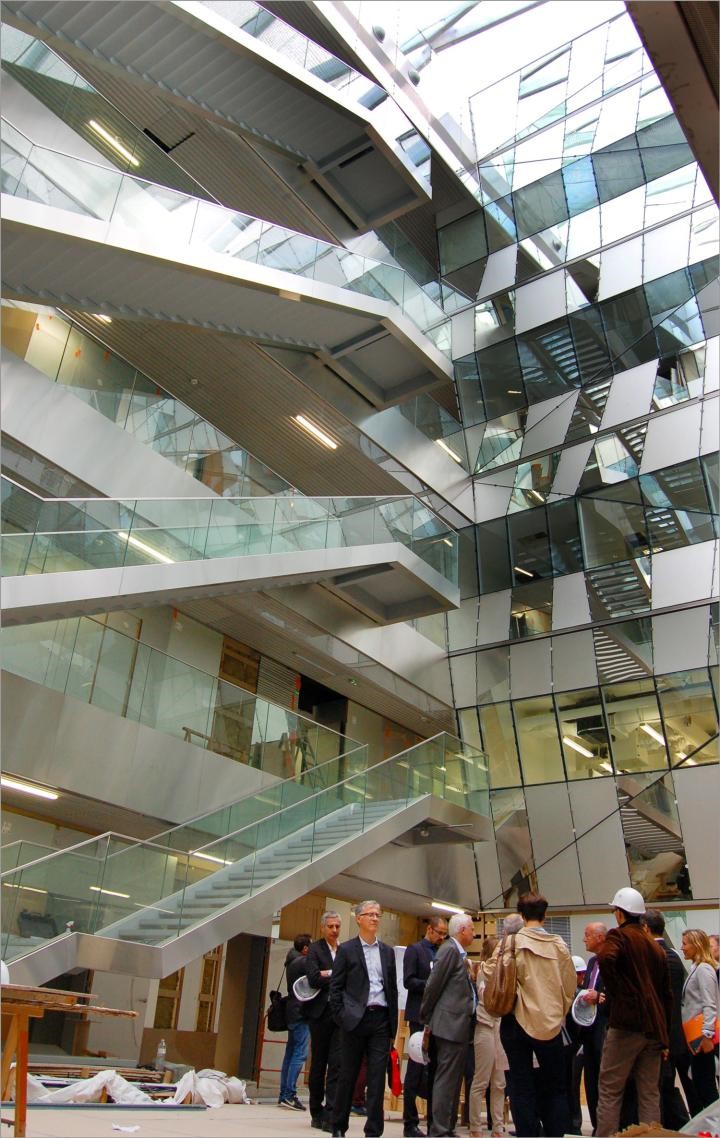
In the last weeks of October 2016, the BIC has settled in a brand new building, constructed by the Regional Council of Aquitaine as part of the Neurocampus project. This building, of around 13 000 m2, is shared with the Interdisciplinary Institute for Neuroscience (IINS) and the Institute for Neurodegenerative Disorders (IMN). This building, constructed in two years, cost 47 M€ and is part of a large project to develop Neuroscience and imaging in Aquitaine. The new building is conveniently located and connected by footbridges between the Magendie Neuroscience center and the Center for functional genomics (CGFB) that hosts several core facilities.
In total, the BIC will occupy 1000 m2, split between the CGFB and the new building. The major part in the new building is dedicated to photonic microscopy. Electron microscopy instruments, including two brand new ones coming in 2017, will be dispatched between the CGFB and Neurocampus building. In these new spaces, users have access to a culture room and also a room with analysis stations. Other rooms are dedicated to each kind of microscopy (one room for live cells imaging, one room for multiphoton, one room for confocal, one room for new scanning electron microscope etc…). Special rooms are dedicated to host R&D projects as well as confidential collaborations with industry.
Development of training capacities at the BIC – joint projects with the Cajal School of Neuroscience
The BIC has engaged for many years in active training programs for imaging at all levels (beginners to advanced training) for local, national and transnational users. The BIC personnel also participates extensively to various theoretical and hands on training/showcase activities in France and abroad (MifoBio, NeuBias, etc…). Within the strategy to develop the BIC-FBI training, we are engaging a partnership with the Cajal Advanced Neuroscience Training Program to develop special ima ging training for Neuroscience. The Cajal school is a European FENS and IBRO initiative in partnership with Bordeaux Neurocampus and the Champalimaud Foundation, which offers state-of-the-art hands-on training courses in neuroscience.
Construction of a light sheet microscope for super resolution imaging inside living samples
Fast and non-damaging imaging of single molecules inside live organisms is essential to study physiologically relevant biochemical mechanisms occurring at the subcellular level. For example, the dynamic organization of transmitter receptors at the membrane of excitatory neurons should, ideally, be studied in vivo in the brain of animal models. Unfortunately super resolution techniques such as PALM1, STORM23 and uPAINT4 are mostly restricted to the sample external surfaces and are unable to image inside live samples.
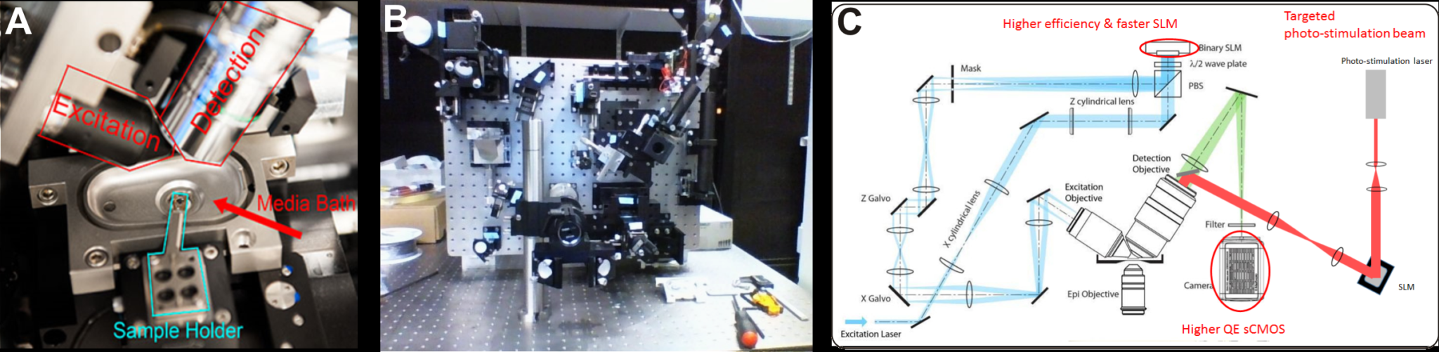
For these reasons the Bordeaux Imaging Center is developing a new light sheet microscope specially dedicated to image single molecules into live samples. Light sheet fluorescence microscopy (LSFM) is recognized as the method of choice to image thick live samples. Compared to other fluorescence imaging modalities such as wide field, confocal, structured illumination, two-photon or STED, LSFM strongly reduces out of focus fluorescence, decreases photobleaching and phototoxicity, and improves temporal resolution. Among the numerous technical implementations of LSFM 5, we decided to build a lattice light sheet microscope (LLS) because it has been specifically designed to perform super resolution imaging in thick live samples 6. Indeed In LLS the illumination beam is shaped by a spatial light modulator (SLM) to produce a < 1 µm thick excitation plane over a length of > 50 µm at the sample. A 1.1 NA detection objective ensures efficient light collection required for high localization precision. Illumination and detection objectives are both long working distance and water immersion, thus allowing observation of live samples up to 5 mm in diameter. (Fig 1 A)
Our LLS microscope is mostly based on the documentation freely and kindly shared by Eric Betzig’ group (HHMI Janelia Farms, USA).
Photo Credits: Mathieu Ducros
Fig 1. (A) The sample is placed at the intersection of the excitation and detection objective optic axes in a temperature controlled perfusion chamber. It is held at the tip of motorized arm on a 5 mm diameter cover slip (from 6). (B) The LLS microscope under construction in June 2016. (C) In blue and green the optical path of the excitation and detection beams respectively (from 6). A higher efficiency SLM, higher QE camera should improve the light budget compared to the original specifications. In addition, a targeted laser beam (red) will allow precise photo-conversion of light sensitive molecules.
We made a few modifications compared to the original specifications of the LLS as described in 6 : our microscope will be equipped with a laser combiner including 4 high power lasers at 405 nm (300mW), 488 nm (1 W), 560 nm (2 W), 642 nm (2W), a higher efficiency SLM (Fourth Dimension DD QXGA) and a sCMOS camera with improved quantum efficiency (Hamamatsu ORCA Flash V2). These improvements should mitigate the weak throughput of the LLS beam path, and, in turn, improve molecule localization precision and/or time resolution. In addition, a targeted photostimulation beam will be coupled through the detection objective to photo stimulate or photoconvert with a high spatial and temporal resolution photosensitive molecules.
STORM, PALM and PAINT imaging modalities will be fully compatible with the constructed LLS.
The microscope construction by Mathieu Ducros, INSERM research Engineer on the BIC, started in April (Fig 1B). First images are expected by the end of 2016. Once our LLS is fully operational and running, it will be accessible to all BIC users under the supervision of a local engineer.
For this project we are supported financially by the GIS IBiSA, LABEX brain and FBI.
References
Betzig, E. et al. Imaging intracellular fluorescent proteins at nanometer resolution. Science 313, 1642–1645 (2006).
Rust, M. J., Bates, M. & Zhuang, X. Sub-diffraction-limit imaging by stochastic optical reconstruction microscopy (STORM). Nat. Methods 3, 793–795 (2006).
van de Linde, S. et al. Direct stochastic optical reconstruction microscopy with standard fluorescent probes. Nat. Protoc. 6, 991–1009 (2011).
Giannone, G. et al. Dynamic superresolution imaging of endogenous proteins on living cells at ultra-high density. Biophys. J. 99, 1303–1310 (2010).
Santi, P. a. Light sheet fluorescence microscopy: a review. J. Histochem. Cytochem. 59, 129–138 (2011).
Chen, B.-C. et al. Lattice light-sheet microscopy: Imaging molecules to embryos at high spatiotemporal resolution. Science (80-. ). (2014). doi:10.1126/science.1257998
On September 22nd, 2016, a joint meeting took place between the FBI coordination, IPDM node and other partners, to discuss the implementation of data management services within FBI. Please find below the main points discussed during the meeting.
Objective – Setting the required elements for public access to different types of Image Data within FBI.
Purpose and expectations –to have, in one year’s time, a successful proof of concept for FBI. This topic is of particular importance for FBI, as it will soon be required of any project funded with public money to provide data management plans. In the very short term, infrastructures will likely be expected to guarantee the safe conservation and fast access to any data supporting a publication (see for example the Netherlands protocol).
Audience – two different audiences were considered as targets of the project: users within the FBI perimeter, and the general public (fully public access); the former being more feasible in a year’s time than the latter.
Proposals
WP1a Support
Support the work of WP1a in their benchmarking action (Contact: Orestis Faklaris);
Being workflow-oriented, so that the methods defined to support WP1a can be applied to other WPs.
Metrology-oriented database
Instrument-based metrology database: asking each site to share metrology benchmarking of microscopes from 2016, including software used when any;
At midterm: go to real biological object to associate metrology data as quality metrics;
Before the end of the project: include metrology oriented annotations to WP1a support;
Long term: include metrology-oriented annotations to open image data repositories.
Open Image Data Repository:
Hosting 2016 publications acknowledging FBI, uploading raw data at least supporting published figures.
Resources: Remaining funds from Pasteur IPDM could be mobilized to carry out the project (the funds would go towards financing the equipment and staff). Coordination funds will be allocated to the employment of an engineer for a period of approximately 9 months.
Meeting Participants:
Volker Baecker (Montpellier), Anatole Chessel (Paris Sud), Stéphane Dallongeville (IPDM Paris Centre), Anne Danckaert (Paris Centre), Elaine Del Nery (Paris Centre), Nimisha Gupta (OpenImadis Bengalore), Cédric Matthews (Marseille), Perrine Paul-Gilloteaux (IPDM), Jean Salamero (Coordination), Corinne Tessier (Coordination).
The Global BioImaging project entails an international job shadowing program that aims to give the opportunity to the project’s stakeholders to visit imaging facilities across the globe and learn from their peers.
The program will allow both the hosting facilities and their guests to exchange experiences and ideas, while working on innovative imaging technologies and the related technical aspects. It also has the added value to support networking and prepare possible future collaborations between imaging infrastructures.
The call for the first GBI international shadowing program is now open!
Deadline for submission of the applications is 31/10/2016.
NEUBIAS, the network of European BioImage Analysts funded by the EU framework COST, has opened a new activity that will run continuously for 4 years, and is centered on mobility grants, also called Short-Term Scientific Missions.
The first call deadline is the 10th of September, but several other call will be regularly published.
BioImage Analysts and Life Scientists, from Research Labs AND Core facilities, can apply for funds to cover their expenses when visiting a Host-Lab in a different country (Lab or industry) where they will perform a short scientific project strictly focused on BioImage Analysis, and that should enable:
1) collaborations on innovative image analysis methods,
2) access to big data analysis technology and/or image analysis tools for scientists lacking them locally,
3) knowledge transfer to support careers and regional development.
Already before year 2016, FBI was involved in many teaching and training activities. With the care about “not reinventing the wheel”, FBI first brought its support to running activities in this domain at the national level. For this reason, it was decided to bring recurrent help to the MiFoBio school of the CNRS, organized by the GDR MIV since 2004. Moreover and similarly, FBI is co-organizing the FBI-Advanced Training sessions, also with the GDR MIV. These two 5-9 days events are highly valuable and are recognized as such by a large community, far beyond core facility staffs. They are largely focused on theory and hands-one training and on applying this knowledge to specific biological questions. In 2016, FBI was a pushing force to give an international visibility to both actions. Both concepts merit a stronger international impact as other workshops or training series do, like EMBL-EMBO or Woods Hall workshops and courses. Taking advantage on diverse opportunities at the European and International level together with other main international actors in this topic (H2020, EuBI, NEUBIAS, FENS CAJAL), FBI now drafted a strategy at an upper level, with the aim at defining a complete “train the trainers” portfolio.
Let us mention some almost achieved deliverables: organizing surveys on Training Activity and Training Sites for Core Facility Staff and User within the European landscape followed by the proposition of new and necessary “trainings” (H2020 WP7 EuBI PPII) which will be presented during the 1st EuBI Core Facility Staff meeting (30th of October beforehand the MiFobio 2016), including organizing virtual (e-training), soft and management skills courses, hands-on training.
In this respect, France BioImaging co-organizes and will also teach at the 1st International Training Courses for Imaging Core Facility Staff of the Global BioImaging project (deadline 18th September), next November 2016 in Heidelberg (GBI International Training Courses for Imaging Core Facility Staff) which will adress two specific issues:
-“Challenges in image data management and analysis”
-“Management and operation of imaging core facilities”
Our experience in international course activities, expertise, equipment of our Local-Nodes with state of the art imaging techniques, allow us to propose the largest and most advanced portfolio of “à la carte” trainings in BioImaging over Europe. As a future step, France-BioImaging is supporting the opening of a call for “EuBI Training Nodes” in which FBI will propose itself as a “Training Node”. FBI is in a very strong position to propose an integrated Training offer in bioimaging. The Marseille and Bordeaux Nodes are currently building Training Centers. In Paris Centre, an Imaging Training common program is part of the recent official partnership between Institut Pasteur and Institut Curie (February 2016). Joint ventures between Marseille and Montpellier nodes and inclusiveness between Paris Centre and IdF Sud nodes teaching programs are foreseen.
We use cookies on our website to give you the most relevant experience by remembering your preferences and repeat visits. By clicking “Accept All”, you consent to the use of ALL the cookies. However, you may visit "Cookie Settings" to provide a controlled consent.
This website uses cookies to improve your experience while you navigate through the website. Out of these, the cookies that are categorized as necessary are stored on your browser as they are essential for the working of basic functionalities of the website. We also use third-party cookies that help us analyze and understand how you use this website. These cookies will be stored in your browser only with your consent. You also have the option to opt-out of these cookies. But opting out of some of these cookies may affect your browsing experience.
Necessary cookies are absolutely essential for the website to function properly. These cookies ensure basic functionalities and security features of the website, anonymously.
Cookie
Duration
Description
cookielawinfo-checkbox-analytics
11 months
This cookie is set by GDPR Cookie Consent plugin. The cookie is used to store the user consent for the cookies in the category "Analytics".
cookielawinfo-checkbox-functional
11 months
The cookie is set by GDPR cookie consent to record the user consent for the cookies in the category "Functional".
cookielawinfo-checkbox-necessary
11 months
This cookie is set by GDPR Cookie Consent plugin. The cookies is used to store the user consent for the cookies in the category "Necessary".
cookielawinfo-checkbox-others
11 months
This cookie is set by GDPR Cookie Consent plugin. The cookie is used to store the user consent for the cookies in the category "Other.
cookielawinfo-checkbox-performance
11 months
This cookie is set by GDPR Cookie Consent plugin. The cookie is used to store the user consent for the cookies in the category "Performance".
viewed_cookie_policy
11 months
The cookie is set by the GDPR Cookie Consent plugin and is used to store whether or not user has consented to the use of cookies. It does not store any personal data.
Functional cookies help to perform certain functionalities like sharing the content of the website on social media platforms, collect feedbacks, and other third-party features.
Performance cookies are used to understand and analyze the key performance indexes of the website which helps in delivering a better user experience for the visitors.
Analytical cookies are used to understand how visitors interact with the website. These cookies help provide information on metrics the number of visitors, bounce rate, traffic source, etc.
Advertisement cookies are used to provide visitors with relevant ads and marketing campaigns. These cookies track visitors across websites and collect information to provide customized ads.