The Executive Board of the Rhône-Alpin Node of France-BioImaging is pleased to invite you to the event “Imaging & Microscopy Day in Rhône-Alpes – Image Analysis” – pre-program attached.
It will be held on Tuesday, July 1st at the Faculté Rockefeller, 69008 Lyon
As the number of seats is limited, please register as soon as possible to best organize the final program!
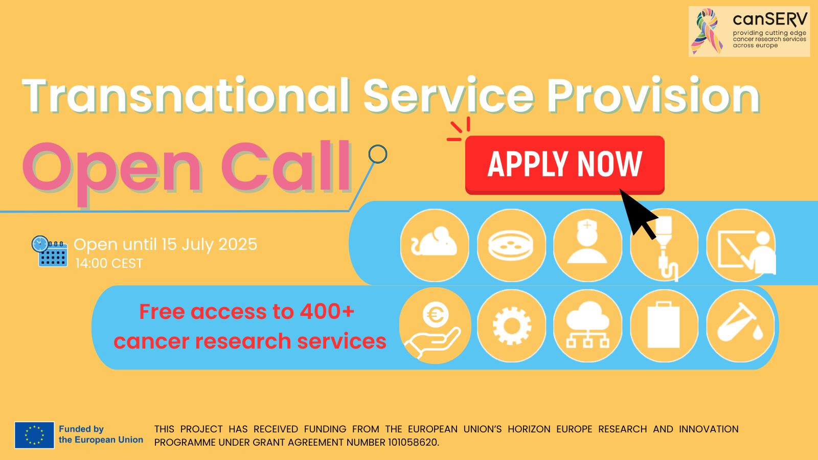
Last weeks to apply to the last canSERV Open Call, a unique opportunity designed specifically for early career cancer researchers worldwide. This initiative aims to provide these researchers with access to cutting-edge services and training, helping them advance their groundbreaking work in cancer research.
Who can apply?
This call is open to:
First-stage researchers: PhD students and junior researchers without a PhD.
Recognised researchers: Postdoctoral fellows, assistant professors or young investigators.
What’s on offer?
Selected applicants will gain free access to imaging services and expertise provided by 36 Euro-BioImaging Nodes, as well as state-of-the-art image data analysis resources. This support empowers researchers to elevate their projects through innovative technologies and expert guidance.
Research focus areas
Your project should address one or more of the following:
Cancer research topics, spanning discovery science, translational research, personalised oncology or clinical studies.
At least one of the four strategic goals of the EU Cancer Mission:
Our colleagues from the National Biomedical Imaging Center (NBIC) in China are organizing a summer school focused on biomedical imaging. As International Research Network partner, France-BioImaging invites you not to miss this opportunity!
Program overview
Five modules covering cutting-edge technologies applied to the biomedical field:
Optical Super-resolution Imaging
Fluorescent Probes and Optogenetics
Illuminating the Mechanobiology
In Vivo Imaging
Computational Imaging & Visualization
General informations
Training Schedule: July 14 – July 27 (2 weeks), combining lectures and hands-on workshops
Application Period: April 1 – June 30
Application Requirements: Personal statement + two recommendation letters + academic transcripts. Please send your application to beiliu@pku.edu.cn
Participants: Graduate students (Master and PhD students), selection of 30 students + 10 internal students
Training Fee: Application fee of 5000 RMB (approximatively 620€). Accommodations and meals throughout the two-week training will be fully covered by the center
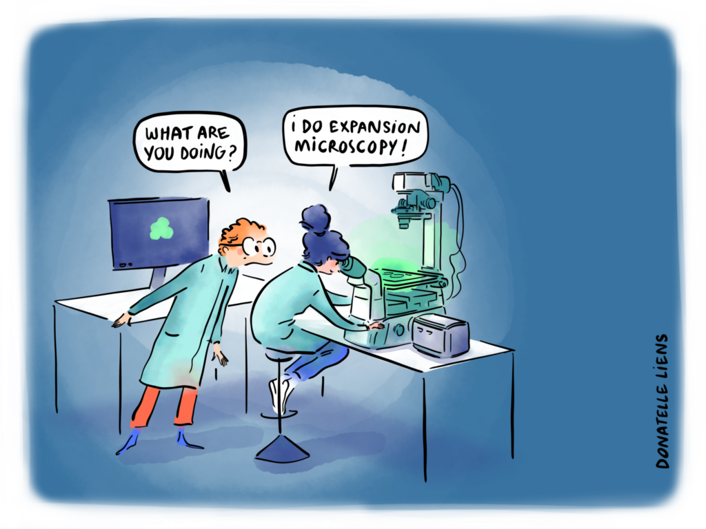
A research team from the Laboratoire de Biogenèse Membranaire (CNRS/University of Bordeaux), in collaboration with the Bordeaux Imaging Center, has recently developed ROOT-ExM, a novel protocol enabling the application of expansion microscopy to plant tissues, specifically the primary root of Arabidopsis thaliana. This method overcomes key limitations that had so far prevented the use of expansion microscopy in plant systems.
Zoom in on expansion microscopy
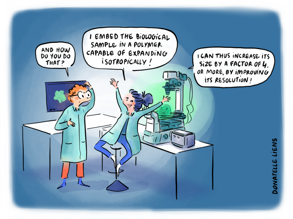
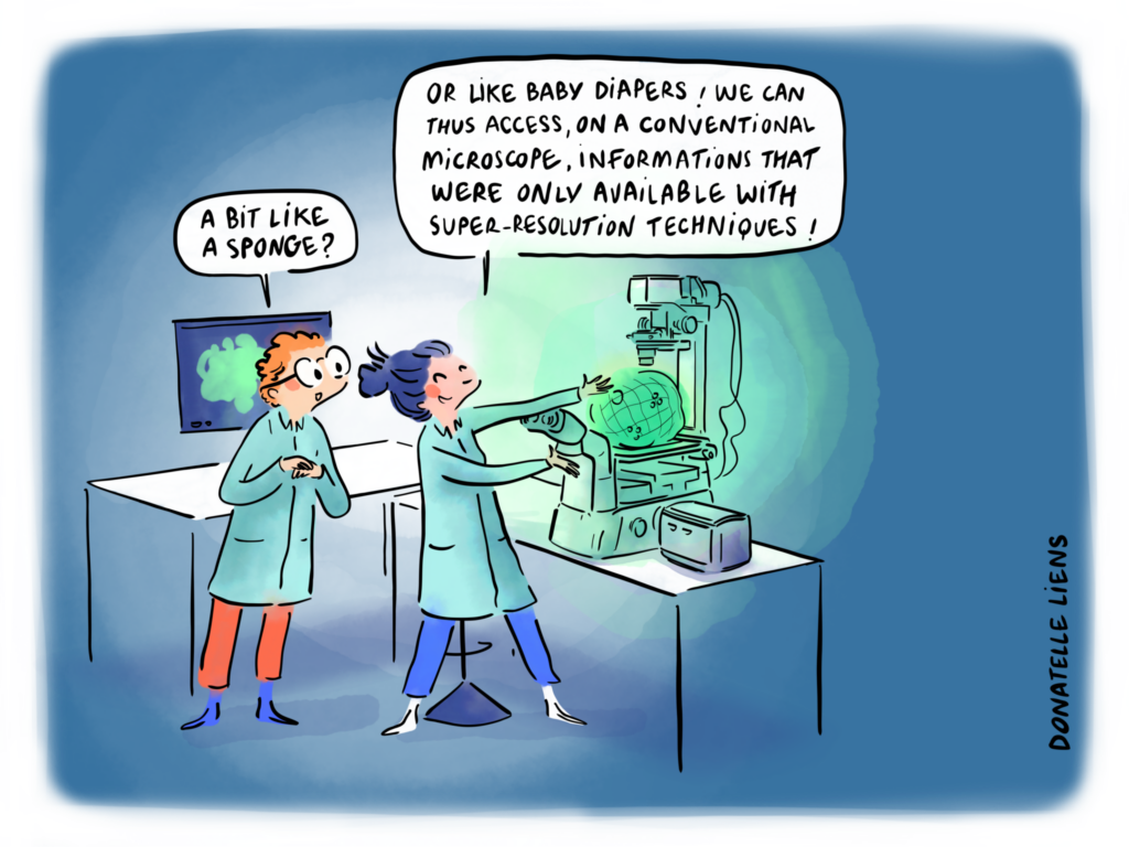
Expansion microscopy (ExM) is a super-resolution imaging technique that relies on the physical enlargement of biological specimens embedded in a swellable hydrogel. By expanding the gel, the distance between fluorescent labels increases, thereby enabling nanoscale resolution with conventional microscopes.
Plant cells are embedded in a dense network of polymers (cell walls) that provide mechanical strength and prevent expansion. These structural barriers are especially problematic for expansion microscopy, as they hinder both the penetration of labeling molecules and the isotropic expansion of the sample.
ROOT-ExM: Tailoring expansion microscopy for plants
To adapt ExM to plant roots, the team developed a two-step strategy combining:
A mild cell wall digestion, sufficient to relax the cell wall structure and facilitate labeling,
And a plant-optimized ExM protocol, compatible with common fluorescent markers and preserving tissue architecture during expansion.
Promising results
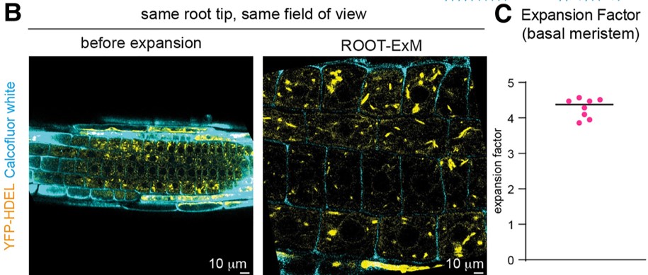
ROOT-ExM achieves an approximately 4-fold linear expansion of Arabidopsis roots with minimal deformation.
B) Confocal microscopy images of the same root with the same field of view before expansion and after ROOT-ExM. – In yellow: Endoplasmic Reticulum – In blue: Cell wall C) Expansion factor quantification. The diameter of nuclei was measured before and after expansion.
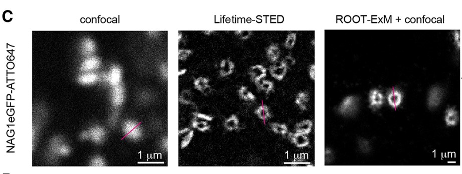
When combined with confocal microscopy, this technique enables nanoscale visualization of intracellular (nuclei, Golgi apparatus) and intercellular (plasmodesmata, microtubules) structures with resolution comparable to advanced super-resolution methods.
Representative images of acquisitions in confocal on nonexpanded samples (left), super-resolution lifetime-STED on nonexpanded samples (middle) and confocal after ROOT-ExM (right).Labelling of Golgi apparatus.
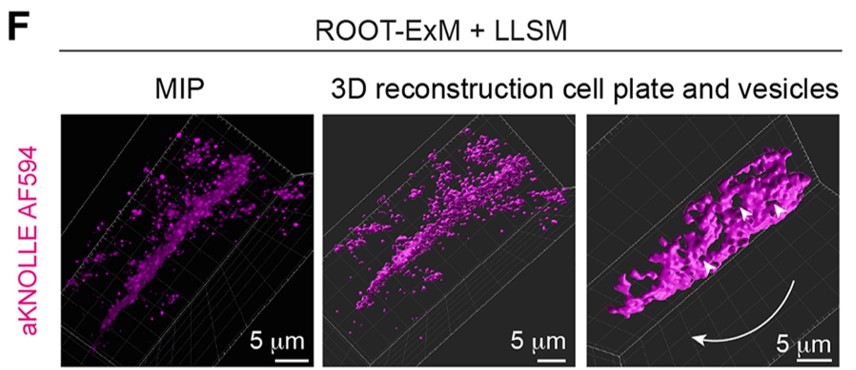
In addition, coupling ROOT-ExM with lattice light-sheet microscopy (LLSM) allows 3D reconstructions of cellular processes at nanometric resolution and across large tissue volumes.
Volume acquisition of a root cell labeled with anti-KNOLLE (membrane marker during cell division) after ROOT-ExM and imaged by LLSM.
Looking ahead
ROOT-ExM demonstrates that super-resolution imaging of plant tissues is possible using conventional microscopes and accessible labels. While currently limited to the primary root of Arabidopsis thaliana, this protocol lays the groundwork for expanding ExM to more complex plant organs and other species. Rather than replacing high-end super-resolution techniques, ROOT-ExM stands as a complementary approach, providing an accessible, scalable alternative for plant imaging at the nanoscale.
These proteins, which promote cohesion between cells, appear to facilitate the transmission of mechanical stress from one cell to another. The researchers demonstrated that colonies of cells capable of transmitting these mechanical forces are more likely to “win the cellular competition” compared to other colonies with similar characteristics but fewer cadherins — and therefore less ability to share the stress caused by mechanical forces.
These new findings open up new perspectives for understanding how cells compete within healthy tissues, and also during the proliferation of cancer cells.
As the journalists at Le Monde nicely put it: “unity is strength”!
For this second edition of Challenge, “Fuse My Cells”, we had 136 registrations and 6 participating teams. After a preliminary test phase, teams submitted their methods for evaluation on 32 images, with just one unique submission allowed!
We’re thrilled to reveal the top 3 winners of the challenge
1st place: Marek Wodzinski – FuseMyCells algorithm
Dorian Kauffmann, our Challenge Project Engineer, will present the results at the ISBI conference on April 17th, joined by challenge winner Marek Wodzinski!
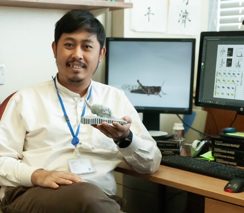
We are pleased to introduce Atitheb Chaiyasitdhi, one of the winners of the “FBI Call for User Access Projects 2024.” Atitheb received a grant to access imaging services at one of the France-BioImaging facilities.
In this interview, discover his research project focused on auditory mechanotransduction and hearing loss in locusts.
To start, could you tell us a bit about yourself? What has been your academic journey so far, and what is your current role or area of work?
My name is Atitheb Chaiyasitdhi. I am a research fellow in Benjamin Warren’s lab at the University of Leicester in the United Kingdom. At the lab, we focus on auditory mechanotransduction and hearing loss in insects. Before joining the lab, I did my PhD at the Institut Curie in Paris, where I studied the biophysics of hearing in vertebrates.
What are you currently working on in your research? What is the main topic or challenge you’re exploring?
I have always been fascinated by how hearing works. Currently, I am investigating how insect ears operate. Unlike humans, insects possess ears in a wide variety of shapes and locations—sometimes on their legs, antennae, or abdomens. Despite this diversity, they all rely on a similar mechanosensitive structure known as a chordotonal organ. Interestingly, insects also use chordotonal organs to detect other mechanical cues, such as gravity or body position (proprioception), and these organs are located in different parts of the body as well.
This raises two key questions:
How does the chordotonal organ convert such a broad range of mechanical forces into electrical signals? And do these organs share a common underlying mechanism?
To explore these questions, I am focusing on the chordotonal organ in the locust ear. On one front, I am using a high-speed camera to capture sound-evoked motion of the organ and electrophysiology techniquesto measure electrical current in the chordotonal neurons evoked by the sound.
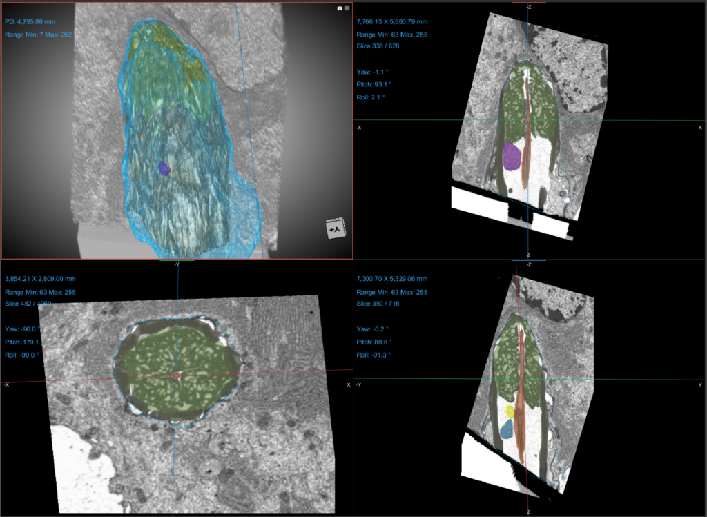
On another front, I am collaborating with Claire Boulogne at Plateforme Imagerie-Gif, using Focused Ion Beam Electron Microscopy (FIB-EM) to reconstruct the organ’s three-dimensional structure. These complementary approaches will provide insights into the mechanics of insect auditory transduction and bring us closer to solving the two questions I previously talked about.
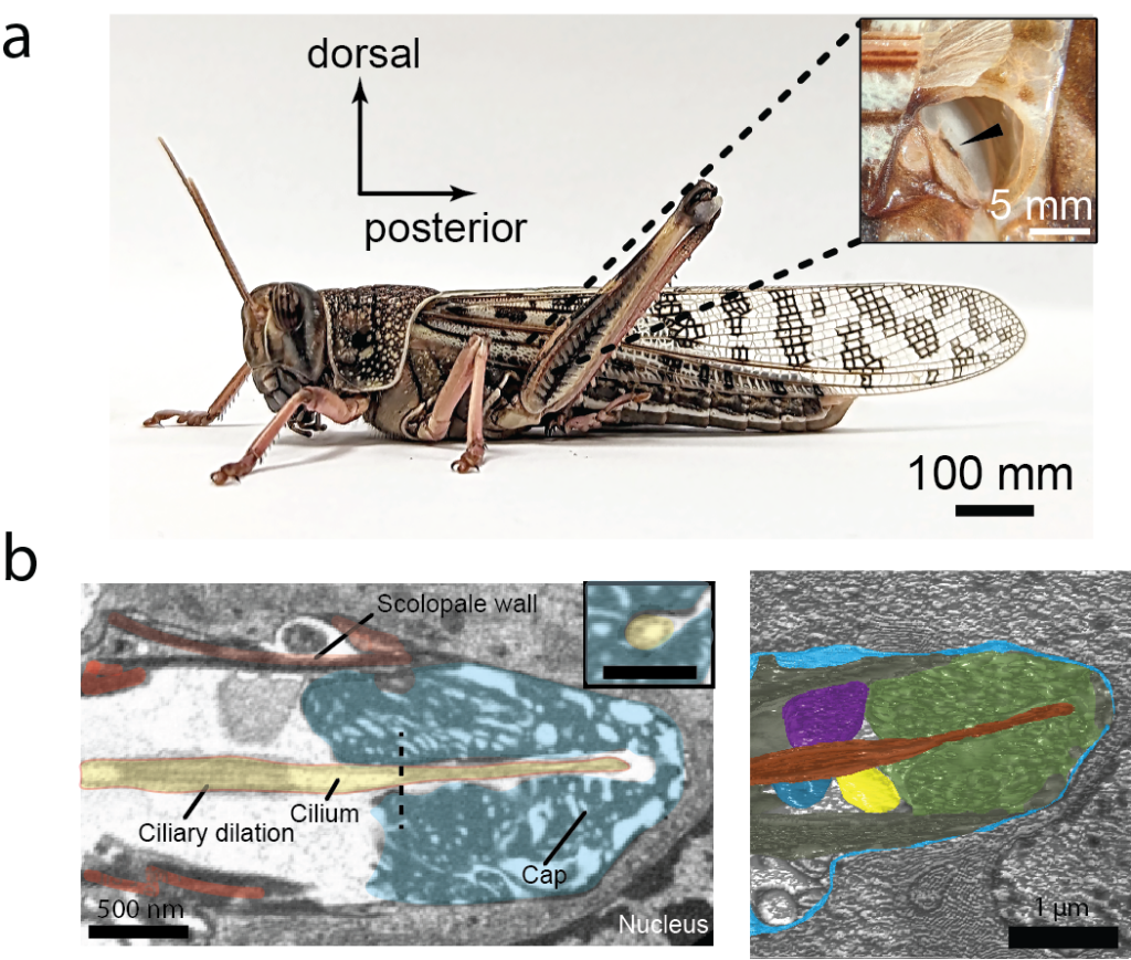
(a) The locust’s ears are on the first abdominal segment.. Each ear consists of a tympanic membrane with a hearing organ, Müller’s organ, directly attached to it. When viewed externally, the tympanic membrane (inset) shows a dark ridge (arrowhead). (b-lef) An electron micrograph from FIB-EM showing the distal region of the scolopidium where the sensory cilium (yellow) inserts into the attachment cap (blue). The inset shows a cross-section of the cilium at the dashed line in (b), displaying a 9×2+0 axonemal structure. The inset scale bar is 500 nm. (b-right) The 3-dimensional reconstruction of the scolopidium.
At what point did you come across France-BioImaging, and what made you want to use its services or connect with the infrastructure?
I first learned about France-BioImaging during my PhD in France. At that time, I had the opportunity to collaborate with and use the excellent resources provided by one of the France-BioImaging Nodes—the Cell and Tissue Imaging Platform (PICT-IBiSA) at the Institut Curie. That experience left a strong impression on me, highlighting the expertise and support that France-BioImaging offers. Since then, I have been interested in collaborating with France-BioImaging platforms again.
Could you walk us through your experience accessing France-BioImaging? Which facility did you work with, how did the process go, and what stood out to you during your time there?
I worked with the Plateforme Imagerie-Gif in Gif-sur-Yvette, near Paris, using Focused Ion Beam Electron Microscopy (FIB-EM) with the support of Claire Boulogne, the lead engineer. Since I am based in the UK, and thanks to modern communication technology, our collaboration could start remotely. We discussed the experimental plan online, and I prepared the preliminary samples here before shipping them to the platform in France. Claire then carried out further sample preparation and image acquisition.
We worked closely, going back and forth to establish an optimal workflow, and soon began obtaining promising results afterwards. Despite the platform’s tight schedule, I always received prompt responses and consistent support from Claire.
What did microscopy bring to your project specifically? Were there insights or results you couldn’t have obtained otherwise?
The first ultrastructural image of a chordotonal organ was captured over 60 years ago, and it provided key insights into how the organ functions. In our lab, we have been able to resolve the mechanics of the chordotonal organ with unprecedented temporal resolution using high-speed cameras.
However, we still lack the spatial resolution to fully understand its structure. FIB-EM enables us to visualize the ultrastructure in three dimensions at a level of detail that has not been previously achieved. Seeing how each cellular component connects in 3D allows us to answer questions that arise from our high-speed recordings of sound-evoked motion in the organ.
At the same time, it opens up new questions and research opportunities. While there are other techniques, such as array tomography, they cannot match the spatial resolution of FIB-EM and would require hundreds of hours of manual effort.
Example of a 3D reconstruction of the scolopidium from FIB-EM stacks obtained at Imagerie Gif.
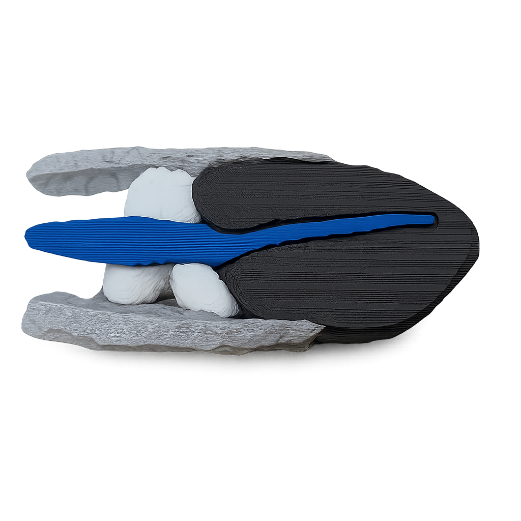
A 3D plastic model of the distal end of the scolopidium (see b-left of the 2nd figure)
Looking back, would you encourage other researchers to use France-BioImaging’s platforms and access program? What would you say to someone considering it?
Yes, I would. I would tell them that I have extensive experience working with France-BioImaging platforms, and the collaborations have always delivered reliable results. I trust their expertise and the quality of their support. In fact, I’ve already recommended France-BioImaging to colleagues here in the UK, as well as from the US and Germany. We are currently exploring the possibility of continuing our collaboration with Imagerie-Gif to investigate the diversity and evolution of chordotonal organs in various insect ears, even those involved in proprioception in the legs.
Celebrate the four seasons through imaging! Submit your best scientific images that capture the essence of each season, from electron micrographs of flu viruses in autumn to heat shock proteins in summer!
This contest is open to all members of the Euro-BioImaging community and anyone involved in scientific imaging and research worldwide. The contest has four quarterly sessions, each linked to a season.
How to participate?
Register, submit as many entries as you like, and upload your images. Only images captured with a microscope or other imaging devices are eligible (macrophotography is not allowed).
What can you win?
Quarterly winners will receive reimbursement for travel expenses (up to 1000€) to attend a scientific conference or event. Runner-ups get up to 500€. Selected images will be featured on Euro-BioImaging’s website and social media.
Contest timeline:
Spring: 31 Mar – 20 Jun 2025
Summer: 21 Jun – 21 Sep 2025
Autumn: 22 Sep – 20 Dec 2025
Winter: 21 Dec 2025 – 20 Mar 2026
You are interested? Find more information and detailed rules here.
Are you ready to expand your professional network, exchange ideas, and boost your expertise? The second call for the Cross-Node Job Shadowing Program is now open! This initiative, part of the EU-funded EVOLVE project, offers staff at Euro-BioImaging Nodes a chance to visit and learn from other Nodes across Europe.
This program is open to all Euro-BioImaging Node staff, including technicians, administrators, and managers. It’s an excellent opportunity for learning and growth through hands-on experience.
Key benefits:
Gain insights into instruments, techniques, and data management practices
Learn about facility operations, Node administration, and soft skills
Application Deadline:
Submit your application by May 16th, 2025. If you work at a Euro-BioImaging Node, you should have received the official application link. For any inquiries, contact info@eurobioimaging.eu.
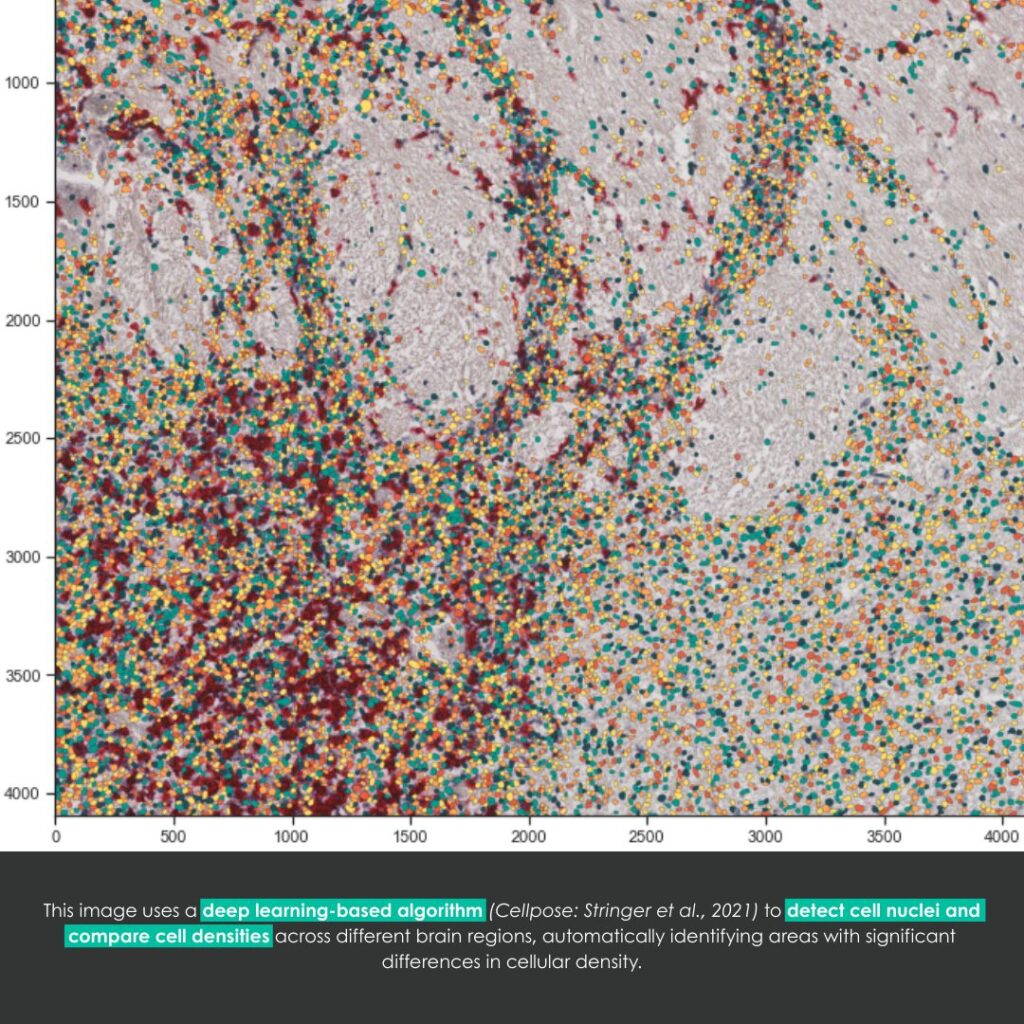
A European interdisciplinary research project, coordinated by Thibault Lagache (BioImage Analysis Unit, Institut Pasteur – France-BioImaging’s platform), David Menassa (University of Oxford), and David Holcman (University of Cambridge), has recently led to the creation of an innovative tool: DeepCellMap. This tool significantly improves the mapping of microglia, brain cells that remain widely unknown to the general public.
Microglia belong to the family of glial cells, which form the environment around neurons. They provide immune protection for the nervous system and play a crucial role in brain development.
What does DeepCellMap bring?
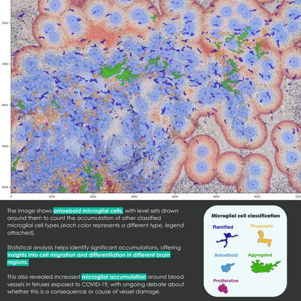
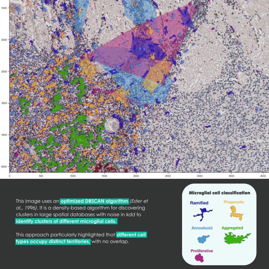
The tool can classify microglia into five categories based on their shape and location in the brain. This classification allows researchers to track their roles throughout brain development.
DeepCellMap also revealed a dynamic spatial organization of microglia. These cells occupy distinct territories and reorganize as the brain grows. Clusters of cells appear and later disperse, following specific patterns.
An unexpected finding: DeepCellMap uncovered a strong association between microglia and blood vessels in the cerebral cortex of fetuses exposed to SARS-CoV-2 during pregnancy. This discovery raises a major question: Are microglia reacting to vascular alterations, or do they themselves contribute to these changes? This observation could pave the way for new research into the impact of prenatal infections on the developing brain.
How does DeepCellMap work?
This tool relies on a deep learning algorithm (artificial intelligence) capable of detecting and classifying microglia based on their morphology, using bright-field or fluorescence microscopy images.
The analysis of microglial spatial organization is made possible by the use of advanced statistical models. In conclusion, DeepCellMap is a groundbreaking image analysis tool for biology. By enabling large-scale studies of brain cells, it opens new research perspectives in neuroscience to better understand the mechanisms of brain development.
In the long term, DeepCellMap may also help improve our understanding of how prenatal infections affect the developing brain. As an open-source resource, it can be adapted to study other cell types and applied to a wide range of human health research.
Congratulations to all the teams involved! France-BioImaging is proud to support interdisciplinary projects such as the development of DeepCellMap by members of our bioanalysis community. Supporting research through cutting-edge R&D and fostering international collaboration are at the heart of our infrastructure’s missions.
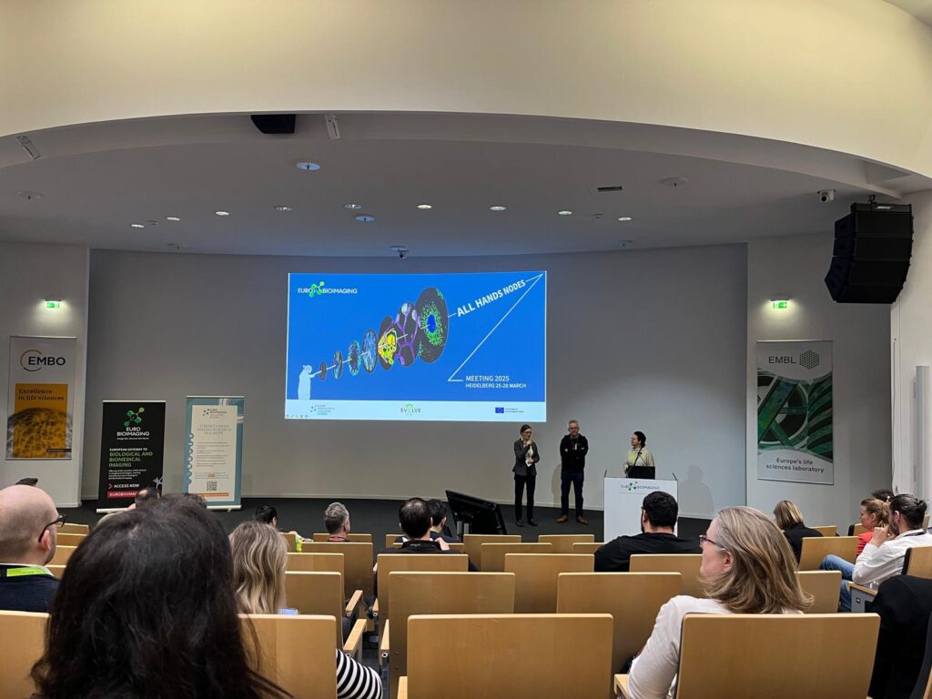
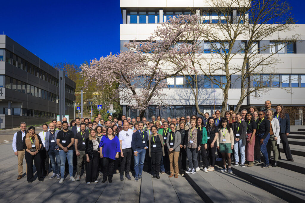
Last week we attended Euro-BioImaging All Hands Meeting at EMBL in Heidelberg (25-28 March 2025). It was a pleasure to gather with all our colleagues from the Euro-BioImaging nodes and discuss imaging technology innovation, data, access, training & international collaboration.
All Hands Meeting recap!
On Tuesday, March 25, the event began with several parallel sessions, including a FLIM workshop by PicoQuant, a networking meeting for staff from preclinical and medical imaging facilities, an AI4Life session, and a session on how to improve infrastructures’ and facilities’ acknowledgement by their users.
The second day, co-organized with the Euro-BioImaging Industry Board (EBIB), featured updates from Euro-BioImaging, several presentations focusing on innovations in imaging technologies, including new approaches to multispectral optoacoustic tomography, FLIM imaging modalities, and advanced 3D multiplex imaging, high-resolution imaging, cryogenic sample preparation, and correlative imaging techniques. The day concluded with a session on Industry User Experience at Euro-BioImaging, where discussions focused on industry collaborations and access to research infrastructures.
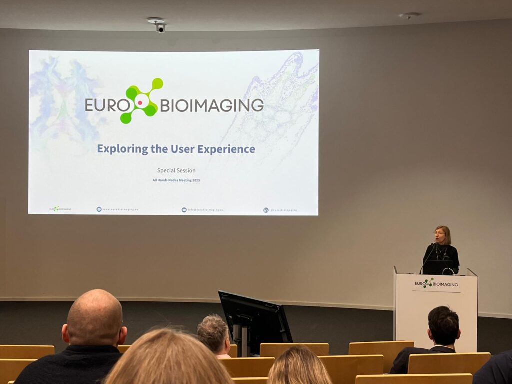
On Thursday, the day started with an inspiring keynote lecture by Lisa Yen from Microscopy Australia on the role and impact of open-access research infrastructures in scientific collaboration. This was followed by sessions on Global BioImaging and the Imaging4All initiative, highlighting international opportunities for Euro-BioImaging Nodes. The morning also featured presentations from various imaging nodes, including insights into new services, methodologies, and applications in imaging research. Later in the day, the focus shifted to Euro-BioImaging’s User Access Experience, featuring an interactive session where Yves Lutz (IGBMC) presented France-BioImaging Alsace Node experience. The afternoon was dedicated to Euro-BioImaging’s data services, with updates on AI-driven image analysis, medical imaging, and data-sharing initiatives. A session on theFoundingGIDE project concluded the day.
The final day of the meeting highlighted training opportunities under the EVOLVE program, with presentations onjob shadowing experiences from different imaging nodes. The morning also included breakout sessions on topics such as training, communication strategies for engaging with funders and the general public, data management, and core facility performance evaluation.
This annual gathering offers an essential platform for the EuBI nodes to exchange on key topics, current challenges and best practices with European colleagues and build on new transnational collaborations.
Thanks to the Euro-BioImaging hub team for organizing and hosting this inspiring event! We are glad to be part of this amazing community and working together as a European Research Infrastructure!
Photo by EMBL/PhotoLab
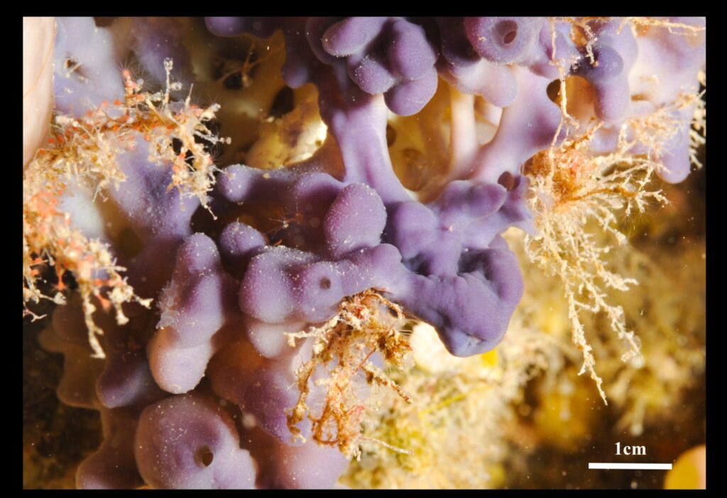
A study conducted by the BEEM team (Molecular Biology, Evolution, and Ecology) of the Mediterranean Institute of Biodiversity and Marine and Continental Ecology, in collaboration with IBDM (Marseille), ENS (Paris) and ISEM (Montpellier) has characterized the buds of Oscarella lobularis as a promising model for studying cell development and sponge evolution.
Researchers successfully induced the in vitro production of these buds and maintained them in culture. These structures are small fragments of the sponge that detach from the adult body and develop into fully independent individuals. The study revealed that they possess remarkable properties from the early stages of their development:
Regeneration ability: When a bud is cut in half, each piece can regenerate into a fully functional new bud. Even when completely broken down into individual cells, they can migrate, reconnect, and self-organize into structured layers.
Autonomous metabolism: They filter water, consume oxygen, and move slightly using tiny cilia.
Complex cellular organization: Their structure resembles that of more evolved organisms, making them a relevant model for understanding the evolution of early animals.
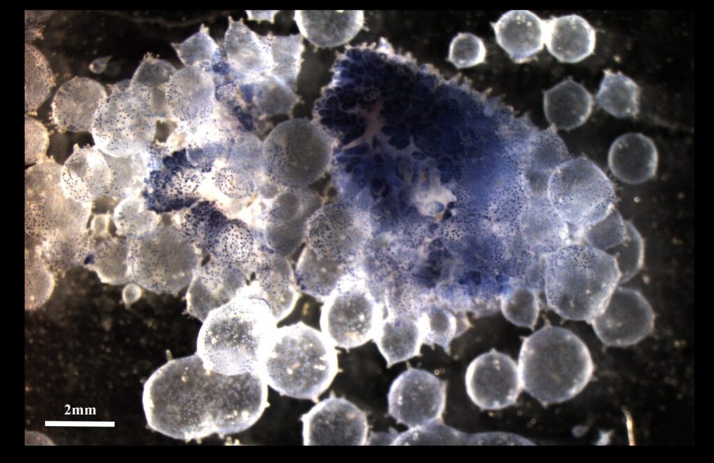
Imaging at the heart of discovery
To observe these buds in detail, researchers used advanced electron and fluorescence microscopy techniques. These analyses were carried out at the PICsl (IBDM, Aix-Marseille University) platform, a member of France-BioImaging.
Thanks to these high-resolution images, scientists were able to examine the buds’ development, cellular organization, and internal functioning, revealing mechanisms still largely unknown in the animal kingdom.
Why study sponges?
If we trace back the phylogenetic tree of animals, it is possible that we share a common ancestor with this species! It may seem hard to believe, but we actually have similarities with sponges.
Sponges are among the oldest organisms on Earth. Studying them could help us understand the origins of animal ancestral features among which the formation of cell layers.
We use cookies on our website to give you the most relevant experience by remembering your preferences and repeat visits. By clicking “Accept All”, you consent to the use of ALL the cookies. However, you may visit "Cookie Settings" to provide a controlled consent.
This website uses cookies to improve your experience while you navigate through the website. Out of these, the cookies that are categorized as necessary are stored on your browser as they are essential for the working of basic functionalities of the website. We also use third-party cookies that help us analyze and understand how you use this website. These cookies will be stored in your browser only with your consent. You also have the option to opt-out of these cookies. But opting out of some of these cookies may affect your browsing experience.
Necessary cookies are absolutely essential for the website to function properly. These cookies ensure basic functionalities and security features of the website, anonymously.
Cookie
Duration
Description
cookielawinfo-checkbox-analytics
11 months
This cookie is set by GDPR Cookie Consent plugin. The cookie is used to store the user consent for the cookies in the category "Analytics".
cookielawinfo-checkbox-functional
11 months
The cookie is set by GDPR cookie consent to record the user consent for the cookies in the category "Functional".
cookielawinfo-checkbox-necessary
11 months
This cookie is set by GDPR Cookie Consent plugin. The cookies is used to store the user consent for the cookies in the category "Necessary".
cookielawinfo-checkbox-others
11 months
This cookie is set by GDPR Cookie Consent plugin. The cookie is used to store the user consent for the cookies in the category "Other.
cookielawinfo-checkbox-performance
11 months
This cookie is set by GDPR Cookie Consent plugin. The cookie is used to store the user consent for the cookies in the category "Performance".
viewed_cookie_policy
11 months
The cookie is set by the GDPR Cookie Consent plugin and is used to store whether or not user has consented to the use of cookies. It does not store any personal data.
Functional cookies help to perform certain functionalities like sharing the content of the website on social media platforms, collect feedbacks, and other third-party features.
Performance cookies are used to understand and analyze the key performance indexes of the website which helps in delivering a better user experience for the visitors.
Analytical cookies are used to understand how visitors interact with the website. These cookies help provide information on metrics the number of visitors, bounce rate, traffic source, etc.
Advertisement cookies are used to provide visitors with relevant ads and marketing campaigns. These cookies track visitors across websites and collect information to provide customized ads.