The second workshop of Holotomography microscopy will be held from April 12th to 14th on the PFIC microscopy facility.
You will test the HT-2H microscope which has more resolution than his bigger brother HT-X1.
The key features are: No label needed, High resolution with one single lens 60X NA 1.2 water immersion (120 nm XY resolution, 356 nm Z resolution, 150 fps T resolution ), quantifiable data, Low phototoxicity, fast imaging.
The microscope has both 3D Holotomograms and 3D fluorescence capabilities in one single unit.
To better prepare your samples for imaging the Tomocube will give us some mounting chambers.
Correlative X-ray imaging and electron microscopy (CXEM) is the combination of X-ray imaging and electron microscopy. It is a correlative approach that makes it possible to characterise a sample of interest and locate a structure of interest in a non-destructive way. Nicolas BROUILLY is in charge of the Electron Microscopy Unit of the PICsL imaging facility on the Marseille node of France BioImaging, where CXEM is used for developmental biology studies. As part of Euro-BioImaging’s Proof-of-Concept study, his facility is now accepting applications from external users for CXEM projects. Learn more about how this approach works and what it can be used for in the interview below.
We are today talking about CXEM imaging. Please provide a short summary of this type of imaging and tell us some applications:
Nicolas Brouilly: It is often very useful to combine 2 imaging modalities to take advantage of each while trying to lower their respective drawbacks. For example, by combining Light Microscopy and Electron Microscopy, we obtain the popular CLEM (for Correlative Light and Electron Microscopy). Visible light can then be used in combination with EM either:
To target a precise region of interest ;
To localize molecules within the ultrastructural information obtained by EM.
Using the same acronym building, CXEM corresponds to Correlative X-ray and Electron Microscopy. X-rays are photons of shorter wavelength than those from visible light, and can again be used to characterize a sample in 2 different ways:
To use their ability to easily go through tissues in order to record the 3D morphology of a sample: either by computed x-ray micro-tomography (or micro-CT) for micrometric resolution of big samples (mm to cm range) or by Soft X ray tomography for nanometric resolution of small samples (100’s of nm to um range);
To use a focused beam of high energy x-rays to analyse the localization of the elements of a sample: X-Ray Fluorescence microscopy (or XRF).
Both modalities can be used to complement the ultrastructural information obtained by electron microscopy. At the Marseille node of France BioImaging, in the Electron Microscopy Unit, we routinely use Correlative Micro-CT and Electron Microscopy to answer developmental biology questions.
What are some advantages of this technique that make it suited to addressing this type of question?
Nicolas Brouilly: The main advantage of Micro-CT (or Computed X-ray Tomography) is its ability to “see through” a sample and to reveal its overall organization in 3D without any labelling. The second advantage of Micro-CT is the fact that it is non-destructive. Thirdly, the contrast we usually give to samples for electron microscopy is compatible and even beneficial for X-ray imaging.
Altogether, this means that we can use X-ray tomography to map the microscale morphology of a sample in order to target a specific region of interest without having to go through the time-consuming and destructive collection of semi-thin sections.
We routinely use the micro-CT tool, not only to target a given organ or a given group of cells, but also to pre-orient the sample in order to cut it under a specific orientation. It is a timesaving tool within the frame of a 2D electron microscopy project, but it really is key within the frame of a 3D electron microscopy project given that Serial BlockFace and Focused Ion Beam techniques are destructive.
Tell us a bit more about a specific project that was done in your facility using this technology? What scientific questions were you addressing?
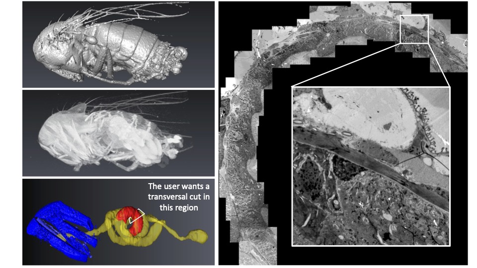
Nicolas Brouilly: Imagine that, first, you have a ball of yarn, second, you cannot untangle it, and third, you want to cut small bits of the thread at 24 cm from the end (not 22, not 26… 24 !). CXEM enabled us to do this on Drosophila gut. The micro-CT gave us the 3D map of the sample within the resin block. We could then use this map to find the best itinerary within the sample to make transverse sections of the portion of interest that was precisely indicated by the user on the micro-CT dataset. At the end of the day, the user was able to look at perfect transverse ultrathin TEM sections, at a precise position of this ball of yarn that Drosophila gut is. He could finally get precise metrics from this precise part of the gut in several samples. None of this could have been achieved without CXEM.
Like a ball of yarn… Above is an example of how CXEM can be used to find the best itinerary within a sample to make transverse sections of the portion of interest. On the left, the micro-CT provided a 3D map of the sample within the resin block. On the right, a transverse ultrathin TEM section of the drosphilia gut.Image courtesy of Nuno Luis (Schnorrer lab, IBDM) & Nicolas Brouilly (Electron Microscopy Facility, IBDM AMU/CNRS, France BioImaging).
For another example, you can have a look at the following paper where we used CXEM to map platelet aggregates within arteries in order to explore them by Serial BlockFace SEM, another example of “Find a needle in a haystack”. Have a look at movie S1, it is a wonder that we could not obtain without CXEM:
CXEM is part of the Euro-BioImagingProof-of-Concept study. The Proof-of-Concept study makes it possible to introduce exciting, new imaging technologies to our portfolio that were previously unavailable via our network. We are currently accepting applications to use these technologies at participating Nodes as part of the Proof-of-Concept study. Be part of this study – and contribute to community-wide continuous technological innovation!
All scientists, regardless of their affiliation, area of expertise or field of activity can benefit from Euro-BioImaging’s pan-European open access services. Potential users of these new technologies are encouraged to submit project proposals via our website. To do so, you can Login to access our application platform, choose the technology you want to use and the facility you wish to visit, then submit your proposal. All applications will be processed by the Euro-BioImaging Hub. As usual, users will benefit from advice and guidance by technical experts working at the Nodes, training opportunities, and data management services.
Two projects recently received funding from the Chan-Zuckerberg Initiative (CZI), in which France-BioImaging members take part actively: COMULIS and NEUBIAS. Two community building activities breaking up frontiers to gather scientists around one goal: developing biological imaging.
COMULIS received a 2-year funding from CZI to expand their network both globally and sustainably. Being designed to harness the power of multimodal imaging (MMI) across scales, from basic to clinical diagnostics, this European initiative aims at facilitating access and training a new generation of scientists for whom multimodal imaging will be the new norm. Thanks to this grant, the project will be consolidated and it will help extend the collaborative and innovative network to establish a global multimodal imaging association (COMULISglobe) and ensure long term sustainability.
MMI integrates the best features of combined techniques and overcomes limitations faced when applying single modalities independently. MMI relies on the joint expertise from biologists, physicists, chemists, clinicians, and computer scientists, and depends on coordinated activities and knowledge transfer between technology developers and users. To achieve this inherently interdisciplinary goal, the ultimate goal is toestablish a network of scientists across continents and disciplines, from academia to industry, including transnational research facilities (e.g. synchrotrons, Euro-BioImaging ERIC), to foster and market MMI as a versatile tool in biomedical research and diagnostics.
COMULISglobe will help bridge the gap between biological and clinical imaging, identify, fund, and showcase novel multimodal pipelines, and develop, evaluate, and publish correlation software through dedicated networking activities, including conferences, training schools, open databases, and fellowships for lab exchanges, access to research infrastructures, and conference attendance. And, of course, all outputs of the project will be open access!
Please do not hesitate to join the community and help organize activities or publications – and please share the news, mobility and access grants available at: https://www.comulis.eu/comulisglobe-czi
Thanks to Perrine Paul-Gilloteaux, Bretagne-Loire node of France-BioImaging, for taking part of this amazing project!
The international Network of European BioImage Analysts (NEUBIAS), hosted by German BioImaging has also received a 2-year funding from the Chan Zuckerberg Initiative (CZI) as part of their Advancing Imaging through Collaborative Projects program. This grant will secure the sustainability of NEUBIAS, establish strong connections to similar initiatives, and share knowledge about state-of-the-art bioimage analysis tools and methods globally.
Spreading the profession of bioimage analysts and bioimage analysis knowledge internationally are the major aims of NEUBIAS. Modern life-sciences are unthinkable without advanced microscopy imaging techniques and quantitative bioimage analysis. This grant will help ensure novices and experts can access cutting edge techniques, reduce duplications of effort, and support everyone who is working to making new discoveries possible.
NEUBIAS had a tremendous impact on the community by training a powerful generation of bioimage analysts across Europe and beyond. The next step of this project will expand the network internationally and connect to related imaging and image analysis societies around the globe. With that in mind, the project includes travel grant opportunities for early-career bioimage analysts who seek to join NEUBIAS activities, explicitly including scientists outside central Europe. Besides, a dedicated team will work on collecting bioimage analysis teaching materials and make them accessible to the global imaging and life science community.
Great news for both projects that – we hope – will continue to write the great story of bioimaging!
Thanks to Florian Levet, Bordeaux node of France-BioImaging, for being a member of this fantastic project!
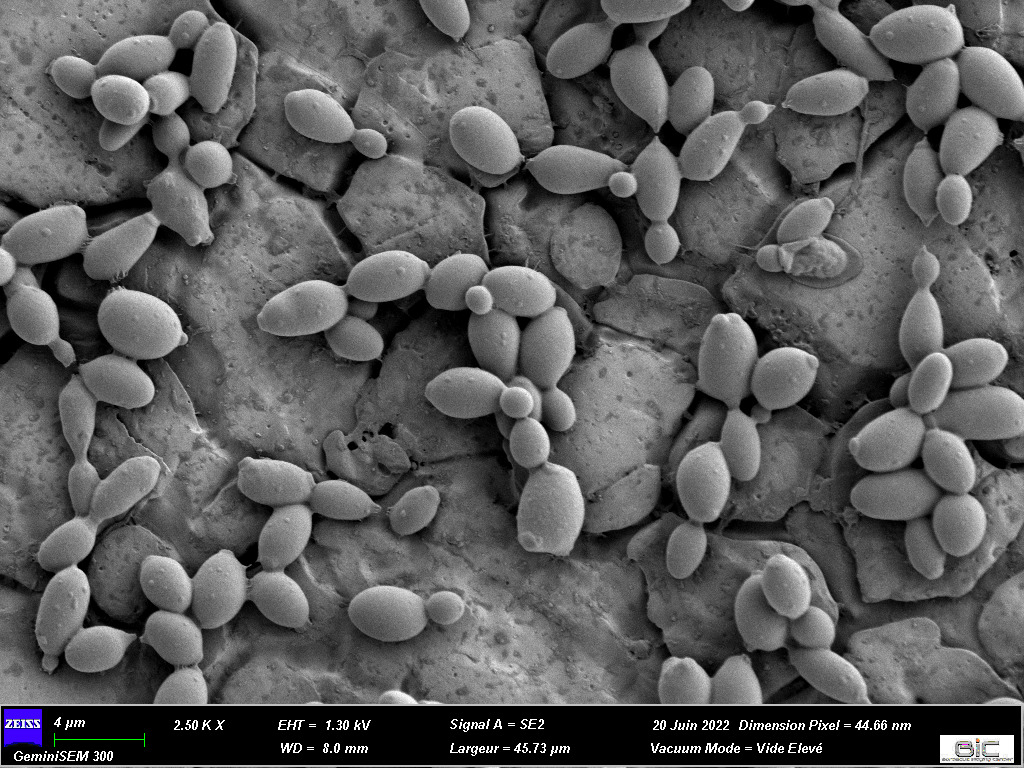
Brettanomyces bruxellensis is one of the most damaging spoilage yeasts in the wine industry because of its impact on the beverage’s flavor. Lysiane Brocard, research engineer specialised in plant biology at the Bordeaux Imaging Center (FBI Bordeaux node), recently co-published an article on this yeast cell surface and bioadhesion properties.
Fruits are transformed into beverages through fermentation processes carried out by microorganisms naturally present in the environment. In wine, yeasts and bacteria play this role and contribute to the development of volatile compounds. Scientists targeted Brettanomyces bruxellensis in this study, a yeast famous for the production of volatile phenols, characterized by horse sweat odors which is – usually – not very enjoyable for the consumer.
A yeast characterized by bioadhesion abilities
This specific odor comes from volatile compounds, the 4-ethylgaïacol (4EG) and 4-ethylphenol (4 EP), that winemakers try to avoid. Beside adding an unpleasable flavor to the beverage, the issue is that the spoilage yeast is persistent in cellars over several years, resulting in recurrent wine contamination. This suggests a bioadhesion process that helps the microorganism to survive in its environment. To put it simply, bioadhesion is the ability of an organism to adhere on a surface to, then, participate in the formation of a biofilm (which is defined as “a structured community of microorganisms adhered to a surface and producing an extracellular matrix”).
Here, 54 strains of B. bruxellensis were characterized for their cell surface physico-chemical and bioadhesion properties. And all of them have shown bioadhesion abilities (after only three hours on stainless steel) both on synthetic medium and wine. Enough to highlight the persistence of our favorite horse sweat flavored yeast.
How did bioimaging help in this project?
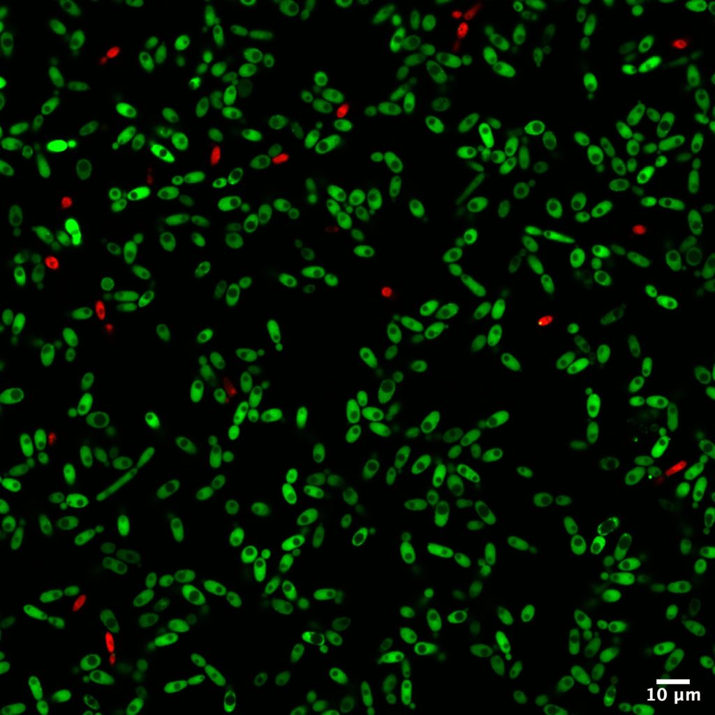
Among all the analytical methods used in this study, microscopy helped identify the structure of the biofilms formed with B. bruxellensis. Two imaging techniques were used: confocal microscopy and scanning electron microscopy. The first one offers the advantage of realizing live imaging without being too time-consuming. With fluorescent dyes, the status of cells can be easily determined at the same time as the cell repartitions and concentrations.
Moreover, comparisons have been made thanks to confocal microscopy to determine if some strains of B. bruxellensis could form biofilm with only one cell layer or if they proliferate in three dimensions. To complete these observations, scanning electron microscopy was performed at the Bordeaux Imaging Center (FBI Bordeaux node) with the help of Isabelle Svahn, expert of this type of microscopy. It was a great addition to this study as these observations validated the morphological variability among Brettanomyces strains.
Bioimaging helped a broader project about Brettanomyces led by Isabelle Masneuf-Pomarède who works in the Institut des Sciences de la Vigne et du Vin of Bordeaux. Isabelle studies the persistence and proliferation of Brettanomyces over the years. Isabelle’s PhD student, Paul Le Montagner, carried out most of the experiments published in this paper. Thanks to them for this amazing paper!
Confocal microscopy observations after 3h of bioadhesion of cells on stainless steel
SEM observation of 3h-aged cells adhered on stainless steel
Original article: Paul Le Montagner, Morgan Guilbaud, Cécile Miot-Sertier, Lysiane Brocard, Warren Albertin, Patricia Ballestra, Marguerite Dols-Lafargue, Vincent Renouf, Virginie Moine, Marie-Noëlle Bellon-Fontaine, Isabelle Masneuf-Pomarède, High intraspecific variation of the cell surface physico-chemical and bioadhesion properties in Brettanomyces bruxellensis, Food Microbiology, Volume 112, 2023, 104217, ISSN 0740-0020, https://doi.org/10.1016/j.fm.2023.104217
You are interested in our bioimaging services, including technologies and expertise?
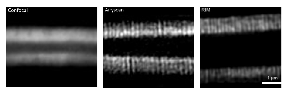
An innovative technology to look at thick samples at high resolution? Marc Tramier, a group leader at the Institute of Genetics & Development of the University of Rennes/INSERM/CNRS, and scientific director of MRic (Microscopy Rennes Imaging Centre), is currently working with his team on Random Illumination Microscopy (RIM), a fast and easy to use microscopy technique with low phototoxicity. His facility, which is part of the Bretagne-Loire Node of France-BioImaging, offers RIM as a Euro-BioImaging Proof-of-Concept study, and is now accepting applications for projects. He explains the ideas behind RIM in the article below.
The idea of Random Illumination Microscopy is to use the speckle of the illumination laser in wide field to create a structured illumination pattern at the diffraction limit. By varying the pattern from image to image using a diffracting element (in our case a SLM), scientists are able to acquire a stack of images (around 100 images) on a camera which corresponds to a cumulative homogeneous illumination. By resolving the inverse problem, a super-resolved image is, then, reconstructed, at the focal plane with unprecedented optical sectioning. In comparison to conventional SIM, RIM is able to work in depth inside diffusive samples as the speckle is insensitive to diffusion.
A transfer full of advantages
The method was first implemented by Thomas Mangeat – that we are happy to welcome in our new Toulouse node! – and collaborators in Toulouse (Mangeat et al., 2021. doi: 10.1016/j.crmeth.2021.100009). In the MRic, after the transfer of the prototype, the facility was able to image microvilli of intestine in c-elegans (depth > 50µm) having a spatial resolution of around 100 nm. This structure is impossible to be revealed by conventional confocal microscopy. Before the use of RIM, only the airyscan approach allowed us to resolve the microvilli but with higher illumination power (photobleaching of the sample) and longer acquisition time (around 10 times more). Now with RIM, we are able to follow microvilli in the living C–elegans at the second time-scale during several minutes.
RIM is one of the powerful methods to achieve super-resolved images in depth at high speed with very low phototoxicity. This makes a very nice compromise of z-sectioning and super-resolution with wide field illumination particularly adapted to thick live samples.Beside reconstruction and data analysis, MRic is offering a user-friendly system, with a complete set of microscopy methods for live sample investigation, from wide-field to light sheet including spinning disk, confocal and airyscan. And of course, this technology is available in open access through France-BioImaging and Euro-BioImaging!
How to apply to use RIM:
Random Illumination Microscopy is part of the Euro-BioImaging Proof-of-Concept study, in collaboration with our Nodes. The Proof-of-Concept study makes it possible to introduce exciting, new imaging technologies to our portfolio that were previously unavailable via our network. We are currently accepting applications to use these technologies as part of the Proof-of-Concept study. Be part of this study – and contribute to community-wide continuous technological innovation!
All scientists, regardless of their affiliation, area of expertise or field of activity can benefit from Euro-BioImaging’s pan-European open access services.Potential users of these new technologies are encouraged to submit project proposals via our website. To do so, you can Login to access our application platform, choose the technology you want to use and the facility you wish to visit, then submit your proposal. All applications will be processed by the Euro-BioImaging Hub. As usual, users will benefit from advice and guidance by technical experts working at the Nodes, training opportunities, and data management services.
Thank you Marc Tramier and Marianna Childress, communication officer of Euro-BioImaging, for the original article.
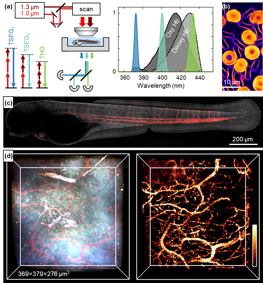
Researchers from the Laboratory for Optics and Biosciences (LOB, CNRS / École Polytechnique / INSERM), a member of the Ile-de-France Sud node of France-BioImaging, and from the Developmental Biology and Stem Cells Department (UMR3738, CNRS / Institut Pasteur), developed a new form of multiphoton microscopy providing label-free imaging of red blood cells and oxygenation. This technique is called color third-order sum-frequency generation microscopy, and is described in an article recently published in Light: Science & Applications.
Keeping resolution, saving time
Current methods used to map microcirculation and blood oxygenation at high resolution typically require the injection of fluorescent or phosphorescent markers. In addition, they usually require relatively long pixel times and thus are limited in spatio-temporal resolution. The novel approach is based on label-free third-order sum-frequency generation (TSFG) and third-harmonic generation (THG) contrasts. In practice, this microscopy is based on the illumination of samples by two pulsed infrared lasers and the simultaneous detection of several TSFG and THG signals emitted at different colors. This method has the advantage of providing simultaneous measurements at several wavelengths spanning the hemoglobin absorption spectrum. This simultaneity makes TSFG microscopy appropriate for studying dynamic samples…
To better understand brain and tissue physiology
…such as biological tissues! Biological tissues are supplied with oxygen by red blood cells, responsible for circulating hemoglobin through the body. Scientists have shown that the intensity of the different signals detected by TSFG microscopy depends on their spectral proximity to the absorption wavelength of hemoglobin. As the color of hemoglobin depends on its oxygenation state, the TSFG makes it possible to image red blood cells circulating in live zebrafish larvae – and even to probe their oxygenation state. Researchers also demonstrated that this contrast modality is also compatible with deep-tissue microscopy and can be used to observe the brain of a live adult zebrafish. An example that confirms the broad range of application of this novel imaging technique.
Reference: Ferrer Ortas, J., Mahou, P., Escot, S. et al. Label-free imaging of red blood cells and oxygenation with color third-order sum-frequency generation microscopy. Light Sci Appl12, 29 (2023). https://doi.org/10.1038/s41377-022-01064-4
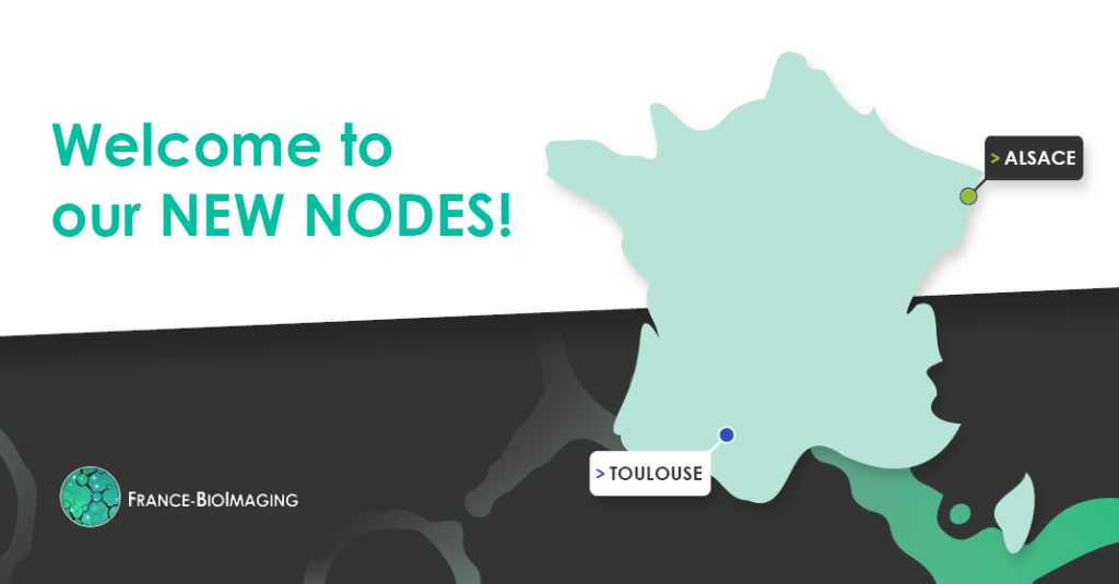
Following the final decision of France-BioImaging Institutional Committee on February 21th, 2023, we are delighted to announce that two new nodes are joining France-BioImaging: the Alsace Node and the Toulouse Node.
The new Alsace Node is composed of six imaging facilities: QuEST, the Cell Imaging Facility from IBMP, PIV, PI2, PIC-STRA and PMC. The node has also six highly visible R&D teams (from IRIMAS, LBP, ICube, IGBMC and IPHC) expert in microscopy techniques and tools.
Located in Strasbourg, Illkirch and Mulhouse, the Alsace node is offering high level technical and innovative methodological expertise in multi-scale imaging at the interface between biology, chemistry, optics and physics, from the atom to the small animal/plant.
The Alsace Node has a strong expertise in probes with the development of highly innovative fluorescent probes and luminescent nanoparticles.
The node provides cutting-edge technologies and methodologies, with among others:
Tomographic diffractive microscopy, for 3D label-free imaging at cellular level, with an improved resolution compared to conventional microscopes, and, not being limited by possibly weak fluorescence, potentially at high speed (several 3D images/s)
Single particle tracking and time-resolved luminescence microscopy, to image upconverting nanoparticles (UCNPs). Due to their anti-Stokes emission, UCNPs allow imaging applications with exceptional signal to noise ratio.
Single-shot full-field optical coherence tomography, extracts an FF-OCT image from a single interference acquisition, enabling single-shot high-resolution imaging within turbid media such as biomedical samples.
SharpViSu & ClusterViSu, development of an integrated software for image reconstruction, correction, co-localization, resolution estimation, segmentation and clustering of labelled complexes
A large range of molecular and nanoparticle probes for SR and advanced microscopy techniques as well as molecular and supramolecular complexes for anti-Stokes imaging at the molecular level have been developed.
The new Toulouse node is composed of a large nationally recognized multi-site core facility: Toulouse Reseau Imagerie. Distributed on Toulouse greater area, the facility is divided between medical science, fondamental science, cancer and rejuvenation, and agro-bio science. Moreover, eight R&D teams complete the node and supports the facility (from LAAS, CBI, IPBS, IRSD and IMT).
The Toulouse node aims to maintain the level of scientific excellence in response to the needs of users and the concerns of host laboratories. Four scientific axes are conducted: mechano-biology, molecules and single cells, whole organisms, image processing and quantitative data analysis.
The mission of the node is also to develop original devices to explore biophysical properties in living samples, to work at the interface between machine and sample and to develop artificial intelligence applied to bioimaging.
The node provides cutting-edge technologies and methodologies, with among others:
Random illumination microscopy, a super-resolution microscopy technique, interesting for its robustness
Protrusion force microscopy, combination of micro-fabrication, imaging approaches, including Atomic Force Microscopy, dSTORM coupled to supercritical angle fluorescence, and random illumination
Cryomethods for electron microscopy, includes sample preparation of frozen samples for cryo-microscopy, in view of bridging structural biology and cell biology.
ANchOR technology, a labeling system based on ParR/ParB labeling system from bacteria, which allowed the strong fluorescent labeling of genomic site with small DNA insert.
Microfabrication for organoids, 3D bioprinted scaffolds have been adapted to permit 3D characterization at the organoid and tissue scale
Welcome to the FBI Alsace Node and the FBI Toulouse Node!
From February 6th to February 10th, France-BioImaging organised a group meeting on the project “FBI.data” in Bordeaux. For a week, participants focused on the architecture and the implementation of image data management tools. A user-friendly response to the challenges of never-ending data production.
New imaging technologies are very greedy in terms of image processing and data management. Beside the image itself, biological imaging generates a huge amount of metadata. The FBI.data project, one of the key missions of France-BioImaging, addresses the questions related to the computational analysis and handling of image data.
Speeding up the implementation of tools across the infrastructure
Although the distributed FBI.data team meets once per week, the FBI.data Sprint aims at only focusing on data management scenarii and accelerating the project. Two essential aspects have been discussed:
First of all, the data management system architecture must be simple for them to be implemented across France-BioImaging nodes. It also has to be compatible with long-term data storage and of course, to be user-friendly – we want to keep it easy for our users!
The second point is all about anticipating as many data management cases as possible. Running through all the needs of bioimaging experts and users, the team lists the specific features of each case and considers the perfect solutions for all of them.
Working for the FAIRisation of data
The FAIRisation of data for Open Science is an initiative fully endorsed by France-BioImaging. Meaning that data are Findable, Accessible, Interoperable and Reusable, the benefits for the bioimaging community are numerous. It improves transparency and reproducibility, enhances quality of results, accelerates scientific progress and method development and finally boosts collaboration within the scientific community.
OMERO, developed by the University of Dundee & Open Microscopy Environment teams, on which the FBI.data group is working, is one of the software making user data FAIR. Being a microscopy image data management decentralised platform, it helps organise, access and archive data. Besides, it combines image and metadata storage, a viewer and data analysis resources. Furthermore, OMERO is linked to the most valuable tools for bioimaging experts (ImageJ, Napari, QPath, etc.). And users can access their data from anywhere and keep them safe.
But much more has to be set up to have functional solutions: to ease user authentication and management, manage big data transfer, and have an adequate metadata scheme. Accompanying users is one of the mission of the FBI.data team, and the FBI.data Sprint is also the occasion to join efforts from the training working group led by the training mission officer Fabrice Cordelières (Bordeaux Imaging Center) to produce adequate training material on data management.
Sharing efforts and helping the community
The FBI.data working group is composed of:
Perrine Paul-Gilloteaux, research engineer at MicroPICell core facility and FBI.data mission officer
Thierry Pecot, research engineer in image analysis at Rennes (Bretagne Loire Node)
Further recruitments are on-going and will be reinforcing the team
By joining their skills and experience, they are working together on setting up tools and good practices for the management and FAIRisation of data inside France-BioImaging nodes but also for the entire bioimaging community. With this in mind, the project has collaboration with, among others, the Institut Français de Bioinformatique (IFB) and the Centre National de Ressources Biologiques Marines (EMBRC), and other infrastructure through the MUDIS4LS Equipex+ project. Moreover, the FBI.data project has an open GitLab, providing image data management codes in open source, and a blog with tutorials, recommendations and so much more!
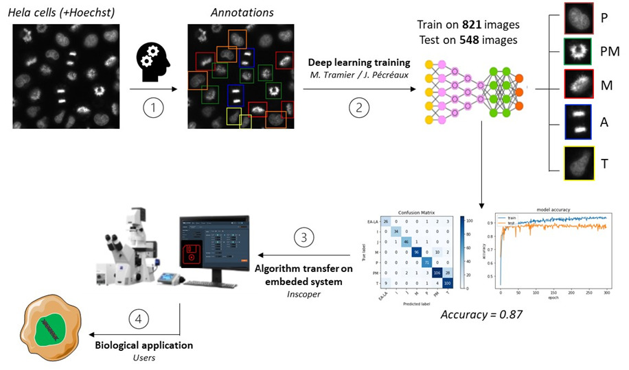
Being designed in response to imaging challenges, the Roboscope is the product of a collaboration between Marc Tramier’s team (FBI Bretagne-Loire node) with Julia Bonnet-Gélébart, research engineer, Jacques Pécréaux’s team of the Institut Génétique & Développement de Rennes (IGDR), and the Inscoper company, spin-off of the lab. This technology could become a great timesaver for fluorescence microscopy.
Allowing the automation of fluorescence microscope acquisitions, the Roboscope is an embedded technology based on a deep learning algorithm. To be precise, it is a predesigned event-driven acquisition (PEDA) based on a learning automatization of any cellular changes tracked by fluorescence. Catching rare and fast cellular events then becomes possible!
The use of the Roboscope would also save precious time of research, providing users with results without the need to stand by the microscope during acquisition. This technology goes beyond as they will be able to recover the data already classified and with only the specific points of illumination that they have previously triggered.
A broad range of applications
The teams have almost finished to develop an entire algorithm monitoring the cell cycle progression in mitosis. These events specific to the cellular division correspond to major challenges in the control and treatment of cancer progression (Kops, 2005). As the cell cycle study is needed to understand several biological processes helping the development of targeted drugs, the technology aims to monitor efficiently and automatically simple cell models through their division cycle.
And this is not its only benefit: this automatized fluorescence microscopy acquisition can be adapted in very diverse fields. From a cell cycle progression analysis to specific analysis, organelles, proteins and biological events can be tracked or activated within cells. A noteworthy advantage of the integrated device that – we hope – will be deployed widely in the future.
Workflow of a Roboscope experiment. 1. The user annotate a bench of images with different class of interest to be detected. 2. The pre-trained Convolutional Neural Network is adjusted for the experiment by fine tuning and/or transfer learning. 3. The algorithm is transfered on embedded systems to perform real-time image analysis during microscopy acquisition. 4. The biological application with event-driven acquisition is defined and started by the user in order to start, interrupt and parametrize different acquisition sequence following real-time image analysis and event classification.
Deadline: February 28th, 2023
For users from a unit attached to the INSB who do not usually use Research Infrastructures’ services for their projects
For access to technologies and related expertise
For access to expertise in data analysis
The scientific project must be a project in the process of being finalized
The field of Life Sciences has undergone major developments over the past two decades. The change of scales, both in spatial and temporal resolution, and the integration of data from a wide variety of sources, as induced by the development of technologies, have revolutionized the exploration of life. These technologies call for expensive investments and specific knowhow, carried out by highly qualified personnel, having led to the creation of common infrastructures such as research infrastructures (IRs) open to the entire scientific community.
The Institut des Sciences Biologiques (INSB) from Centre National de la Recherche Scientifique (CNRS) is launching the second edition of a call for projects to fund full access to the national infrastructures in health and biology “INBS Access to national research infrastructures”. This call aims to encourage teams to get a first access to the services offered by Research Infrastructures to help their research project. The objective is to demonstrate the significant impact of these services to improve the quality of their results (resolution, reproducibility, change of scale, etc.) or to help remove technological barriers. We also aim to augment the awareness of our teams to the cutting-edge technologies, methods and expertise offered by these national infrastructures.
Program description
Access to Research Infrastructures is open to the entire French and international scientific community, with contribution to the running costs of equipment being charged to the users on quote, after project feasibility confirmation by the infrastructure. The purpose of this program is to facilitate and finance access to these infrastructures. Target audience are INSB teams new to a technology or method offered by these infrastructures, seeking to validate the contribution that the infrastructures could make to their research topic by removing barriers that impair the completion or finalization of an ongoing project of the team. A new project is not eligible to this call. The list of eligible Research Infrastructures, to which the research team could apply, is available here: https://www.insb.cnrs.fr/fr/infrastructures-nationales. Other infrastructures may be considered depending on the proven needs of the project. Several types of access can be supported by this call for projects:
Access to technologies and related expertise, upon quote from the infrastructures and confirmation of feasibility in 2023
Access to expertise in data analysis, upon quote from the infrastructures and confirmation of feasibility in 2023
In addition to the costs of access to infrastructure, upon a quote emitted by the infrastructure at the second stage of the call, travel/mission costs, as well as consumables that are not covered by the costs of access to infrastructure, may be covered. The funding for each project will be in the range of 10 to 30 K€.
Instructions for submission
This is two-stage submission.
The first stage consists of a letter of intent, prepared by the scientific leader of the project, outlining the project in finalization in which the application fits and highlighting the barriers that could be removed by access to IRs. The novelty for the team of the usage of the technology and methods to which this application would give access has to be underlined. This letter of intent must also be signed by the director of the applicant’s unit. The identification of the infrastructures to access is possible at this stage but not mandatory and the application can focus on the technology and methods needed. An estimated timeline is however welcome.
The letter of intent (maximum of three pages including figures and references, in French or English), signed by the unit director, accompanied by a CV of the project leader (maximum of two pages) must be sent before Tuesday 28th of February 2023 to the following address: insb.ain@cnrs.fr. These proposals will be screened for eligibility criteria and selected projects will go to the second phase on 8th of March: The second phase consists of a consultation between the INSB and the national Research Infrastructures. This will lead to putting the applicant in contact with the infrastructures that are able to meet the expressed needs, in order to evaluate the feasibility and to establish a quote from their official invoicing cost models.
This stage will lead to the final submission of the project, including the requested budget, signed by the heads of the involved IR(s), no later than Wednesday 22th of march 2023. Funding decision will be sent to the scientific leader beginning of April.
Project eligibility and selection criteria
Access to the infrastructure must be fully implemented before 31/12/2023 (deadline for the engagements of funds by the teams). A scientific and financial report will be asked to the granted team in June 2024.
The scientific project must be a project in the process of being finalized. Access to infrastructure must unlock or accelerate the project. The start of a scientific project is not eligible for this call.
The access to the infrastructure technology or expertise required should be new to the scientific leader.
A project can call to different infrastructures.
In the first stage, budget is not expected, but the project should demonstrate its feasibility from the team side before the end of 2023 if access to the required technologies or expertise is granted (for example identified human resource from the team to undertake the project) and by providing an estimated timeline. The infrastructure(s) will confirm it in the second stage.
Project will be screened for scientific quality of the scientific leader and of the project in finalization, the interest of the targeted method and technology to finalize the project, the novelty of access to the infrastructure. In the second stage, the quote produced by the infrastructure will assess the feasibility and adequacy to the budget.
Eligible expenses are:
Billing from the infrastructure based on quote presented at the second stage
Mission fees to access the infrastructures
Consumables needed to prepare samples for access if not provided by the infrastructures
We use cookies on our website to give you the most relevant experience by remembering your preferences and repeat visits. By clicking “Accept All”, you consent to the use of ALL the cookies. However, you may visit "Cookie Settings" to provide a controlled consent.
This website uses cookies to improve your experience while you navigate through the website. Out of these, the cookies that are categorized as necessary are stored on your browser as they are essential for the working of basic functionalities of the website. We also use third-party cookies that help us analyze and understand how you use this website. These cookies will be stored in your browser only with your consent. You also have the option to opt-out of these cookies. But opting out of some of these cookies may affect your browsing experience.
Necessary cookies are absolutely essential for the website to function properly. These cookies ensure basic functionalities and security features of the website, anonymously.
Cookie
Duration
Description
cookielawinfo-checkbox-analytics
11 months
This cookie is set by GDPR Cookie Consent plugin. The cookie is used to store the user consent for the cookies in the category "Analytics".
cookielawinfo-checkbox-functional
11 months
The cookie is set by GDPR cookie consent to record the user consent for the cookies in the category "Functional".
cookielawinfo-checkbox-necessary
11 months
This cookie is set by GDPR Cookie Consent plugin. The cookies is used to store the user consent for the cookies in the category "Necessary".
cookielawinfo-checkbox-others
11 months
This cookie is set by GDPR Cookie Consent plugin. The cookie is used to store the user consent for the cookies in the category "Other.
cookielawinfo-checkbox-performance
11 months
This cookie is set by GDPR Cookie Consent plugin. The cookie is used to store the user consent for the cookies in the category "Performance".
viewed_cookie_policy
11 months
The cookie is set by the GDPR Cookie Consent plugin and is used to store whether or not user has consented to the use of cookies. It does not store any personal data.
Functional cookies help to perform certain functionalities like sharing the content of the website on social media platforms, collect feedbacks, and other third-party features.
Performance cookies are used to understand and analyze the key performance indexes of the website which helps in delivering a better user experience for the visitors.
Analytical cookies are used to understand how visitors interact with the website. These cookies help provide information on metrics the number of visitors, bounce rate, traffic source, etc.
Advertisement cookies are used to provide visitors with relevant ads and marketing campaigns. These cookies track visitors across websites and collect information to provide customized ads.