FBI launches a call for its future Scientific Director!
▽ Scroll down
Category: Announcement
Starts: November 10, 2023 • Ends: December 31, 2023
FBI opens a call for the recruitment of its next Scientific Director(2024-2028)
(Deadline is 31st of December 2023)
France BioImaging-FBI (laureate of the INBS program of the PIA in 2011) is the National Research Infrastructure in Biological Imaging. FBI is built on 8 geographical Nodes identified on the basis of strong relationships between R&D labs and imaging Core Facilities. Each Node shows a specialization of a local expertise in methods and biological topics. This crossover between imaging technologies and expertise in scientific topics is a characteristic of the complementarity between FBI Nodes. A 9th Transversal Node gathers FBI strengths and resources in Image Analysis and DATA management. The Operating Coordination is done under the umbrella of the UAR 3426.
Our motto is “Innovation-Training-Access”
(i) Invent and disseminate new imaging technologies (ii) Training users and facility staff on existing and new technologies (iii) Make them accessible to as many people as possible.
Role of the Scientific Director of FBI
-He/She is the Strategic Manager of the Infrastructure
–He/She leads the National Coordination(NC), composed of an adjunct director for international affairs, a manager of internal affairs and a manager of external affairs. The Infrastructure also benefits from several support functions: a communication officer, a business developer and an accounting officer. The NC leads the Executive Committee(EC) to manage the Infrastructure. With the Help of an international Scientific Advisory Board (SAB), he/she reports the overall Infrastructure policy and strategy to the Institutional Committee (Steering Committee, SC) which is the “decision maker”.
–He/She is responsible for the arbitration of recruitment proposals (in interaction with the other governance bodies), equipment investments (PIA, TGIR, other National and International common actions…) and new service opening, in relation to the development objectives (R&D, service offers…) and the overall infrastructure strategy at national and international levels.
–He/She is responsible, with the staff concerned, for the inventory of the different activities/tasks of the infrastructure: links with Europe and International, work with the different committees, web site/communication, training, animation of the “FBI-community”, scientific and financial reports…
– He/She is responsible, in interaction with the EC, for managing interactions/collaborations (R&D, service providing, partnerships, technology watch…) with other PIA Research Infrastructures.
Entry into function is planned for the 1st of July 2024.
If you are interested, please send a short CV and a letter of intent (2 pages) indicating your motivation, vision and strategy, at direction@france-bioimaging.org BEFORE the 31st of December 2023
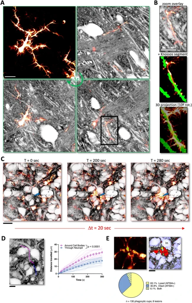
In 2022, in a project funded by the Euro-BioImaging pilot User Access Fund, Andrew Boyce, postdoctoral fellow at the University of Calgary, used state-of-the-art microscopy techniquesat the Bordeaux Imaging Center, part of the Bordeaux node of France-BioImaging, for panoptical visualization of brain tissue in vivo. His work just got published in a Nature Communications article!
The advantages that shadow imaging has in visualizing brain tissue
Progress in neuroscience research hinges on technical advances in visualizing living brain tissue with high fidelity and facility. Unfortunately, current neuroanatomical imaging approaches either require tissue fixation (electron microscopy), do not have cellular resolution (magnetic resonance imaging) or only give a fragmented view (fluorescence microscopy).
In this study, scientists have shown how regular light microscopy together with fluorescence labeling of the interstitial fluid in the extracellular space provide the information lacking in the previous techniques. Basically, it’s like looking at the negative of a photo! Called Shadow Imaging, they have demonstrated the power of this approach on revealing neurons, microglia, tumor cells and blood capillaries together with their complete anatomical tissue contexts.
The perfect combination between the right technology and the right expertise
How did Andrew Boyce contribute to this study? Interested in blebbing neuron dendrites during cell death caused by a stroke, he only had, at that time, results from how cells in culture reacted during this kind of events but no information about the neighboring cells.
Thanks to Euro-BioImaging pilot User Access Fund, he came to Bordeaux to use a combination of live cell 3D stimulated emission depletion (3D-STED) microscopy and super-resolution shadow imaging (SUSHI), and adapt shadow imaging approaches to conventional confocal microscopy (COSHI). Shadow imaging allows you to visualize fine details of cell-cell interactions and the extracellular space in an unbiased manner. Brain tissue is complex and shadow imaging is a technique that allows researchers to visualize all of the cells in this very complex architecture in an unbiased manner so it was the perfect match for Andrew’s research project!
A career-boosting experience
“This has really been an incredible experience. It’s amazing to be trained by the person and team who pioneered this technique. I’m very thankful for this opportunity and humbled to be over here,”reflects Andrew from Bordeaux.
When asked how his visit to Bordeaux might impact his career, Andrew explains that he can certainly imagine doing shadow imaging in the future. He would eventually like to run his own lab at an academic institution in Canada. Being able to bring an exciting and novel technique like this back to Canada would be really impactful for his career, and for science in general.
“Shadow imaging is a very exciting technique for studying brain tissue, but getting started on sample preparation and adapting this technique to your research question can be daunting. Coming to the Bordeaux Imaging Center to work with the person who developed this approach and get expert training is a dream come true and will make a big difference to my research. This is an amazing opportunity made possible by the Euro-BioImaging Pilot User Access fund with the goal of making shadow imaging more accessible across diverse platforms,” concludes Andrew.
You want to be the next user to get access to state-of-the-art technologies that France-BioImaging provides? Please login via Euro-BioImaging website! You just have to choose the technology you want to use, then submit your proposal. All applications will be processed by the Euro-BioImaging Hub in close relation with France-BioImaging. And of course, all scientists regardless of their affiliation, area of expertise or field of activity can benefit from open access services! Users whose projects will be validated by Euro-BioImaging will benefit from a waiver for the access cost on France-BioImaging core facilities (france-bioimaging.org/access)
Thanks to Marianna Childress and Andrew Boyce for the original article!
Dembitskaya, Y., Boyce, A.K.J., Idziak, A. et al. Shadow imaging for panoptical visualization of brain tissue in vivo. Nat Commun14, 6411 (2023). https://doi.org/10.1038/s41467-023-42055-2
As the 2023 edition of the France-BioImaging Image Contest admissions is still running, we wanted to highlight our previous winners and their projects. Here is a quick throwback to our 2022 winners.
Before getting to the heart of the matter, we want to remind you that you still have time (before November 10th) to submit your best images and try to win your registration fees for one 2024 microscopy-related event! Please make sure you upload your images on the following link:
Last year, we enjoyed the winning images submitted for their artistic take and their quality. Thanks to Carole SIRET, Magalie BENARD and Frédéric FERCOQ for their beautiful images!
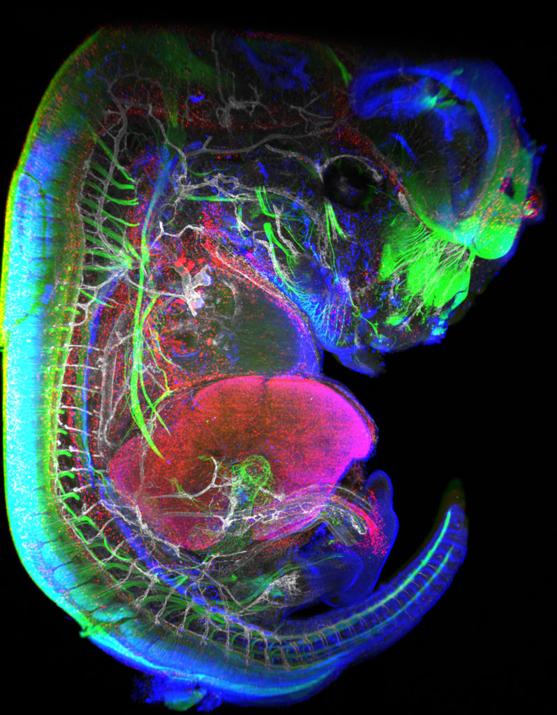
1st Place: Carole SIRET, Van de Pavert Team, Centre d'Immunologie de Marseille-Luminy
"Little Monster"
The embryonic formation of lymph nodes, small organs essential for the immune response, is now known. Using light sheet microscopy, scientists were able to determine the dynamics at work in this 13.5-day-old mouse embryo. In blue, the lymphoid cells (LTi), derived from the haematogenous endothelium, a specific tissue of the embryo. They pass into the liver where they proliferate before migrating through the body to give rise to lymph nodes. The 3D information obtained thus makes it possible to follow the interactions of lymph nodes with their environment, in particular with nerve cells, in green, and blood vessels, in white. The lymphatic endothelial cells and some macrophages are visible in red.
Lightsheet Microscopy
Carole Siret is a Research engineer, expert in Lightsheet microscopy, at the Centre d’Immunology Marseille Luminy (CIML) since 2018. She is working in Dr Serge van de Pavert team where they study immune system development. They are particularly interested in the lymph nodes (LN) formation during mouse embryogenesis.
The image she submitted is a projection from a lightsheet acquisition on the UMII (Miltenyi). This image illustrates an E13.5 mouse embryo stained for neurons, LTi (Tissue inducer cells which are the precursor cells for the lymph node), lymphatic and blood vessels. This acquisition was done in the context of the study of the role of Cxcl12 in embryonic LN formation. From previous work it is clear that Cxcl13 and Ccl21 are not expressed present near blood vessels, but it likely that some chemokines, possibly Cxcl12, could be expressed on the endothelial cells. We focus on Cxcl12 since this chemokine has shown to be important for the attraction of several hematopoietic cells. Although it was shown that the receptor for Cxcl12, Cxcr4, is expressed by the mature hematopoietic inducer cells, it is not clear whether it also expressed by the progenitor hematopoietic inducer cells. Next to the possible attraction of hematopoietic cells towards the lymph node anlagen, Cxcl12 is involved in the attraction of nerve fibers. Therefore, the possible role of Cxcl12 could be to both attract hematopoietic cells as well as nerve fibers to initiate a region which is permissive to form lymph nodes.
Thanks to the France-Bioimaging Image Contest, Carole participated to the SFI Congress, where, this year, it was a special joint conference both between the Société Française d’Immunologie (SFI) and the Deutsche Gesellschaft für Immunologie (DGfI). It was a great opportunity to exchange with people at the cutting edge of the immunology field.
2nd Place:Magalie BENARD, Plateforme de Recherche en IMAgerie CEllulaire de Normandie (PRIMACEN), Research infrastructure HeRacLeS, Inserm US 51, CNRS UAR 2026,
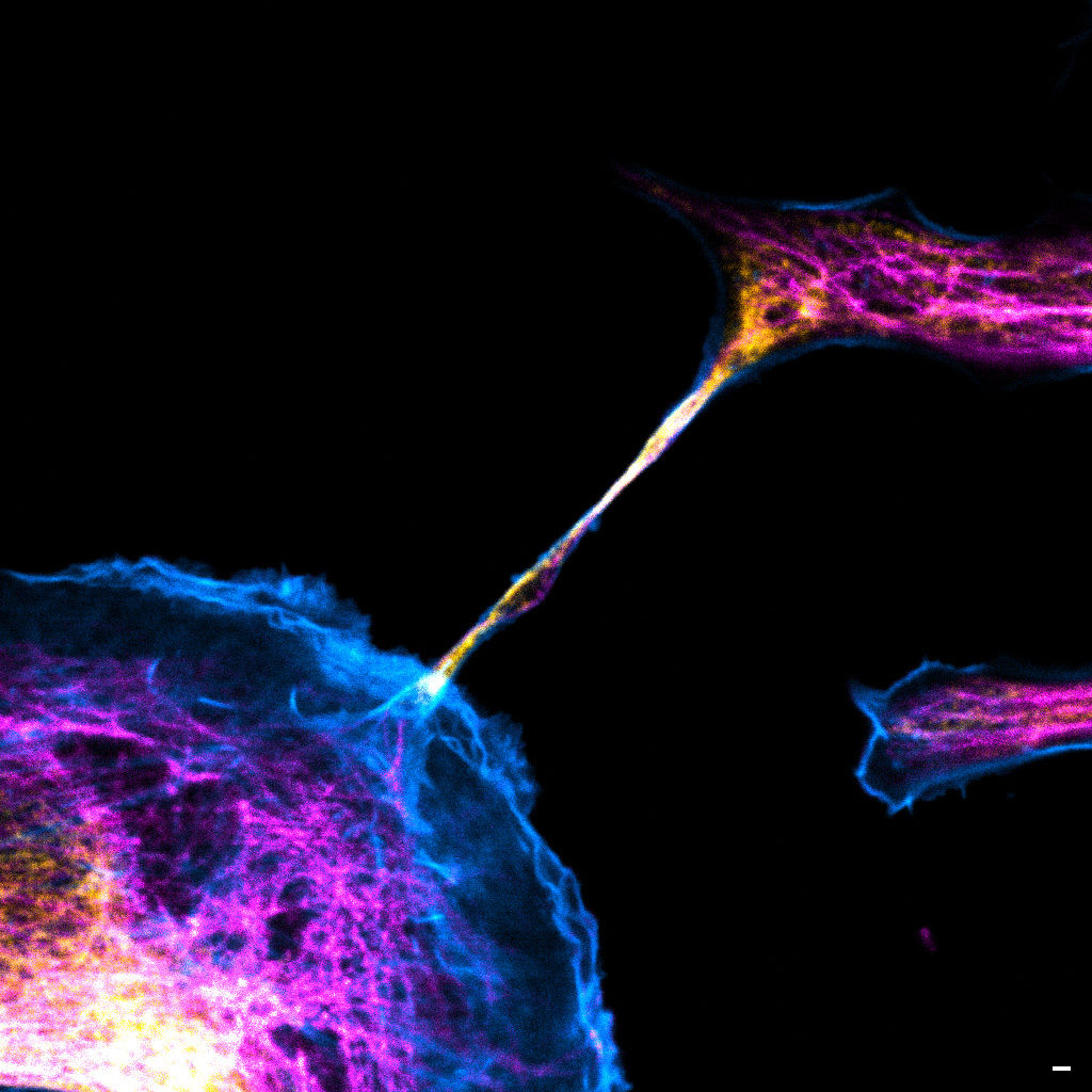
"The communication link with others"
Image of a cellular interconnection between two human tumor cells whose cytoskeleton has been labeled with anti-tubulin (ATTO-647N), anti-vimentin (AlexaFluor594) antibodies and with Phalloidin probe (AlexaFluor488). Scale bar 1µm.
Confocal microscopy
Magalie Bénard is a Research Engineer and the Technical Manager at the Cellular Imaging Facilty PRIMACEN (Plate-forme de Recherche en IMAgerie CEllulaire de Normandie).
The image she submitted is a confocal image representing a cellular interconnection tunneling nanotube (TNTs) between two human tumour cells. In a cancer case, some cells are able to express spontaneously TNTs with cytoskeleton protein composition corresponding to specific role of this communication mechanism. In the winning image, the TNT is composed of tubulin (magenta), actin (cyan) and vimentin (yellow) proteins. Called TNT1, this nanotube allows the transfer of intracellular elements such as RNA, proteins or organelles. Moreover, due to the thinness of TNTs, their photo-sensitivity and their fragility, live-cell imaging is technically challenging with regards to potentially damaging methods. Magalie and her team have developed an adapted method to observe TNTs in living cell with high resolution imaging (STED) enhanced by FLIM by using red and near infrared probes.
France-Bioimaging sponsored her participation to the ELMI (European Light Microscopy Initiative Meeting June 6-9, 2023) congress. During this event, she had the chance to present her project through a poster. This congress also offered a great opportunity to have an overview and the last updates on state-of-the-art imaging techniques.
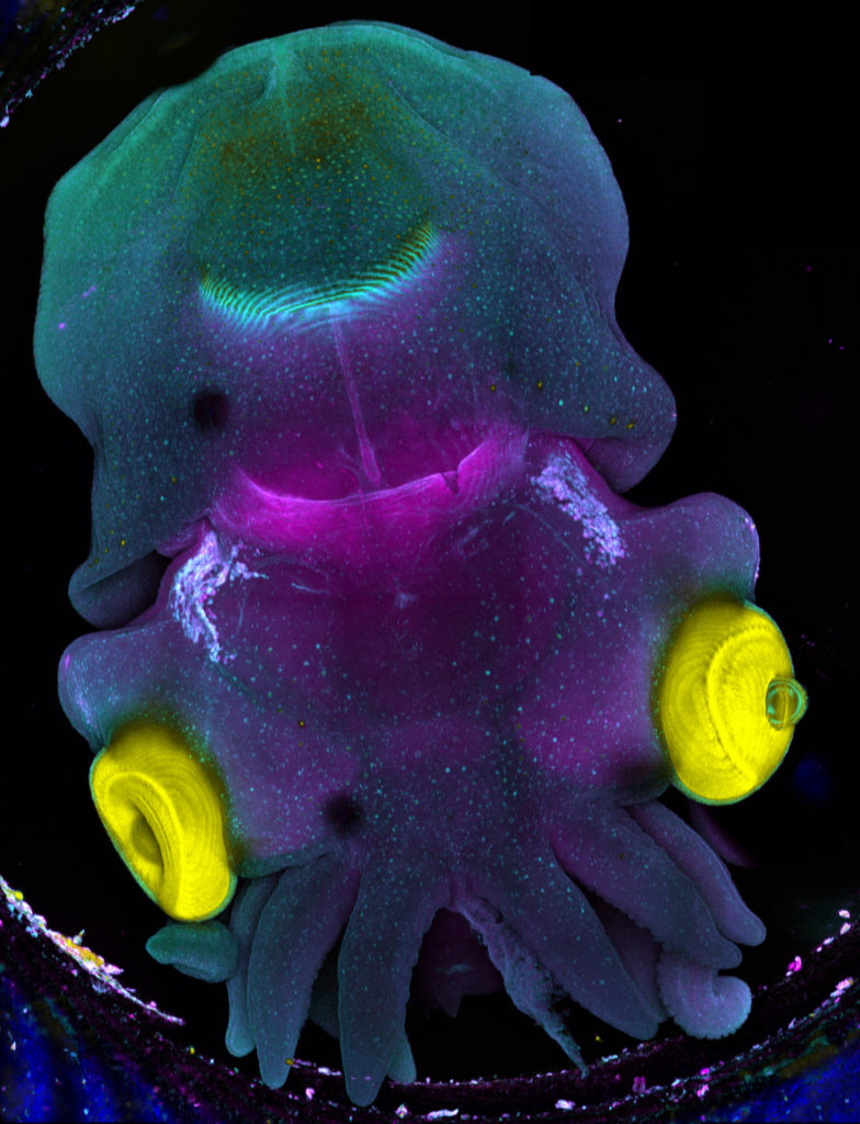
3rd Place:Frédéric FERCOQ, Parasites et Protistes Libres (PPL), Museum National d'Histoire Naturelle
"Sepia"
Stage 25 cuttlefish embryo (Sepia officinalis) observed under a confocal microscope. The cuttlefish was cleared and the tissue autofluorescence was captured.
This image was produced in collaboration with Laure BONNAUD-PONTICELLI and Luis MOLINA from the BOREA laboratory.
Confocal microscopy
Frédéric Fercoq is a postdoc scientist in the Parasitology laboratory of the Muséum National d’Histoire Naturelle (MNHN) in Paris. My main interest is on how myeloid cells participate to the control of parasitic infections, but sometimes at the price of collateral tissue damage. This project involves a lot of microscopy of immune cells, parasites and host tissues to analyse the complex interactions taking place at the site of infection.
The image he submitted has nothing to do with his main project! As he has the chance to work on very different topics and models, this image was acquired as a proof of concept for imaging full embryos of the cuttlefish Sepia officinalis for Frédéric's colleagues Laure BONNAUD-PONTICELLI and Luis MOLINA (BOREA laboratory, MNHN). They work on the nervous system of cephalopod and on the influence of environmental factors during its development. They are now optimizing fluorescent staining for neuronal markers to test the effect of light on the nervous system in situ.
France-Bioimaging sponsored his participation to the FOM (Focus on Microscopy) 2023 congress in Porto. He had the chance to be granted the opportunity to both present his current project through a poster and to give an oral presentation. He was also amazed by the new avenues opened up by the cutting-edge imaging techniques presented throughout the conference.
You can now apply for a new call to get free open access to instruments and services at one of our nodes’ core facility!
We are happy to announce the first Open Call from the Horizon Europe-funded canSERV project! Cancer Researchers are invited to apply for FREE state-of-the-art services and training at several European Research Infrastructures, including Euro-BioImaging ERIC. Within this project, 28 Euro-Bioimaging Nodes (- Yes, France-BioImaging is part of it too! -) offer access to their expertise. It’s an amazing opportunity for the cancer research community to access a wide-ranging portfolio of services.
CanSERV invites applications covering the entire range of the oncology developmental pipeline, from supporting discoveries in fundamental research to translational science and personalized oncology, addressing at least one of the four strategic goals of the Cancer Mission:
Understanding of Cancer
Prevention and Early Detection
Diagnosis and treatment
Quality of life for patients and their families
Researchers from academia, industry and SMEs may apply for several service categories including:
disease models,
cutting-edge imaging and structural biology technologies,
biomarker research and development,
new therapeutic solutions,
complex clinical trial design and support,
personalized oncology implementation pipelines and recommendations and
regulatory support and tools to analyse the socioeconomic dimension of research activities.
Launch of the first call is today, Thursday, October 12, 2023.
Submission deadline is January 4, 2024 at 14.00 CEST.
To know which Euro-Imaging’s technologies can help your research project, visit their website www.eurobioimaging.eu/service
In order to answer image data analysis demands, France-BioImaging is launching its first data machine learning competition! The idea? To develop powerful methods that will then end up in creating standards & benchmarks in the field of bioimaging.
The FBI challenge is hinged on a double contribution: from core facilities engineers and from data scientists. The first group will acquire a large number of images to build a dataset, that will later be used by the algorithms. These images will be produced by microscopy engineers & technicians from FBI’s platforms. As for the second contribution, this is where the challenge starts! The challenge will be later published to have a maximum of data scientists to work on the algorithms that best fulfill the analysis task.
The first project is also based on four pillars:
Open source + FAIR (Findable, Accessible, Interoperable, Reusable)
Supervised learning, it involves annotated datasets to maintain control over performances.
In silico annotations, a computer labeling method to avoid manual annotation and its drawbacks.
Image-to-image analysis tasks, an image analysis tasks which aim to predict an output image from the input one.
We need you!
The first challenge is still under preparation. It will predict the fluorescence image of cell culture from a bright field, phase contrast and/or DIC microscopy label-free image. Nevertheless, we need you to contribute to the dataset that will supply the machine learning! The more images we have, the better the prediction algorithm will be.
Going to Rendez-Vous Carnot 2023? Drop by our booth U10 and say hello! 18 & 19 October – Lyon
In a few days, we will be travelling to Lyon for the Rendez-Vous Carnot 2023! This is the fifth time that we will attend the forum as an exhibitor, in the Research Infrastructures Village. We are going to present France-BioImaging R&D ecosystem and the multiple advanced biological imaging technology developments taking place on FBI imaging platforms and R&D teams.
If you are in Lyon between October 18 and October 19 attending the Rendez-Vous Carnot as well, be sure to drop by our booth U10 (at the Research Infrastructure Village) and meet some of our colleagues at the venue:
Etienne Henry, France-BioImaging R&D and Tech-Transfer mission Officer
Alban Belloir, France-BioImaging Communication Officer
We will be happy to discuss with you!
FBI opens a call for the recruitment of its next Deputy Director for Internal Affairs
Mission
The Deputy Director for Internal Affairs will help infrastructure management team to oversee several of its key missions: implement a strategic vision for France-BioImaging, encompassing our three pillars, Innovation, Training, Access; develop a roadmap for next generation imaging technologies and services; increase funding opportunities and develop a sustainable cost model; update and improve infrastructure communication goals and actions; develop ours links with the private sector. The Deputy Director will work closely with the Manager of Internal Affairs to tailor specific activities, prepare strategic decision-making and prioritize needs for initiatives.
Mandate of the Deputy Director for Internal Affairs
Help to develop and implement the strategic vision of FBI’s core activities, in Innovation, Training and Services; synthesize and prepare elements to assist in decision-making
Advise the management team to improve cooperation across FBI geographical Nodes; promote the scientific and structuring activities of the infrastructure at the national level, to seek out and federate potential partners, initiate and manage national cooperation programs, and provide expertise and advice.
Develop a roadmap for technological developments and their implementation as FBI services
Help to develop funding opportunities and develop a sustainable cost model for Innovation and Services
Develop links with the private sector, as users of FBI services or partners in FBI core activities including methodological innovations
Organize a prospective reflection on the development of new initiatives to be carried out to improve FBI communication
Skills
Senior scientist (DR or equivalent) with a strong motivation and willingness to strengthen FBI core activities in Innovation, Service and Training;
Knowledge of the national landscape in biological imaging, including in innovation and services;
Experience in the management of research units or facilities, and projects in the science and innovation domain;
Demonstrated experience in fund raising operations for research and innovation projects in biological imaging;
Knowledge of the national and international research infrastructures in Biology and Health environment
Experience in interdisciplinary scientific and stakeholder networking;
Demonstrated ability to work collaboratively with diverse stakeholders;
Excellent communications and public relations skills;
High proficiency in English language
Organisation
France-BioImaging is a national research infrastructure distributed throughout France that provides researchers with access to the latest innovations in biological imaging and aims to accelerate the transfer of technological and methodological innovations in biological imaging to the 22 platforms that constitute the infrastructure. The Deputy Director for Internal Affairs will contribute to the development and strengthening of France-Bioimaging. The Deputy Director for Internal Affairs will work within the national coordination of the France BioImaging infrastructure. He/she will work within a team distributed between Montpellier and Bordeaux, composed of 4 people: the Scientific Director and the Deputy Director for International Affairs, the Internal Affairs Manager, and the External Affairs Manager.
Time Commitment
FBI’s Deputy Director for Internal Affairs should commit approximately 20% of his overall working time to this mission. Virtual coordination meetings (Directors + Managers) occur weekly, and the FBI Executive Board meets virtually monthly. FBI steering committee meets bi-annually. The Deputy Director is appointed for a 5-year mandate and is eligible to renew his service.
Requirements
Frequent travels in France and abroad.
Entry into function is planned as soon as possible / November 15th, 2023.
You can now apply for two new calls to get free open access to instruments and services at one of our nodes’ core facility!
AgroServ is a transdisciplinary project funded by the European Union under the Horizon Europe programme, running until 2027. It supports the research community by funding interdisciplinary agroecology research projects. The supported research will contribute towards sustainable and resilient agri-food systems, exploring topics like plant biology, water, soil, and microorganisms. Euro-BioImaging is a partner in the AgroServ consortium along with 12 other research infrastructures and partners in 23 countries. Nine Euro-BioImaging Nodes offer access – including France-BioImaging – to their expertise and services within this project.
AgroServ has launched the pre-proposal phase of the first Open Call for projects. Selected projects will receive funding for access to integrated research infrastructure services & expertise.
We encourage all researchers who would like to harness the power of imaging and image data to boost their agroecology research project to look at AgroServ’s impressive Catalogue of Services.
Only core facilities from our Ile-de-France Sud node are available through this call. The node will be happy to welcome you for your research project! For more information about the microscopy techniques you can use thanks to this call, please send an email to contact@france-bioimaging.org
Within the framework of the COMULISglobe project, Euro-BioImaging will be awarding a total of 4 grants for access to Euro-BioImaging Nodes – including France-BioImaging! -, specifically designed to support correlative and multimodal imaging.
Two grants will be awarded to European researchers (including from non-EU countries that are Euro-BioImaging members) accessing services at Euro-BioImaging Nodes. Each grant will provide up to EUR 2000 in support of imaging projects that tackle important research questions using two or more imaging modalities.
Two grants will be awarded for international (non-European) researchers accessing services at Euro-BioImaging Nodes. Each grant will provide up to EUR 3000 in support of imaging projects employing two or more imaging modalities.
France-BioImaging primary mission is to develop, promote, disseminate and provide access to innovative instruments and imaging technologies in the field of bioimaging to scientists. Fostering the technological transfers is at the heart of this mission, and for this France-BioImaging relies on a strong association of leading R&D research teams with core facilities.
However, several bottlenecks exist and often hamper or prevent successful technology transfer:
A lack of human resource leads to difficulties in transferring and stabilizing the technology which is not enough user-friendly
A technology that is too specific, with not enough user base
A difficulty to contract with industry through institutional offices for industrial valuation
In the context of image analysis: the instability of open software economical model, inter-operability, large data handling and algorithm complexity
As a way to tackle these bottlenecks, France-BioImaging launched in January 2021 its first “FBI Internal Call 2021: Technology transfer from the R&D teams to the core facilities” to promote the transfer of new technologies (instrumentation, probes, staining methods, software, data analysis or data visualization) from the R&D teams to the facilities of France-BioImaging, for access and service to end-users. The outcome of the transfer project had to ensure for the prototype to be usable by the end-users until the interpretation of the data. The project had also to include a sustainability plan and a training plan to guide both facility staff and end-users toward autonomy.
The project selection was organized by the National Coordination of France-BioImaging and applications were assessed according to the following evaluation criteria:
Innovation and originality of the proposal
Scientific quality, implementation, timeline
Competitive positioning
Adequacy of resources with the proposed project
Economic impact and tech transfer potential and perspectives
Estimation of the user market and potential for user adoption
Plan for training and sustainability.
2022 laureates
For the first edition of the “FBI Internal Call 2021: Technology transfer from the R&D teams to the core facilities”, 5 projects were selected:
Icy@FBI: Jean-Christophe Olivo-Marin (IPDM Node), Broadening the scope of applications of Icy (http://icy.bioimageanalysis.org/) by implementing several key new bioimage analysis components
BIC-HCS-SMLM: Jean-Baptiste Sibarita (Bordeaux Node), Technological transfer of a Single-Molecule-based High Content Screening platform to the Bordeaux Imaging Center
CloudFISH: Marcello Nollmann (Montpellier Node), A tool for the analysis of single-molecule RNA and DNA FISH images
MorphoNet: Emmanuel Faure (Montpellier Node), An interactive online morphological browser to explore complex multi-scale data
BioImageIT (https://bioimageit.github.io/#/about): Jean Salamero, Sylvain Prigent (IPDM Node), An open source framework for integration of image data management with analysis
2023 laureates
The call was renewed for another edition in 2022! Here are the 7 laureates:
UV_FLIM NA: Yves Mely (Alsace Node), Microscope for live cell imaging of fully functional nucleic acids by Fluorescence Lifetime Imaging Microscopy coupled to Ultra-Violet excitation
RIM-Ouest: Marc Tramier (Bretagne-Loire Node), Random Illumination Microscopy transfer in Rennes
TempFoCash: Laurent Bourdieu (Paris-Centre Node), 3D-ultrafast optical functional activity recordings based of TEMPoral FOcusing and Custom Access Serial Holography
Side C.A.R.S.: Elric Esposito (Paris-Centre Node), Stand-alone temporal synchronization module for CARS microscopy
3D LIGHTiss: Laurent Malaquin (Toulouse Node), Development of an Embedded Optical Lens MicroPhysiological System for Mesoscopic Live Deep 3D High-Resolution Imaging
MAPS: Cyril Favard (Montpellier Node), Multicolor imaging and Absolute positioning using Planar all dielectric Surface enhanced TIRF microscopy
FBI-MIN: Emmanuel Margeat (Montpellier node), Implementation of a RASTMIN / p-MINFLUX microscope
Each selected project was awarded with a 80k€ grant for salary and/or equipment, and several positions are currently available: https://france-bioimaging.org/jobs/
This event, organized by the Bioimaging axis and the Image analysis working groupof Biogenouest, will take place at Pôle Numérique Rennes Villejean, on October 4th, 2023.
This visit was the perfect occasion to have a look on the broad range of technologies and expertise both nodes are providing in open access to their users. They cover a multitude of imaging techniques in electron microscopy, light microscopy and cytometry and working on various models going from the single cell to small animals. Besides, they both provide excellent services in – of course – imaging expertise, but also in sample preparation and bioimage analysis.
Furthermore, we discussed about the future of the nodes and their promising perspectives. The core facilities and R&D teams are working towards novel innovative imaging technologies that, we hope, will be in open access very soon!
Thank you to all of the following core facilities and R&D teams for the warm welcome:
We use cookies on our website to give you the most relevant experience by remembering your preferences and repeat visits. By clicking “Accept All”, you consent to the use of ALL the cookies. However, you may visit "Cookie Settings" to provide a controlled consent.
This website uses cookies to improve your experience while you navigate through the website. Out of these, the cookies that are categorized as necessary are stored on your browser as they are essential for the working of basic functionalities of the website. We also use third-party cookies that help us analyze and understand how you use this website. These cookies will be stored in your browser only with your consent. You also have the option to opt-out of these cookies. But opting out of some of these cookies may affect your browsing experience.
Necessary cookies are absolutely essential for the website to function properly. These cookies ensure basic functionalities and security features of the website, anonymously.
Cookie
Duration
Description
cookielawinfo-checkbox-analytics
11 months
This cookie is set by GDPR Cookie Consent plugin. The cookie is used to store the user consent for the cookies in the category "Analytics".
cookielawinfo-checkbox-functional
11 months
The cookie is set by GDPR cookie consent to record the user consent for the cookies in the category "Functional".
cookielawinfo-checkbox-necessary
11 months
This cookie is set by GDPR Cookie Consent plugin. The cookies is used to store the user consent for the cookies in the category "Necessary".
cookielawinfo-checkbox-others
11 months
This cookie is set by GDPR Cookie Consent plugin. The cookie is used to store the user consent for the cookies in the category "Other.
cookielawinfo-checkbox-performance
11 months
This cookie is set by GDPR Cookie Consent plugin. The cookie is used to store the user consent for the cookies in the category "Performance".
viewed_cookie_policy
11 months
The cookie is set by the GDPR Cookie Consent plugin and is used to store whether or not user has consented to the use of cookies. It does not store any personal data.
Functional cookies help to perform certain functionalities like sharing the content of the website on social media platforms, collect feedbacks, and other third-party features.
Performance cookies are used to understand and analyze the key performance indexes of the website which helps in delivering a better user experience for the visitors.
Analytical cookies are used to understand how visitors interact with the website. These cookies help provide information on metrics the number of visitors, bounce rate, traffic source, etc.
Advertisement cookies are used to provide visitors with relevant ads and marketing campaigns. These cookies track visitors across websites and collect information to provide customized ads.

 FBI opens a call for the recruitment of its next Deputy Director for Internal Affairs