Researchers from the University of Rouen (INSERM UMR1096, EnVI Laboratory), in collaboration with engineers from the Normandy microscopy platform PRIMACEN, both members of France-BioImaging, have identified genetic and cellular remodelling mechanisms of the cardiac lymphatic system in mice with cardiovascular diseases. This work provides new insights into the mechanisms underlying cardiac lymphatic dysfunction(1).
PRIMACEN, a platform at the heart of the project
As part of a research project dedicated to cardiovascular diseases, the PRIMACEN microscopy platform played a central role in the study of cardiac lymphatic vessel remodelling. The platform was selected for its expertise in microscopy applied to complex biological tissues and for its ability to support advanced imaging strategies.
Seeing to understand: the key contribution of light microscopy
While molecular approaches, including transcriptomics, revealed disease-associated genetic changes, microscopy was essential to visualise and validate these findings at the cellular and tissue levels. Light microscopy enabled direct observation of cardiac lymphatic structures and their organisation.
3D imaging to uncover cardiac lymphatic remodelling
Using advanced microscopy techniques, including light-sheet microscopy and deep confocal imaging, researchers accessed a three-dimensional view of the cardiac lymphatic network, not achievable with conventional histological sections. This approach demonstrated, for the first time, the presence of valves within cardiac lymphatic capillaries and loss of these structures in mice with cardiovascular disease.
Figure 4: Modification of cardiac LEC [Lymphatic endothelial cells] subpopulations post-TAC in BALB/c. (D) Examples of cardiac lymphatic valves in healthy versus post-TAC mice (Lyve1 [lymphatic marker], gray; Podocalyxin [blood capillaries marker], red; yellow arrows: lymphatic valves in capillaries, white arrowheads: valved precollectors. Scale bar, 200 µm. (E) Quantification of lymphatic capillary valves (n = 5 mice/group) in sham (white circles) and TAC (black circles), and assessment of average lymphatic intervalve distances. ##P < 0.0079 Mann–Whitney U test. Data shown as mean ± s.e.m.(Heron, C., Lemarcis, T., Laguerre, O. et al. Molecular determinants of cardiac lymphatic dysfunction in a chronic pressure-overload model. EMBO Mol Med 18, 325–355 (2026). https://doi.org/10.1038/s44321-025-00345-w)
Towards a better understanding of cardiac lymphatic dysfunction
These results highlight a link between lymphatic valve loss and impaired cardiac lymphatic drainage. By combining tailored 3D imaging and complementary transcriptomic analyses, the Rouen branch of France-BioImaging node contributed to the identification of new markers of cardiac lymphatic remodelling, opening new avenues for research into cardiovascular diseases.
Schematic view of the cardiac lymphatic dysfunction mechanism. (Heron, C., Lemarcis, T., Laguerre, O. et al. Molecular determinants of cardiac lymphatic dysfunction in a chronic pressure-overload model. EMBO Mol Med 18, 325–355 (2026). https://doi.org/10.1038/s44321-025-00345-w)
(1) Heron, C., Lemarcis, T., Laguerre, O. et al. Molecular determinants of cardiac lymphatic dysfunction in a chronic pressure-overload model. EMBO Mol Med 18, 325–355 (2026). https://doi.org/10.1038/s44321-025-00345-w
A recent research project led by Margaux Delaporte, Céline Raguénès-Nicol and Michel Samson (collaboration between Irset Institute and H2P2 platform) has introduced a new imaging protocol to explore the immune microenvironment of human hepatocellular carcinoma using multiplex immunofluorescence. Let’s take a closer look!
Understanding tumor heterogeneity through the immune microenvironment
Hepatocellular carcinoma (HCC) is characterized by pronounced intra- and inter-tumor heterogeneity, which represents a major challenge for the development and efficacy of targeted therapies. The immune microenvironment plays a central role in disease pathogenesis and in the response to treatment. Gaining a better understanding of this complexity requires approaches that can identify immune cell populations, their functional states, and their spatial organization within tumor tissue.
A multiplex immunofluorescence strategy based on Cell DIVE
In this study, Delaporte et al.1 present a multiplex immunofluorescence (mIF) protocol based on the Cell DIVE technology, enabling the simultaneous detection of multiple protein markers on a single section of human hepatocellular carcinoma. The aim of this approach is to perform detailed immunophenotyping of the tumor microenvironment while preserving tissue architecture.
Cell DIVE relies on successive cycles of immunofluorescent staining, high-resolution image acquisition, and chemical inactivation of fluorochromes. Images from each cycle are aligned using nuclear DAPI staining as a common reference and assembled to generate a final multiparametric image of the entire tissue section. Image analysis is performed using open-source tools, notably QuPath, for cell segmentation and phenotyping.
Schematic summary of the workflow for tissue processing, antibody preparation, image acquisition, and treatment
A multiparametric view of the tumor immune ecosystem
The protocol is based on a 20-marker panel combining cellular positioning markers, structural markers of normal and pathological liver tissue, vascular markers, and markers identifying major myeloid and lymphoid populations. Markers of lymphocyte activation, exhaustion, and immune checkpoints are also included to explore the functional status of immune cells within the tumor microenvironment.
This approach enables the generation of a multiparametric, single-cell-resolution spatial map of the immune microenvironment in HCC from a single histological section. It allows the spatial distribution of immune cells and their relationships with tumor and vascular structures to be investigated, while remaining compatible with human samples commonly available in translational and clinical research. The authors also indicate that the methodology can be applied to the study of liver immune infiltration in other pathological contexts and adapted to tissues beyond the liver.
Example of final mIF image of human hepatocellular carcinoma whole slide. For visualization purposes, only 5 markers are displayed: WGA (yellow), CD4 (orange), CD20 (green), CD31 (red), CD68 (blue) and DAPI (gray). Scale bar represents 2 mm.
Technical constraints and practical considerations
The main limitation of this protocol is the requirement for access to a Cell DIVE imaging system, which is essential for multiplex image acquisition. The repetition of staining cycles may also pose challenges for fragile tissues, as tissue detachment can compromise image alignment. In addition, the cell segmentation approaches used are primarily optimized for nuclei and may be less suitable for cells with complex or non-rounded morphologies.
Expanding the applications of multiplex spatial imaging
Overall, this protocol highlights the potential of multiplex imaging technologies to overcome the limitations of conventional histology. By combining immunophenotyping with spatial information at single-cell resolution, it provides a powerful framework for studying the immune microenvironment of hepatocellular carcinoma and contributes to a deeper understanding of tumor heterogeneity in human tissues.
1 Delaporte M, Guillout M, Bellaud P, Sébillot A, Turlin B, Pécot T, Samson M, Raguénès-Nicol C. Protocol for studying the immune microenvironment of human hepatocellular carcinoma by Cell DIVE multiplex immunofluorescence imaging. STAR Protoc. 2025 Sep 19;6(3):103946. doi: 10.1016/j.xpro.2025.103946. Epub 2025 Jul 11. PMID: 40652508; PMCID: PMC12274908.
Read their scientific article and access to the detailed protocol here.
DNA-PAINT is a super-resolution imaging technique that relies on the transient binding of short fluorescent DNA “imager” strands to complementary “docking” strands attached to the target structure. Each binding event produces a localized burst of fluorescence that can be precisely detected and accumulated to reconstruct the image at nanometer resolution.
However, one major limitation remains: imager strands that are not bound continue to diffuse in the sample and emit fluorescence, creating background signal. This prevents researchers from using high imager concentrations and significantly slows down the acquisition process.
To overcome these limitations, a research team led by Yves Mely at the Laboratory of Bioimaging and Pathology (Strasbourg University) in collaboration with a team led by Alain Burger (Nice Institute of Chemistry) developed a new approach that incorporates a dark donor dye into the imager strand. A dark donor is a dye that remains almost non-fluorescent on its own but can transfer its energy to a nearby fluorescent acceptor when the two are brought together. In this system, the modified nucleobase X acts as the dark donor: it stays essentially dark in solution, but when the imager hybridizes with the docking strandlabelled with ATTO 647N, X activates the acceptor’s fluorescence. As the signal appears only during true binding events, this fluorogenic behaviour markedly reduces background noise and enables the use of higher imager concentrations.
Schematic of the DRET-DNA PAINT concept. Oligonucleotides containing the dark donor X as a nucleoside substitute act as imager strands and transiently bind to the docking strands labeled with the acceptor dye ATTO 647N. This leads to DRET from X to ATTO 647N and thus to the turn-on of the acceptor emission
Single-molecule experiments confirm that the system maintains binding kinetics compatible with DNA-PAINT, and that fluorescence increases roughly 50-fold upon duplex formation. The method was then applied to fixed HeLa cells: microtubules were reconstructed in around 30 seconds, with a resolution of ~50 nm and a median localization precision of 18 nm. By comparison, classical DNA-PAINT required 30 minutes to reach a similar result.
When compared to FRET-PAINT, a variant of DNA-PAINT in which fluorescence is generated through energy transfer between a donor and an acceptor dye brought together during hybridization, the dark-donor strategy showed a clear advantage. FRET-PAINT can suffer from signal leakage, as the donor dye may emit light in the acceptor detection channel. In contrast, the dark-donor system produced far less leakage, leading to cleaner images while preserving a similar acquisition speed.
Composite of TIRF projection and super-resolution image reconstruction of microtubules in HeLa cells. a) DRET-PAINT with 100 nM S-Im imager strand and 30 seconds of imaging time. b) FRET-DNA PAINT with 100 nM S-Im imager strand and 30 seconds of imaging time c) DNA-PAINT with 1 nM of imager stand and 30 min of acquisition time. d) DNA-PAINT with 100 nM S-Im imager strand and 30 seconds of imaging time. Scale bar is 5 µm.
The main limitation of this first-generation system lies in the photobleaching of ATTO 647N, which shortens the usable imaging time. The authors suggest possible improvements, including the use of more photostable acceptor dyes or the development of new donor–acceptor pairs with enhanced brightness to support longer and higher-resolution acquisitions.
Overall, this work provides the first proof of concept that dark-donor DNA-PAINT can deliver fast, low-background super-resolution imaging and could become a valuable addition to the growing set of DNA-based nanoscopy tools.
The NeurImag cellular and molecular imaging Facility, member of the Paris Centre Node of France-BioImaging, has initiated the development of a new tool called ExoJ, in collaboration with the teams of Guillaume Van Niel (CRCI2NA, Nantes University), Frederik Verweij (Utrecht University), Thierry Galli (IPNP, Inserm, Université Paris Cité) and Junjun Liu (Shandong First Medical University).
What is ExoJ?
ExoJ is a plugin developed for the Fiji/ImageJ2 software, specifically designed to automate the reliable detection and analysis of exocytosis events from fluorescence microscopy images. Exocytosis is a cellular process where molecules or substances contained within a cell are released to the extracellular environment. This process involves the fusion of a vesicle, a membrane-bound sac, with the cell membrane. Once fused, the contents of the vesicle are expelled into the extracellular space.
How does ExoJ work?
ExoJ automatically identifies user-defined exocytosis events. It extracts key quantitative information such as the intensity, apparent size and duration of each event. ExoJ is fully parameterizable and configurable, making it suitable for studying different types of exocytosis, whatever the imaging modality (TIRF [1] and/or spinning disk [2]). ExoJ is a robust and reliable tool for analyzing large datasets!
What are the benefits of ExoJ?
ExoJ automates the detection of exocytosis events, considerably reducing analysis time compared with manual annotation. Moreover, the results obtained are reproducible, facilitating comparisons between different experiments. Finally, ExoJ is based on Fiji/ImageJ2, an open-source software widely used in the scientific community.
F-BIAS (France-BioImaging Analysts) is a France-BioImaging support initiative that brings together bioimage analysts across France to deliver national-level image analysis services for the life science community. This distributed network addresses two major challenges: reducing the isolation of analysts, who are often the sole experts in their institutions, and expanding access to high-level image analysis expertise.
Responding to the challenges of bioimage analysis
With the rapid growth of imaging technologies and the increasing complexity of image data and analysis tools (deep learning, AI, custom pipelines…), many researchers lack local support to analyze their datasets. F-BIAS was created to fill this gap by federating analysts embedded within imaging core facilities that are part of France-BioImaging. Launched in 2021, the network now includes about 20 members.
Fig.1 The geographical repartition of F-BIAS members and their host structure (here in 2024).
A network designed by and for analysts
F-BIAS operates as a collaborative and horizontal structure. Each analyst, employed within a host institution, dedicates part of their time to the network. Monthly meetings, an active chat system, and annual hackathons foster knowledge sharing, peer support, and collaborative tool development. This model enhances both motivation and skill development across the team.
Image analysis services for the scientific community
F-BIAS offers two main services for researchers in France:
Consultations (Open Desks):Free one-hour online sessions held every two months. Analysts advise users on tools and workflows, demonstrate solutions, or provide quick analysis prototypes. Over 35 sessions have been conducted since 2022, with highly positive feedback.
Collaborative Projects: When a consultation is not sufficient, users can request longer-term collaborations. These projects involve tailored image analysis developments and are carried out by a dedicated analyst. They are billed at a subsidized rate and may include technical mentorship between analysts.
Fig.2 Overview of the bioimage analysis services provided by F-BIAS.
A flexible infrastructure complementing local platforms
As a virtual and distributed structure, F-BIAS complements local imaging core facilities. It pools expertise, tests and disseminates tools, and supports researchers without local access to analysts. In return, host platforms benefit from the enhanced skills of their F-BIAS-affiliated analysts. The network also serves as a relay for other France-BioImaging initiatives, such as FBI.Data or the development of tools like Icy, BioImageIT, and MorphoNet.
A reproducible model
F-BIAS’s success lies in its strong scientific value for members and the strategic support of France-BioImaging. Its flexible model is transferable to other countries. It demonstrates the power of distributed approaches to provide high-quality, accessible image analysis services at the national level.
Fluorescence microscopy allows researchers to explore the living world at the cellular and subcellular scales with remarkable precision. However, as time passes, microscopes inevitably degrade: detectors become noisy, optical systems lose alignment, and image quality declines. This aging process can hinder long-term biological studies and quantitative analysis.
To address this challenge, a team of Engineers from IBDMand LIS (France-BioImaging Marseille node) developed μPIX, a new deep-learning algorithm based on generative artificial intelligence.
A smarter way to restore microscopy images
μPIX uses a specific type of AI called a Pix2Pix conditional Generative Adversarial Network (cGAN): this algorithm learns how to transform low-quality or noisy images into clean and high-quality ones, based on examples.
Figure: µPIX architecture is based on a Pix2Pix generative network. µPIX consists of two subnetworks: a generator, based on a UNet architecture with an EfficientNet-b0 backbone, and a discriminator (PatchGAN). During supervised training, a noisy image is input to the generator, which generate an image. This output is compared to the real clean image using a pixel-wise loss function (MSE). Pairs of real and generated images are then passed to the discriminator, which classifies them as real or fake using a binary cross-entropy loss (BCE). Both subnetworks are progressively refined through adversarial loss during training. In the inference phase, only the trained generator is used to generate clean images. (Bon, Gabriel, Sapède, Daniel, Matthews, Cédric and Daian, Fabrice. “μPIX: leveraging generative AI for enhanced, personalized and sustainable microscopy” Methods in Microscopy, 2025. https://doi.org/10.1515/mim-2024-0024)
Unlike conventional image processing algorithms, μPIX adapts its training to the characteristics of the microscope, making it personalized and highly precise.
It improves image quality while preserving fine structures and intensity relationships, which is essential for quantitative imaging.
Thanks to its capacities, it extends the usefulness of old equipment, offering a cost-effective and sustainable alternative to replacement.
Better results than existing tools
In their publication, the authors show that μPIX outperforms both traditional denoising methods and popular deep learning tools such as CARE or Cellpose3.
It also improves downstream applications: using μPIX as a pre-processing step enhances segmentation accuracy by up to 3% compared to existing pipelines.
Reviving aging detectors
The team went one step further and applied μPIX to an ambitious task: restoring images from an outdated Multi-Alkali photodetector so they resemble those acquired with a high-performance GaAsP detector.
The results are impressive: μPIX manages to compensate for signal loss along the z-axis (represents the depth), recover structural information, and maintain a near-linear relationship between the predicted and original intensities, enabling quantitative analysis on images that would otherwise be considered obsolete.
From user-centered to hardware-centered AI
Unlike most AI tools that require users to train their own models, μPIX proposes a platform-centered paradigm: platforms train one model, tailored to their equipment, and provide it to their users. This approach reduces redundancy, improves consistency, and aligns with the principles of frugal and shared AI development.
The code and models are freely available on GitLab, and μPIX is already proving to be a useful asset for microscopy platforms seeking long-term performance with limited hardware budgets.
Recently, Pierre Bourdoncle, head of the IMAG’IC platform at the Cochin Institute (Paris Centre Node), and his team published a new protocol for intravital imaging of calvarial bone marrow. Today, he tells us more about their research and how it can enhance the study of diseases like leukemia.
Could you tell us a little about yourself and the project?
As the head of the IMAG’IC platform at the Cochin Institute, we have consistently advanced intravital imaging through multiphoton microscopy. For the past 25 years, we have been dedicated to enhancing intravital imaging at the Cochin Institute, with a focus on improving synchronization, laser technology, and OPO (Optical Parametric Oscillator, ed.) systems.
Why is the calvarial bone marrow such an interesting model to study hematopoiesis and vascular dynamics?
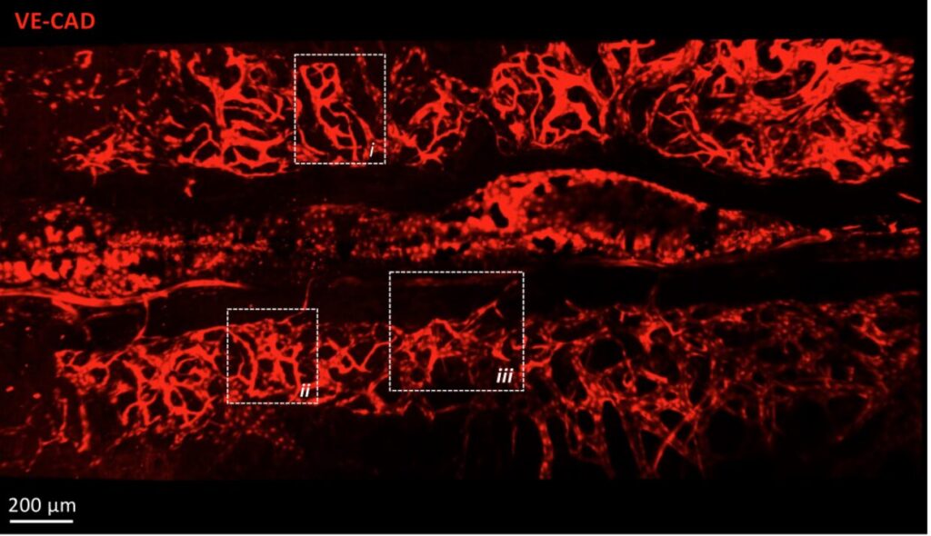
The calvarial bone marrow is an interesting model for studying hematopoiesis and vascular dynamics due to its unique anatomical features. Its thin structure allows for high-resolution imaging, facilitating the observation of cellular interactions and vascular networks. Additionally, it is easily accessible, making it ideal for experimental manipulations and real-time monitoring. This model provides valuable insights into the complex processes of blood cell formation and vascular development.
z-projection of tile scan view of the calvaria vasculature labeled by cdh5-DSRED – 2-photon microscope
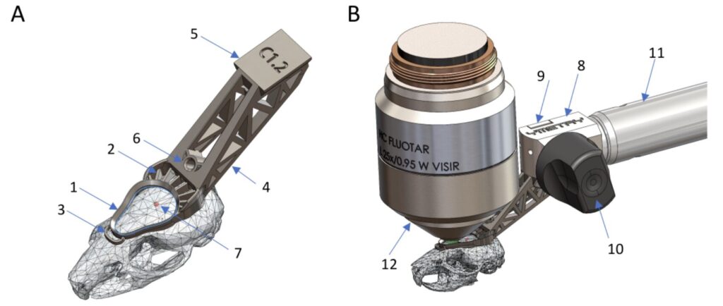
Your team has developed a custom-made titanium cranial implant. What advantages does it offer compared to existing methods?
The stability of the imaging area has always been a major challenge in intravital microscopy. Indeed, the animal’s breathing and temperature variations complicate long-term acquisitions. Moreover, precise repositioning of the acquisition area over several days is essential for observing the evolution of the cellular environment. The development of titanium implants, as opposed to traditional resin 3D printing, allows for more robust fixation of the system to the microscope stage and, most importantly, limits the deformation of the implant.
(A) Parts of the implant in situ: 1 observation ring, 2 cementing feature, 3 stabilizing anchor, 4 tail, 5 dovetail, 6 threaded hole, 7 Bregma. (B) Connection of the head implant to the holder: 8 fixation body, 9 clamp, 10 eccentric lever, 11 structure, 12 microscope objective.
What perspectives does this method open for the understanding of hematological diseases, such as leukemia?
This method opens significant perspectives for understanding hematological diseases like leukemia by enabling detailed visualization of disease progression and cellular interactions. It allows researchers to study the impact of treatments in real-time, enhancing the development of targeted therapies. Additionally, it facilitates the exploration of the bone marrow microenvironment’s role in disease pathogenesis.
What are your upcoming projects?
Following the same principle, we are collaborating with the company Ymetry to develop similar appendages adapted for soft organs. Our goal remains to maintain the acquisition area for as long as possible without any drift.
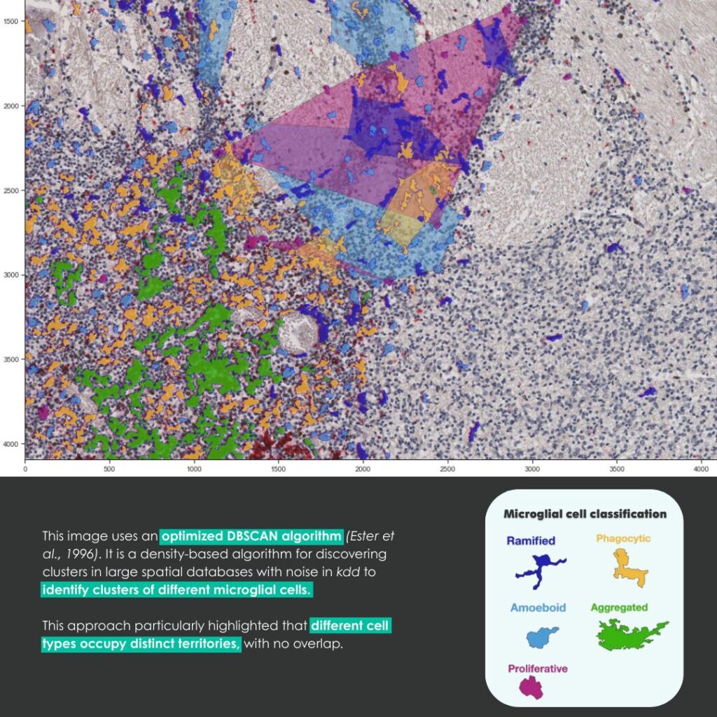
A European interdisciplinary research project, coordinated by Thibault Lagache (BioImage Analysis Unit, Institut Pasteur – France-BioImaging’s platform), David Menassa (University of Oxford), and David Holcman (University of Cambridge), has recently led to the creation of an innovative tool: DeepCellMap. This tool significantly improves the mapping of microglia, brain cells that remain widely unknown to the general public.
Microglia belong to the family of glial cells, which form the environment around neurons. They provide immune protection for the nervous system and play a crucial role in brain development.
What does DeepCellMap bring?
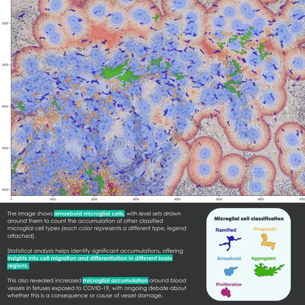
The tool can classify microglia into five categories based on their shape and location in the brain. This classification allows researchers to track their roles throughout brain development.
DeepCellMap also revealed a dynamic spatial organization of microglia. These cells occupy distinct territories and reorganize as the brain grows. Clusters of cells appear and later disperse, following specific patterns.
An unexpected finding: DeepCellMap uncovered a strong association between microglia and blood vessels in the cerebral cortex of fetuses exposed to SARS-CoV-2 during pregnancy. This discovery raises a major question: Are microglia reacting to vascular alterations, or do they themselves contribute to these changes? This observation could pave the way for new research into the impact of prenatal infections on the developing brain.
How does DeepCellMap work?
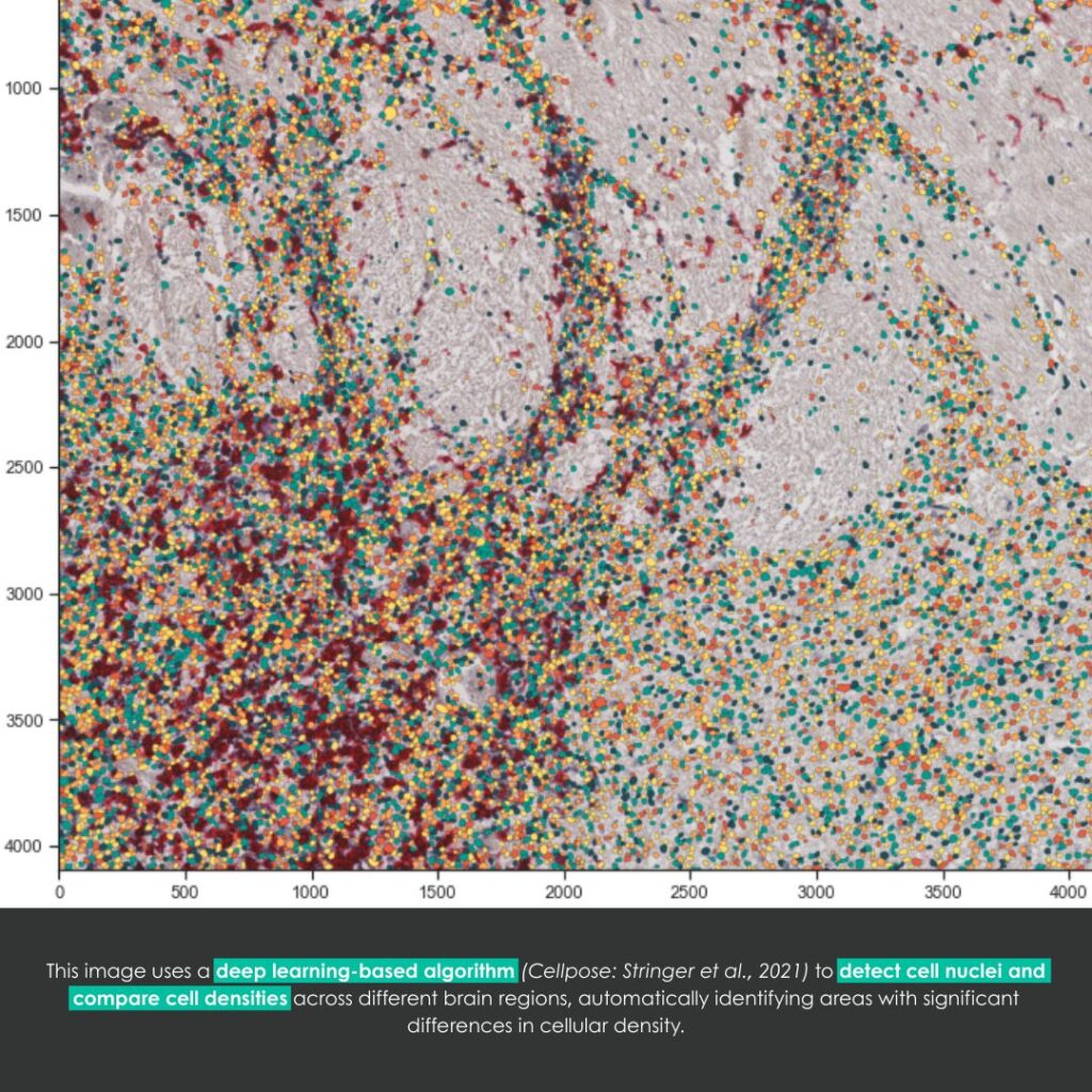
This tool relies on a deep learning algorithm (artificial intelligence) capable of detecting and classifying microglia based on their morphology, using bright-field or fluorescence microscopy images.
The analysis of microglial spatial organization is made possible by the use of advanced statistical models. In conclusion, DeepCellMap is a groundbreaking image analysis tool for biology. By enabling large-scale studies of brain cells, it opens new research perspectives in neuroscience to better understand the mechanisms of brain development.
In the long term, DeepCellMap may also help improve our understanding of how prenatal infections affect the developing brain. As an open-source resource, it can be adapted to study other cell types and applied to a wide range of human health research.
Congratulations to all the teams involved! France-BioImaging is proud to support interdisciplinary projects such as the development of DeepCellMap by members of our bioanalysis community. Supporting research through cutting-edge R&D and fostering international collaboration are at the heart of our infrastructure’s missions.
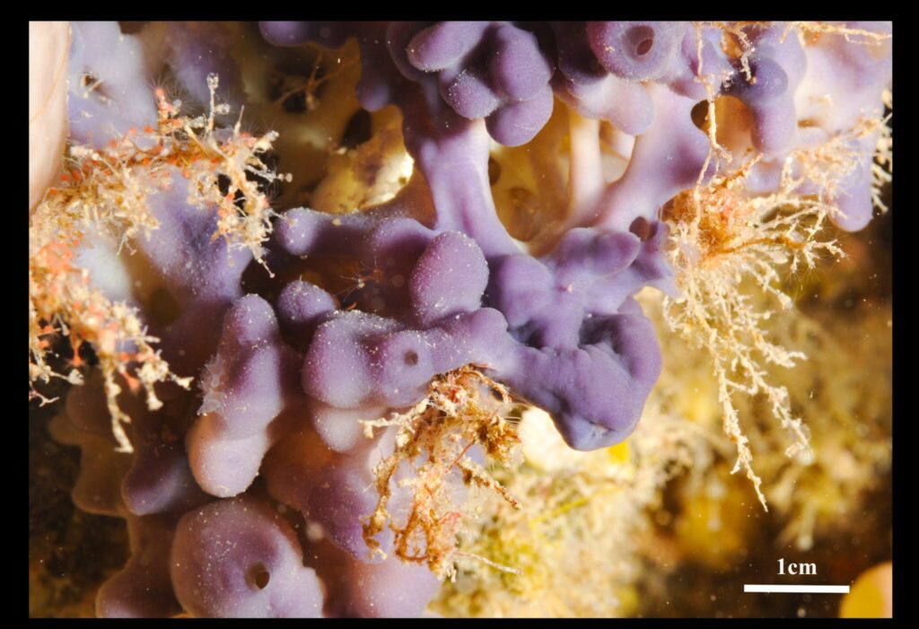
A study conducted by the BEEM team (Molecular Biology, Evolution, and Ecology) of the Mediterranean Institute of Biodiversity and Marine and Continental Ecology, in collaboration with IBDM (Marseille), ENS (Paris) and ISEM (Montpellier) has characterized the buds of Oscarella lobularis as a promising model for studying cell development and sponge evolution.
Researchers successfully induced the in vitro production of these buds and maintained them in culture. These structures are small fragments of the sponge that detach from the adult body and develop into fully independent individuals. The study revealed that they possess remarkable properties from the early stages of their development:
Regeneration ability: When a bud is cut in half, each piece can regenerate into a fully functional new bud. Even when completely broken down into individual cells, they can migrate, reconnect, and self-organize into structured layers.
Autonomous metabolism: They filter water, consume oxygen, and move slightly using tiny cilia.
Complex cellular organization: Their structure resembles that of more evolved organisms, making them a relevant model for understanding the evolution of early animals.
Imaging at the heart of discovery
To observe these buds in detail, researchers used advanced electron and fluorescence microscopy techniques. These analyses were carried out at the PICsl (IBDM, Aix-Marseille University) platform, a member of France-BioImaging.
Thanks to these high-resolution images, scientists were able to examine the buds’ development, cellular organization, and internal functioning, revealing mechanisms still largely unknown in the animal kingdom.
Why study sponges?
If we trace back the phylogenetic tree of animals, it is possible that we share a common ancestor with this species! It may seem hard to believe, but we actually have similarities with sponges.
Sponges are among the oldest organisms on Earth. Studying them could help us understand the origins of animal ancestral features among which the formation of cell layers.
A recent study, led by the Advanced Molecular Virology Unit(Institut Pasteur) and combining virology, structural biology, and immunology, has uncovered a key mechanism involved in the establishment of an efficient infection and evasion of innate immunity of HIV-1 virus (responsible for AIDS disease) in the organism.
The authors focused their work on HIV-1 condensates, called HIV-1 membraneless organelles (HIV-1-MLOs) which are tiny structures composed of viral genetic material and proteins that form in the nucleus of infected cells.
Key results
Researchers have discovered HIV-1 MLOs exist in vivo and act as a shield, allowing viral DNA to hide from cellular DNA sensors that trigger a pathway of our antiviral immune response when they detect a foreign DNA.
These structures can regulate the temporal and spatial conditions to create the perfect environment for reverse transcription (RNA to DNA conversion), a crucial step for the virus to replicate and spread in the organism.
New perspectives
These discoveries open new perspectives for:
Enhancing our understanding of how HIV-1 escapes immune detection.
Developing treatments targeting the formation of HIV-1 MLOs in the early stages of infection.
Exploring similar mechanisms in other viruses.
Microscopy imaging: a key player
Advanced microscopy techniques were essential in visualizing the formation and function of these structures, including:
Confocal microscopy (Pasteur PBI platform) – to study host-pathogen interactions in live-cell imaging and visualize the existence of HIV-1-MLOs in vivo.
France-BioImaging’s Pasteur PBI and UBI platforms (Paris-Centre Node) in collaboration with the Electron Microscopy Platform of the University of Tours provided cutting-edge expertise and equipment, playing a central role in unraveling this viral strategy. By enabling researchers to see what was once invisible, imaging technologies continue to be at the heart of discoveries that push the boundaries of infectious disease research!
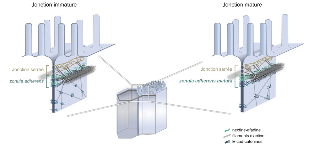
Cellular junctions are essential to the integrity of epithelia, which cover most of our organs. In an article published in the journal PNAS, scientists, with among them members of our FBI Marseille node, reveal the existence of a new category of cell junctions. Using Stimulated emission depletion (STED) microscopy, they have put forward the need to reconsider the organization of intestinal cell junction described as such for more than 40 years.
Imaging intestine with STED
Stimulated emission depletion (STED) microscopy is a super-resolution technique that bypasses the diffraction limit of light microscopy to increase resolution. In our case, scientists were able to resolve the organization of complexes located at cell junctions with a resolution of a few tens of nanometers thanks to STED. Moreover, STED tripled the spatial resolution in the junctional plane and, using cryosections, they achieved imaging with a seven times greater spatial resolution compared to approaches that would use confocal microscopy and thus, without physical sectioning.
Although the resolution of STED is at least an order of magnitude lower than that of electron microscopy, the combination of STED with immunostaining reveals organization up to then unknown as multiple proteins can be efficiently labeled at the same time.
Three types of intestinal cell junctions
The intestine is covered with cells, most of which absorb the nutrients we ingest. These cells are joined together by three types of junctions which coexist and provide different functions, ranging from the selective filtration of certain ions to the mechanical maintenance of the epithelial layer. These junctions, the tight junction, the adherens junction, also called zonula adherens, and the desmosomes, were discovered in the 1960s and their constituent elements as well as their organization were proposed during the 1980s and 1990s.
The adherens junction in particular is established as being organized into a belt of adhesion proteins anchored to the membrane, the cadherins, and supported by filaments, the actin filaments. This junction has an important mechanical role in the cell, for example by impacting the shape of the cell. The zonula adherens (ZA), a fundamental module of epithelial cell–cell adhesion initially observed in intestinal cells, is believed to comprise a single contractile actin belt linked via E-cadherin-catenin to the ones of neighboring cells.
How did microscopy help reevaluate our current knowledge?
By observing the adherens junction of epithelial cells obtained from human intestinal biopsies, or from human cells in culture using STED super-resolution microscopy, scientists have made a very surprising discovery. They show that the ZA consists of two distinct belts of adhesive complexes, a basal one with E-cad-catenin and an apical one with nectin–afadin. Contrary to the prevailing view, the major actin belt aligns with nectin and afadin, not E-cad-catenin.
The authors further demonstrate that this organization depends on the cell maturation state and that the classical ZA found in textbooks corresponds to a less mature state of the intestinal junction. Therefore, they decided to call the junction found in mature cells the zonula adherens matura. Genetic and physical perturbations show that afadin is essential for force transmission across cell junctions. This work redefines the intestinal ZA architecture and prompts a reevaluation of how forces propagate within an epithelial sheet.
Not only, these results are important to better understand the adhesion and mechanics of epithelial cells, but these two essential characteristics of the epithelia are particularly affected in cancers of epithelial origin, which represent 80% to 90% of current cancers. This discovery is, thus, a step forward to the comprehension of cancers and to their treatment.
Get access to one of our services!
You need FRAP, two photon FLIM-FRET, PALM/dSTORM at France-BioImaging? To get open access, please login via Euro-BioImaging website! You just have to choose the technology you want to use, then submit your proposal. All applications will be processed by the Euro-BioImaging Hub in close relation with France-BioImaging. And of course, all scientists regardless of their affiliation, area of expertise or field of activity can benefit from open access services! Users whose projects will be validated by Euro-BioImaging will benefit from a waiver for the access cost on France-BioImaging core facilities (https://france-bioimaging.org/access/).
Mangeol, P., Massey-Harroche, D., Sebbagh, M., Richard, F., Le Bivic, A., & Lenne, P. F. (2024). The zonula adherens matura redefines the apical junction of intestinal epithelia. Proceedings of the National Academy of Sciences, 121(9), e2316722121. https://doi.org/10.1073/pnas.2316722121
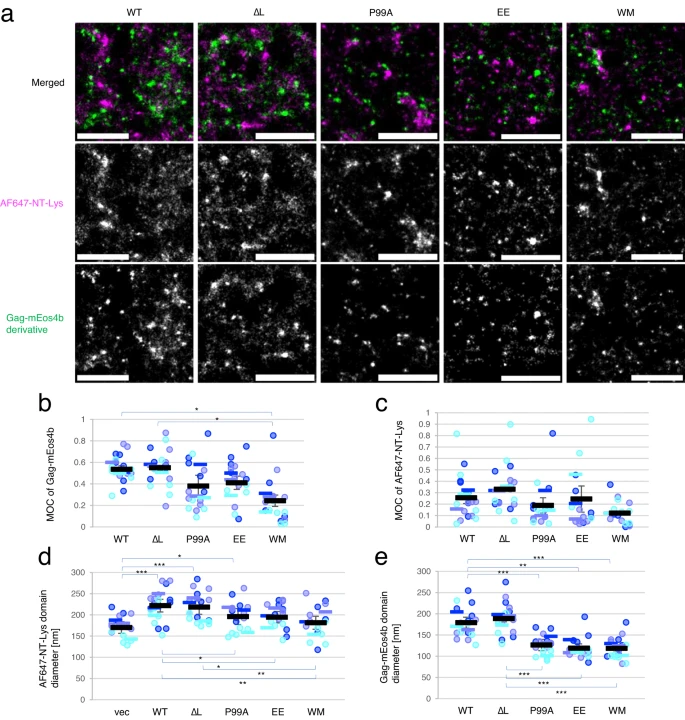
HIV type 1 virus has a lipid envelope enriched with host cell sphingomyelin and cholesterol. In order to understand the mechanism of this enrichment, the FBI Alsace node (Laboratoire de Bioimagerie et Pathologies from Université de Strasbourg and the Imaging Center PIQ-QuESt) has participated in a study recently published in Nature Communications about HIV-1 virus assembly. Indeed, they have investigated the interplay between the HIV-1 Gag protein and the host cell lipids at the plasma membrane. This work has greatly benefited from the use of a great combination of different quantitative (FLIM-FRET and FRAP) and super-resolution (PALM/STORM) custom-made microscopes with specific probes.
Using FRAP to characterize mobile and immobile molecules
The Fluorescence recovery after photobleaching (FRAP) quantifies the two-dimensional lateral diffusion of fluorescently labeled molecules of interest. This technique is very useful in biological studies of cell membrane diffusion and protein binding as it not only reports on the diffusion rates of mobile fractions of molecules but also provides information about the proportion of immobile molecules.
In our case, FRAP experiments indicated that the expression of Gag significantly decreased the mobile fraction of sphingomyelin (SM)-rich domains. Besides, the technique showed that cholesterol (Chol)-rich domains were intrinsically immobile, even in the absence of Gag. It is speculated that the association of Gag with SM-rich domains restricts the lateral diffusion of the lipid domains, resulting in an increase of the immobile fraction in FRAP measurements.
Using PALM/dSTORM to localize molecules at high resolution
The Photo-activated localization microscopy (PALM) is a widefield fluorescence microscopy imaging method that provides images with a resolution beyond the diffraction limit. By collecting a large number of images each containing just a few active isolated fluorophores, the collection of these images allows to stochastically activate each fluorophore and thus to obtain a global image of the sample with high resolution.
The Stochastic optical reconstruction microscopy (STORM) works on the activated state of a photo-switchable molecule that leads to the consecutive emission of sufficient photons to enable precise localization before it enters a dark state or becomes deactivated by photobleaching.
Coupling these two techniques, scientists next investigated at high resolution the localization of Gag and SM-rich or Chol-rich domains, both labeled with specific fluorescently labeled lipid binding proteins.
PALM/dSTORM visualized domains of different sizes labeled with the two lipid binding proteins, showing that the expression of Gag induced the formation of larger SM-rich domains but not the formation of larger Chol-rich domains. The main hypothesis is that the formation of large lipid domains may be due to the coalescence of smaller lipid domains.
Using FLIM-FRET to identify molecule proximity and interaction
And last but not least, the Fluorescence-lifetime imaging microscopy (FLIM) is an imaging technique for producing an image based on differences in the fluorescence-lifetime rather than its intensity. By quantifying variations in the exponential decay rate of the fluorescence from a fluorescent sample (fluorescence-lifetime) it is possible to report on molecule proximity. Since the fluorescence-lifetime is insensitive to changes in fluorophore intensity or concentration, it is the most quantitatively precise technique to report on fluorescence resonance energy transfer (FRET).
FRET is a mechanism describing energy transfer between two light-sensitive molecules (chromophores). A donor chromophore, initially in its electronic excited state, may transfer energy to an acceptor chromophore through non-radiative dipole-dipole coupling. FRET is extremely sensitive to small changes in distance and therefore an excellent reporter on molecule proximity and interaction.
In this third and final part, to better understand the possible effect of Gag on the lipid distribution in the plasma membrane, scientists investigated by two-photon FLIM-FRET the interaction of Chol-rich lipid domains with SM-rich lipid domains and its dependence on Gag multimerization. These last results showed that Gag multimerization induces SM-rich and Chol-rich domains to be in close proximity and that membrane curvature affects the apposition of SM-rich and Chol-rich domains.
A great example of application on how a combination of high-end technologies in microscopy can help you understand multiple aspects of a biological mechanism. So, what are you waiting for? Dive into the true potential of microscopy!
You need FRAP, two photon FLIM-FRET, PALM/dSTORM at France-BioImaging? To get open access, please login via Euro-BioImaging website! You just have to choose the technology you want to use, then submit your proposal. All applications will be processed by the Euro-BioImaging Hub in close relation with France-BioImaging. And of course, all scientists regardless of their affiliation, area of expertise or field of activity can benefit from open access services! Users whose projects will be validated by Euro-BioImaging will benefit from a waiver for the access cost on France-BioImaging core facilities (https://france-bioimaging.org/access/).
Tomishige, N., Bin Nasim, M., Murate, M. et al. HIV-1 Gag targeting to the plasma membrane reorganizes sphingomyelin-rich and cholesterol-rich lipid domains. Nat Commun14, 7353 (2023). https://doi.org/10.1038/s41467-023-42994-w
We use cookies on our website to give you the most relevant experience by remembering your preferences and repeat visits. By clicking “Accept All”, you consent to the use of ALL the cookies. However, you may visit "Cookie Settings" to provide a controlled consent.
This website uses cookies to improve your experience while you navigate through the website. Out of these, the cookies that are categorized as necessary are stored on your browser as they are essential for the working of basic functionalities of the website. We also use third-party cookies that help us analyze and understand how you use this website. These cookies will be stored in your browser only with your consent. You also have the option to opt-out of these cookies. But opting out of some of these cookies may affect your browsing experience.
Necessary cookies are absolutely essential for the website to function properly. These cookies ensure basic functionalities and security features of the website, anonymously.
Cookie
Duration
Description
cookielawinfo-checkbox-analytics
11 months
This cookie is set by GDPR Cookie Consent plugin. The cookie is used to store the user consent for the cookies in the category "Analytics".
cookielawinfo-checkbox-functional
11 months
The cookie is set by GDPR cookie consent to record the user consent for the cookies in the category "Functional".
cookielawinfo-checkbox-necessary
11 months
This cookie is set by GDPR Cookie Consent plugin. The cookies is used to store the user consent for the cookies in the category "Necessary".
cookielawinfo-checkbox-others
11 months
This cookie is set by GDPR Cookie Consent plugin. The cookie is used to store the user consent for the cookies in the category "Other.
cookielawinfo-checkbox-performance
11 months
This cookie is set by GDPR Cookie Consent plugin. The cookie is used to store the user consent for the cookies in the category "Performance".
viewed_cookie_policy
11 months
The cookie is set by the GDPR Cookie Consent plugin and is used to store whether or not user has consented to the use of cookies. It does not store any personal data.
Functional cookies help to perform certain functionalities like sharing the content of the website on social media platforms, collect feedbacks, and other third-party features.
Performance cookies are used to understand and analyze the key performance indexes of the website which helps in delivering a better user experience for the visitors.
Analytical cookies are used to understand how visitors interact with the website. These cookies help provide information on metrics the number of visitors, bounce rate, traffic source, etc.
Advertisement cookies are used to provide visitors with relevant ads and marketing campaigns. These cookies track visitors across websites and collect information to provide customized ads.