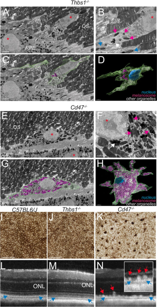
ATP-driven separation of liquid phase condensates in bacteria, Mol Cell 2020
▽ Scroll down
Category: Publications
ATP-driven separation of liquid phase condensates in bacteria
B. Guilhas, J.C. Walter, J. Rech, G. David, N.-O. Walliser, J. Palmeri, C., Mathieu-Demaziere, A. Parmeggiani, J.Y. Bouet, A. Le Gall1, M. Nollmann
Liquid-liquid phase separated (LLPS) states are key to compartmentalise components in the absence of membranes, however it is unclear whether LLPS condensates are actively and specifically organized in the sub-cellular space and by which mechanisms. Here, we address this question by focusing on the ParABS DNA segregation system, composed of a centromeric-like sequence (parS), a DNA-binding protein (ParB) and a motor (ParA). We show that parS-ParB associate to form nanometer-sized, round condensates. ParB molecules diffuse rapidly within the nucleoid volume, but display confined motions when trapped inside ParB condensates. Single ParB molecules are able to rapidly diffuse between different condensates, and nucleation is strongly favoured by parS. Notably, the ParA motor is required to prevent the fusion of ParB condensates. These results describe a novel active mechanism that splits, segregates and localises non-canonical LLPS condensates in the sub-cellular space.
Guilhas et al. revealed that the bacterial DNA segregation apparatus behaves as a non-canonical phase separation system. This apparatus employs an ATP-powered motor that splits nanometer-sized condensates and localizes them robustly within the nucleoid to ensure faithful transmission of genetic material.
The ability to communicate effectively with each other is one of the strongest predictors for our chances to get ahead in life. In their latest publication in Science Advances, scientists and engineers from IGF-Montpellier (CNRS, INSERM, Univ. Montpellier), IPAM platform (BioCampus Montpellier, France-Bioimaging Montpellier Node) and ARO-Israel demonstrated that this also holds true for GnRH neurons.
In humans and all vertebrates, species survival depends on a critical step during embryonic development: the migration of a small subset of GnRH neurons (about 2,000 in humans and less than 100 in fish) from the nose to the brain where they join the hypothalamus to control reproduction. Their latest results unveiled that GnRH neurons make a pause at the nose-brain frontier where they function as an inter-hemispheric network that is isolated from the rest of the brain. Only neurons that integrate into the network and are able to communicate with their neighbors will finally cross the barrier and make their way into the brain, towards their hypothalamic destination.
In other words, these GnRH neurons, that are critical for species persistence, face the same challenges like other immigrants: they must learn to communicate effectively if they are to integrate into their new world.
In this study, in vivo 2-photon microscopy was a key tool for:
Long term imaging with minimal bleaching and phototoxicity
Upright configuration enabling dorsal imaging of the fish in its natural position
Long-distance water-immersion objectives allowing imaging of deep tissue structures without sacrificing image quality
Fast calcium imaging
Imaging of red GECI using the higher wavelengths
Precise cell ablation
Photoactivation of ChR2 while monitoring Ca in the red channel
During embryonic development, cells take on increasingly precise roles in the body as they divide. Be they skin cells, muscle cells or neurons, the different cell types that make up the embryo emerge gradually from a very fine orchestration of their positions and identities, coordinated by the signals they exchange with each other. Like us, the cells need to “talk” to each other to make decisions.
Screaming or whispering: the embryonic cell dilemma
In vertebrate embryos, cells have a very dynamic behaviour. They move around, exchange their neighbours or migrate over long distances. The signals they exchange therefore need to have a long range, which could be characterized as “shouting”. The study of the embryonic development of a sea squirt, a small marine animal with optically transparent embryos, has enabled scientists from several teams at CNRS and INRIA in France, in collaboration with a team from the European Molecular Biology Laboratory (EMBL, Germany), to capture and describe in detail a more discreet mode of cell communication.
The scientists recorded the development of live embryos every two minutes with a new-generation « light-sheet » microscope. They then created software to automatically detect each cell and analyze its position, shape and neighbours up to an advanced stage of development. This work revealed an unusually reproducible mode of development, in which the same cell can be found in the same position across all embryos and where cells move very little in relation to each other. The authors of the study then annotated the films thus made with information on the cell type and the molecular signals emitted by each cell. Using mathematical modelling to integrate the quantitative description of the embryonic geometry with these annotations, their work suggest that cells communicate with very short-range signals. Moreover, the interpretation of these signals is modulated by the area of the contacts between cells. Unlike vertebrates, the cells of ascidian embryos thus have a static and fixed behaviour and the range of their “whispered” signals is very small.
This study indicates that the dynamics of cell movement varies greatly between animals and that these different modalities could be strongly related to the range of signals that the cells exchange with each other. By extending the repertoire of cellular communication mechanisms, this work opens new perspectives on the understanding of self-organization strategies of living forms.
Article: L. Guignard*, U.-M. Fiuza*, B. Leggio, J. Laussu, E. Faure, G. Michelin, K. Biasuz, L. Hufnagel, G. Malandain, C. Godin#, P. Lemaire# (2020) Contact-area dependent cell communications and the morphological invariance of ascidian embryogenesis (Science, July 10 2020 issue, https://science.sciencemag.org/content/369/6500/eaar5663)
2 recent publications using the laser irradiation and photoablation systems available on the MRic facility from the Bretagne-Loire Node are presented here:
Esmangart de Bournonville and Le Borgne (IGDR) characterized the assembly and interactions of tricellular junction components in Drosophila epithelial cytokinesis using laser ablation on a SP5 confocal. Their article entitled “Interplay between Anakonda, Gliotactin, and M6 for Tricellular Junction Assembly and Anchoring of Septate Junctions in Drosophila Epithelium” was published last august in Current Biology (https://doi.org/10.1016/j.cub.2020.07.090).
Rebecca Smith, post-doc in Sébastien Huet’s team (IGDR) in collaboration with Szilvia Juhász from Gyula Timinszky’s team (Szeged, Hungary) used laser irradiation to study chromatin remodeling following DNA damage. Their paper entitled “The chromatin remodeler ALC1 underlies resistance to PARP inhibitor treatment” has just been accepted in Science Advances.
We use cookies on our website to give you the most relevant experience by remembering your preferences and repeat visits. By clicking “Accept All”, you consent to the use of ALL the cookies. However, you may visit "Cookie Settings" to provide a controlled consent.
This website uses cookies to improve your experience while you navigate through the website. Out of these, the cookies that are categorized as necessary are stored on your browser as they are essential for the working of basic functionalities of the website. We also use third-party cookies that help us analyze and understand how you use this website. These cookies will be stored in your browser only with your consent. You also have the option to opt-out of these cookies. But opting out of some of these cookies may affect your browsing experience.
Necessary cookies are absolutely essential for the website to function properly. These cookies ensure basic functionalities and security features of the website, anonymously.
Cookie
Duration
Description
cookielawinfo-checkbox-analytics
11 months
This cookie is set by GDPR Cookie Consent plugin. The cookie is used to store the user consent for the cookies in the category "Analytics".
cookielawinfo-checkbox-functional
11 months
The cookie is set by GDPR cookie consent to record the user consent for the cookies in the category "Functional".
cookielawinfo-checkbox-necessary
11 months
This cookie is set by GDPR Cookie Consent plugin. The cookies is used to store the user consent for the cookies in the category "Necessary".
cookielawinfo-checkbox-others
11 months
This cookie is set by GDPR Cookie Consent plugin. The cookie is used to store the user consent for the cookies in the category "Other.
cookielawinfo-checkbox-performance
11 months
This cookie is set by GDPR Cookie Consent plugin. The cookie is used to store the user consent for the cookies in the category "Performance".
viewed_cookie_policy
11 months
The cookie is set by the GDPR Cookie Consent plugin and is used to store whether or not user has consented to the use of cookies. It does not store any personal data.
Functional cookies help to perform certain functionalities like sharing the content of the website on social media platforms, collect feedbacks, and other third-party features.
Performance cookies are used to understand and analyze the key performance indexes of the website which helps in delivering a better user experience for the visitors.
Analytical cookies are used to understand how visitors interact with the website. These cookies help provide information on metrics the number of visitors, bounce rate, traffic source, etc.
Advertisement cookies are used to provide visitors with relevant ads and marketing campaigns. These cookies track visitors across websites and collect information to provide customized ads.