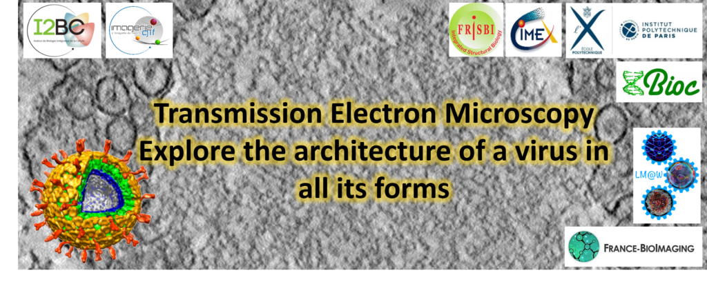
Save the date! The Electron Microscopy facility of Imagerie-Gif (I2BC, France-BioImaging), the Cryo-Electron Microscopy facility (I2BC, FRISBI) and the Cimex facility of Ecole Polytechnique are organizing a 5-day workshop from October 3rd to October 7th, 2022 on Transmission Electron Microscopy to explore the architecture of a virus in all its forms.

The aim of this workshop is to propagate knowledge about transmission electron microscopy applications and to outline the advantages of transversal studies combining structural biology and cell biology. Indeed, structural biology and cell biology approaches both use TEM but are rarely merged in the same studies.

The workshop will focus on the advantages of combining both approaches, which can be easily performed with the same equipment. The workshop will focus on a biological object whose study requires such multiscale approaches: a virus. The virus will be studied in vitro to resolve its high-resolution 3D structure, and will be observed in infected cells to determine the infection and replication mechanisms in situ.

The workshop targets students and young researchers. The training will focus on a given biological object, a virus, which will be studied by two complementary approaches:

  • Single particle analysis by cryo-electron microscopy, allowing high-resolution 3D reconstruction of particles purified in vitro. This part will be performed on a 200kV TEM on the Cimex facility.
  • Cellular tomography of infected cells with observation of the virus replication sites in situ and analysis of its interaction with cellular membranes. This workshop will cover the workflow from sample preparation and resin sections realisation, to acquisition and analysis of tomograms with a 120kV TEM.

Attendees will have a theoretical and practical overview of these two complementary techniques. The practical training will be particularly emphasised, to ensure that attendees will be able to apply the knowledge acquired in the workshop for their further research projects.


Susbscription here: https://www.azur-colloque.fr/DR04/inscription/preinscription/245

Preliminary programme

Open-Neuroscience in collaboration with the Neuroscience department and the Photonic BioImaging Core of the Institute Pasteur and the University of Sussex neuroscience department is organising a FENS satellite event on Open Hardware Microscopy DevelopmentThe aim of this event is to bring into focus excellent open projects that permeate education, ease of access and data collection in advanced research environment.

The symposium will take place on July 8th at the Pasteur Institute campus in Paris (28 rue du Dr Roux). It will be divided into a morning workshop session and an afternoon series of talks. Registration is necessary to access the campus :

 REGISTER

The workshops will be led by specialists in the field, which will also participate in the afternoon session. By the end of the session, participants will be invited to discuss what they have learned, how they could implement Open-Science microscope in their research, and/or how they could improve/modify their own setup by implementing Open tools on it and how their research protocol could benefit from it.

The afternoon session will be dedicated to a series of talks by our invited specialists. It will be split into two distinct topics/sub-sessions:

  1. Open-Source microscopy for education and improving ease of access to research equipment.
  2. Open-Source microscopy for advanced research.

It is our goal that by the end of the day, participants get a full picture of the state of the art for Open-Source Microscopy. It is hoped that development and implementation of such tools and techniques in modern research units promote science accessibility and improve data reproducibility. Furthermore, an earlier and more systematic access to advanced science tools in education could only support shaping the research minds of tomorrow.

Open Microscopy is sponsored by EMBO, TReND in Africa and France-Bioimaging.

The first practical workshop « Imaging Organoids: from the bench to the microscope » will take place in Bordeaux, from Mon 27th Sept to Fri 1st Oct 2021.

The aim of this workshop encompasses most of the workflow steps from the 3D-sample preparation (organoids, spheroids, encapsulated 3D cultures), how to process them (histology, staining), how to mount them (for upright, inverted etc…), and finally how to image them with various microscopy techniques (from super resolution microscopies -liveSR or STED- to microscopies dedicated to thick samples (two-photon, ultramicroscope), from fast optical scanning spinning-disk to High Content Screening -HiTech and LowTech-).

Bench work is possible thanks to the TBM Core Facility, and the microscopes are provided by the BIC facility and the LP2N (home-made setup). 

This workshop is open to all (PhD students, Postdocs, Engineers, Technicians, Researchers and Teaching Assistants) who would like to learn how to prepare their 3D samples for photon microscopy.

The number of attendees will depend on the rules that will prevail at this time. Seats will be limited.

Registration is free but mandatory https://wkshpimageoids.sciencesconf.org/

Registration deadline: 6th Sept at noon. The organisation committee will then select participants and contact them shortly after registration closure.

There will be 4 seminars with the following keynote speakers:

  • Xavier Trepat –link– (IBEC, Barcelone)
  • Fanny Jaulin –link– (Institut Gustave Roussy, Paris)
  • Laura Broutier –link– (CRCL, Lyon)
  • Charlotte Rivière –link– (ILM, Lyon)


Each seminar will be preceded by a talk from our industry partners: CorningStemCellTreeFrog Therapeutics and Idylle. They will be accessible freely on a dedicated streaming platform, upon ad-hoc registration which will be set soon.

This workshop is organized with the support of the GdR ImaBio, the GdR Organoids and France Bio-Imaging.

We are glad to announce the Second International CENN Biennale dedicated to Advanced Microscopy, on May 2021

Such scientific event, focused on Imaging thick samples for biology, will be organized by the Center of Excellence Nikon Nantes (CENN), a French unique collaboration between Nikon company, MicroPICell platform from UMS3556 INSERM CNRS University of Nantes and APEX platform from UMR703 PAnTher INRAE Oniris. It will gather world renowned scientists, expert in clarification, adaptive optics, light sheet and multiphoton imaging techniques.

Cette action nationale de formation regroupe 160 personnes, techniciens, ingénieurs, chercheurs de centres de recherches, voulant approfondir leurs connaissances en microscopie corrélative.

L’objectif de cette formation est de présenter différentes techniques de microscopie corrélative en essayant de présenter les techniques et les appareils utilisés le plus précisément possible.
Les corrélations abordées concerneront les techniques de microscopie optique classique et à fluorescence, la microscopie électronique à balayage et à transmission, le NanoSIMS, la Fluo-RX et le synchrotron.
Des présentations et des ateliers permettront de bien approfondir les outils utilisés de la préparation des échantillons à la corrélation des images.

Date limite pour les inscriptions : 15 Avril 2021

Crédit image header: Cosenza, M. R. et al. Asymmetric Centriole Numbers at Spindle Poles Cause Chromosome Missegregation in Cancer. CellReports 20, 1906–1920 (2017)

 Registration and abstract submission systems are now open ! 

NEUBIAS, the Network of European BioImage Analysts (www.neubias.org), is delighted to announce that the 4th NEUBIAS Conference will take place in Bordeaux, France on February 29 – March 6, 2020.
The conference will include two Training Schools, a Taggathon and will culminate with the Bioimage Analysis Symposium (March 4-6, 2020).

Highlights of the Symposium

Keynote Speakers:

Dr. Suliana Manley
Dr. Emma Lundberg
Dr. Kristin Branson

Discussion topics:

BioImage Analysis & Workflows in Life Science, Current Developments & Applications, Machine Learning, Deep Learning, BioImage Data-Mining, Formats, Management, Object segmentation, Tracking, Atlases, Reconstruction, Visualization, Registration, Correlation, Fusion, Automation, Open Science, and many more.

BioImage Analysis & Workflows in Life Science, Current Developments & Applications, Machine Learning, Deep Learning, BioImage Data-Mining, Formats, Management, Object segmentation, Tracking, Atlases, Reconstruction, Visualization, Registration, Correlation, Fusion, Automation, Open Science, and many more.

Interactive sessions:
Open source Software Lounge, Call4Help, Industry Workshops/Techbites/Digital posters, Panel Discussion, and more

Symposium Electron Microscopy in Bordeaux


The abstract submission site for QBI 2019, Rennes, France, is now open. The conference will be held 9-11 January 2019, with pre-conference workshops on 8 January, 2019.

We seek contributions in any area of quantitative microscopy. Presentations that demonstrate new approaches in detail are particularly welcome, including but not limited to algorithmic and software developments, physical modeling approaches, etc. The use of quantitative imaging techniques in biological applications is also of great interest. For submission details, please see the conference website (www.quantitativebioimaging.com).

Submission deadline: September 15th, 2018

Registration fees for academics: None (supported by external funding)
 
Local accommodation: Dinners and lunches are offered to all participants

In addition to contributed talks we will feature special sessions on:

  • Machine learning and microscopy image analysis
  • New developments in single molecule microscopy
  • Subcellular trafficking in cell biology
  • Microscopy software development
  • Microscopy in biopharma
  • Structured illumination: a review of the state of the art

The pre-conference workshops are:

  • Machine and deep learning in bioimaging and microscopy
  • Adaptive optics
  • 3D single molecule microscopy

Confirmed Speakers

  • Yann LeCun [AI Research – Facebook / NYU Center for Data Science] – Paris, France (Tentative)
  • Pierre Bon [Laboratoire Photonique Numérique et Nanosciences] – Talence, France
  • Edward Cohen [Imperial College] – London, UK
  • Hans-Ulrich Dodt [Medical University of Vienna and Vienna University of Technology] – Vienna, Austria
  • Michael Elad [Technion Israel Institute of Technology] – Haifa, Israel
  • Kevin Elicieri [University of Wisconsin at Madison] – Madison, Wisconsin
  • Seth Flaxman [Imperial College London] – London, UK
  • Spencer Freeman [University of Toronto] – Toronto, Canada
  • Rainer Heintzmann [Institute of Photonic Technology / Friedrich Schiller University] – Jena, Germany
  • Thomas Huser [Biomolecular Photonics Group] – Bielefeld, Germany
  • Khuloud Jaqaman [UT Southwestern Medical Center] – Dallas, Texas
  • Ludger Johannes [Curie Institut] –  Paris, France
  • Florian Jug [Max Planck Institute of Molecular Cell Biology and Genetics] – Dresden, Germany
  • Yannis Kalaidzidis [Max Planck Institute of Molecular Cell Biology and Genetics] – Dresden, Germany
  • Friedemann Kiefer [Max Planck Institute for Molecular Biomedicine] – Muenster, Germany.
  • Judith Klumperman [University Medical Center Utrecht] – Utrecht, The Netherlands
  • Julien Mairal [Inria] – Grenoble, France
  • Molly Maleckar [Allen Institute of Cell Science] –  Seattle, Washington
  • Fred Maxfield [Weill Cornell Medical College] – New York, New York
  • Jean-Christophe Olivo-Marin [Institut Pasteur] – Paris, France
  • Steve Presse [University of Arizona] –  Tucson, Arizona
  • Bernd Rieger [Delft University of Technology] – Delft, Netherlands
  • Jonas Ries [European Molecular Biology Library] – Heidelberg, Germany
  • Daniel Sage [EPFL] – Lausanne, Switzerland
  • Anne Sentenac [Institut Fresnel] – Marseille, France
  • Ernst Stelzer, [Buchmann Institute for Molecular Life Sciences] – Frankfurt, Germany
  • Per Uhlén [Karolinska Institutet] – Stokholm, Sweden
  • Geert van den Bogaart [Groningen Biomolecular Sciences and Biotechnology Institute] – Groningen, Netherlands
  • Simon Walker-Samuel [University College] – London, UK
  • Daniel Wüstner [Syddansk Universitet] – Odense, Denmark
  • Marino Zerial [Max Planck Institute of Molecular Cell Biology and Genetics] – Dresden, Germany
  • Christophe Zimmer [Institut Pasteur] – Paris, France

As in prior years, assuming that our fundraising is again successful, we plan on not having a registration fee for academic attendees.

To obtain email updates, please sign up for membership of the QBI Society at www.quantitativebioimaging.com. Membership is free of charge.

Cette manifestation porte sur l’interprétation des structures tissulaires et cellulaires en microscopie électronique avec une attention particulière sur l’identification des artéfacts de préparation. Cette formation propose aux participants une véritable remise à niveau de leurs connaissances de l’ultrastructure saine versus pathologique. Ce thème a été choisi suite aux retours des questionnaires de satisfaction des journées antérieures et aux nombreuses interrogations posées sur la liste de diffusion du RCCM.

This conference is organized by the “Réseau d’Imagerie Cellulaire Paris-Saclay”.

The two most common techniques of vibrational micro-spectroscopy are infrared (IR) and Raman. These two sophisticated tools enable to visualize the inherent vibrational spectra of biochemical constituents of a cell or a tissue. Therefore, IR and Raman microscopy provide the specific and distinct “fingerprint” spectrum of each cell and offer the powerful possibility of high contrast images without any external labeling.

Recently, significant developments of these approaches provided a better access of these two techniques by the biologist community. Currently, IR and Raman microscopy are used for tissues, biopsies, animal and plant analyses in order to visualize proteins (C-H3 bonds), lipids (C-H2 bonds), water (O-H bonds), membranes, myelin, chromophores such as flavins, etc …

This is a free event.

Registration is mandatory at : specvib@gmail.com

Public

Biologists, doctors, postdoc, engineers, students

Speakers

Christophe SANDT (Synchrotron Soleil)

Alexandre DAZZI (LCP Orsay, UPSud)

Marie-Françoise DEVAUX (INRA Angers-Nantes)

Olivier PIOT (BioSpect / Univ Reims)

How to reach the conference

https://goo.gl/maps/JJoFw7ctc972

More information

www.ric-paris-saclay.fr

Contact

larbi.amazit@u-psud.fr / philippe.leclerc@inserm.fr

Les 15èmes Assises Nationales des Plateformes du réseau RT-mfm
se dérouleront à Rouen en 2018 (PRIMACEN, université de Rouen, Mont Saint Aignan)
du 19 Mars 13h au 21 Mars 13h.

Le formulaire d’inscription aux Assises se trouve à l’adresse :
https://goo.gl/forms/oHBJkdjikkb5TrS83
la date limite d’inscription est le 20 décembre 2017 à 18h00.

Le programme est en cours d’élaboration, faites-nous savoir si vous souhaitez voir développer un thème ou proposer une présentation orale ou un intervenant.

Les propositions sont à envoyer à france.lam@upmc.fr et olivier.renaud@curie.fr.

Votre inscription définitive pour participer aux Assises vous sera confirmée par email fin janvier/début février 2018.
Cordialement,
France & Olivier
Pour le RT-mfm

Le Storage Day est une conférence organisée par l’INRA (Unité Ingenum) avec la participation d’autres Instituts et dispositifs de recherche (CIRAD, IGBMC, Biogenouest) autour des questions de la gestion de la donnée scientifique, et en particulier son stockage.
Cette journée propose des retours d’expérience de différentes communautés (Imagerie, NGS, SHS, etc.) autour de problématiques en lien avec la “vraie vie” sous la forme de présentations synthétiques (15 minutes). Ces retours d’expériences auront pour objet aussi bien la mise en valeur de “success stories” que la mise en évidence des écueils/difficultés auxquelles se trouvent confrontées les communautés.