Service
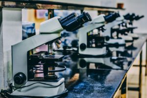
The Montpellier node comprises a tight collaboration of research/development teams and microscopy research platforms. The main objective of this FBI node is to develop, implement and provide access of state-of-the-art optical microscopy systems allowing microscopic imaging from unicellular organisms to whole animals. Specifically, our emphasis is on super-resolution and fluctuation microscopies, high-throughput high-content microscopies, and in vivo cellular imaging and manipulation. Our national specificity resides in the use of high-end microscopies to study different aspects of chromatin biology, such as chromosome organization and gene expression.
FBI-Montpellier is composed of a microscopy research and development facility (MARS of the CBS), two microscopy facilities (MRI and IPAM), and two R&D groups (Nollmann and Margeat labs). Several active and synergistic collaborations exist between these different entities in which R&D groups contribute their expertise in optical/instrument development and platforms contribute their savoir-faire in user service and in general logistics.
The mechanism of force transmission at bacterial focal adhesion complexes. Nature, 2016 Nov 24;539(7630):530-535. doi: 10.1038/nature20121.
Plasmonic Nanoantennas Enable Forbidden Förster Dipole-Dipole Energy Transfer and Enhance the FRET Efficiency. Nano Lett. 2016 Oct 12;16(10):6222-6230.
A single-molecule view of transcription reveals convoys of RNA polymerases and multi-scale bursting. Nat Commun. 2016 Jul 27.
Highly efficient multicolor multifocus microscopy by optimal design of diffraction binary gratings Scientific Reports, in press.
Bacterial partition complexes segregate within the volume of the nucleoid Nature Communications, 7: 12107, 5 July 2016
Condensin- and Replication-Mediated Bacterial Chromosome Folding and Origin Condensation Revealed by Hi-C and Super-resolution Imaging. Molecular Cell 59 (4): 588-602, 20 August 2015
Fine tuning of sub-millisecond conformational dynamics controls metabotropic glutamate receptors agonist efficacy. Nature communications”>Nat Commun. 2014 Oct 17;5:5206.
FISH-quant: automatic counting of transcripts in 3D FISH images. Nat Methods. 2013 Apr;10(4):277-8.