Interdisciplinary access call to Structural biology, Biological imaging and Proteomics facilities
▽ Scroll down
Tag: Microscopy
Deadline: February 13th, 2026
The three national infrastructures ProFi, France-BioImaging and FRISBI along with the GIS IBiSA are pleased to announce a fourth call for a funded access to IBiSA-labelled facilities. Our aim is to promote IBiSA facilities networking through transdisciplinary research projects.
Applications should request access to at least two different IBiSA facilities from two disciplines (structural biology, Biological imaging and proteomics, see below a non-exhaustive list). The call is open to any academic laboratory.
Modalities for application are described in the attached document.
The France-BioImaging Image Contest is back for its 7th edition!
This image contest is open to all within the imaging community: core facility staff and users, R&D labs teams and co-workers, students… Submit your best microscopy images for a chance to showcase your skills, research and creativity to the French bioimaging community and beyond, allowing people to see the visual appeal of the life sciences. Images from the contest will be featured on France-BioImaging communication tools, online and in print.
France-BioImaging and all the French community aims to develop and promote innovative imaging technologies and methods. But microscopy images can also take an artistic, creative look and make the invisible world beautiful.
We are all eager to see your work !
Prizes
First place: France-BioImaging will cover the registration fees for one 2026 microscopy related event of the winner’s choice (FOM, ELMI, EMC, COMULIS conference, etc.).
All participants: Your images will be added to the CNRS Images website, used in our communication materials (digital calendar, flyers, social media posts, etc.), and some of you may also be invited for an interview about your research work.
Important: Only French or foreign participants affiliated to a French institution can enter the contest. Foreign participants non-affiliated to a French institution can submit images and will be featured in the gallery, but will not be evaluated as part of the contest.
Submission deadline extended: Wednesday, November 20th, 2025, 23h59
The France-BioImaging Image Contest is back for its 6th edition!
This image contest is open to all within the imaging community: core facility staff and users, R&D labs teams and co-workers, students… Submit your best microscopy images for a chance to showcase your skills, research and creativity to the French bioimaging community and beyond, allowing people to see the visual appeal of the life sciences. Images from the contest will be featured on France-BioImaging communication tools, online and in print.
France-BioImaging and all the French community aims to develop and promote innovative imaging technologies and methods. But microscopy images can also take an artistic, creative look and make the invisible world beautiful.
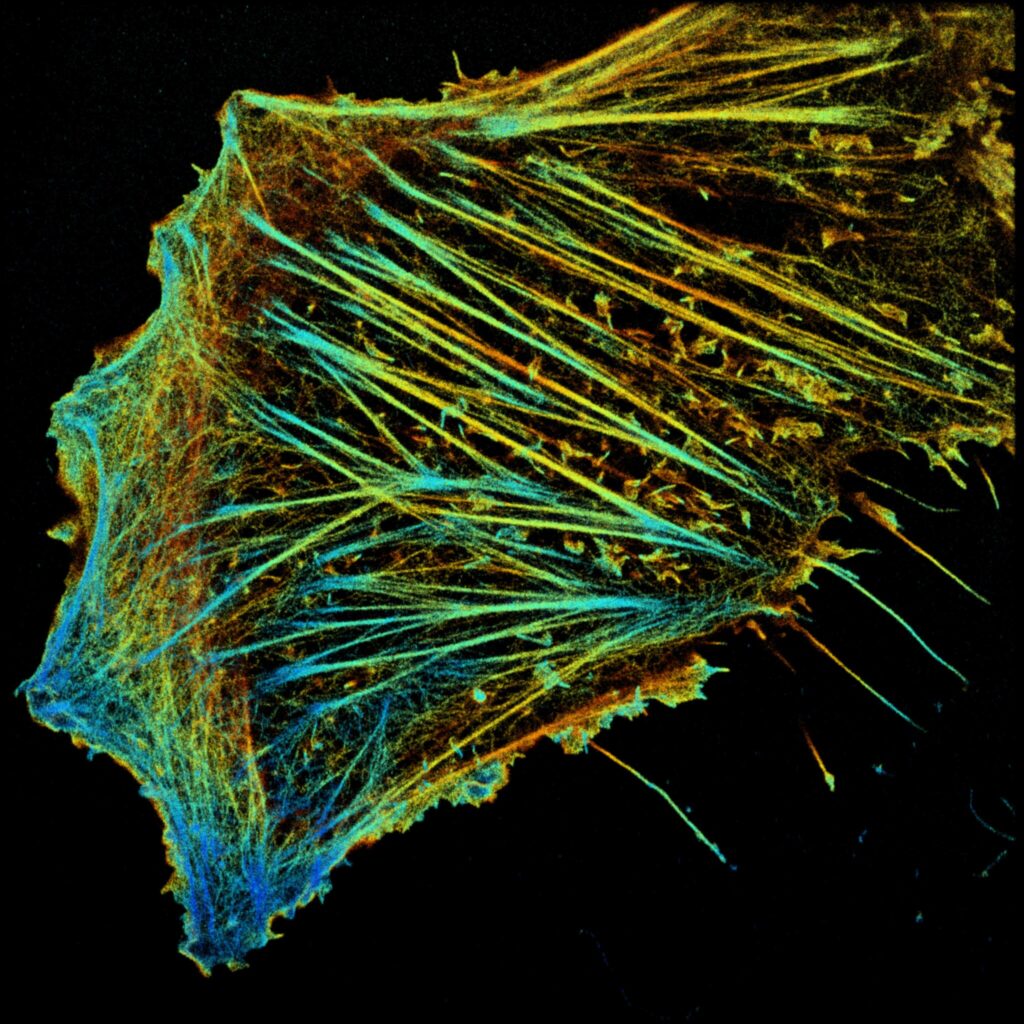
Marine plant collected from the Mediterranean Sea. Actin stained with phalloidin.
2-photon imaging
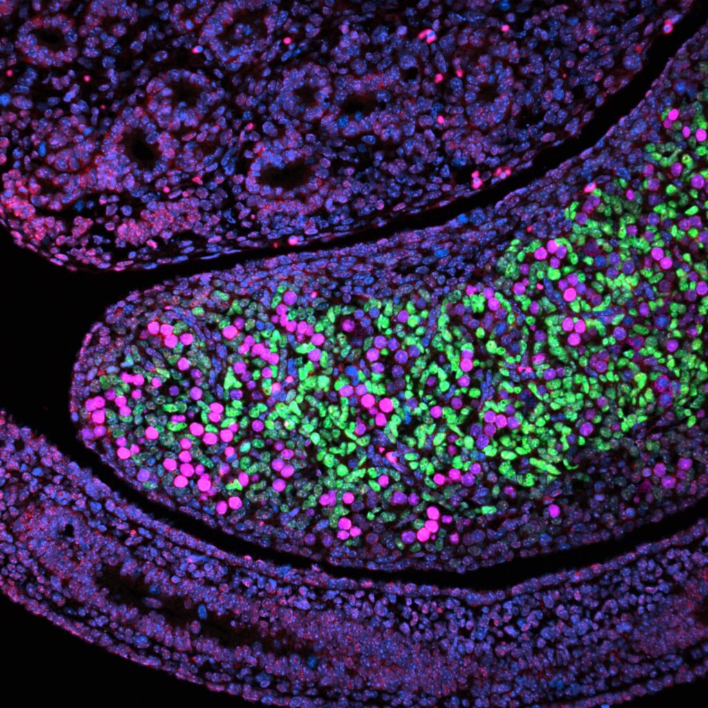
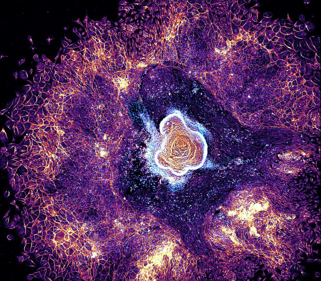
2nd Place:Dalia EL ARAWI, Pierre-François LENNE’s Team, IBDM
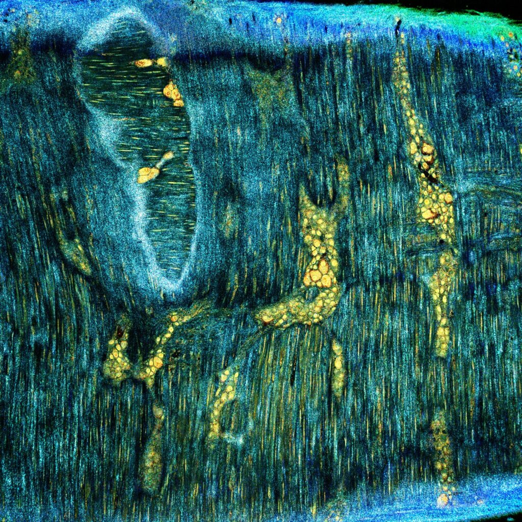
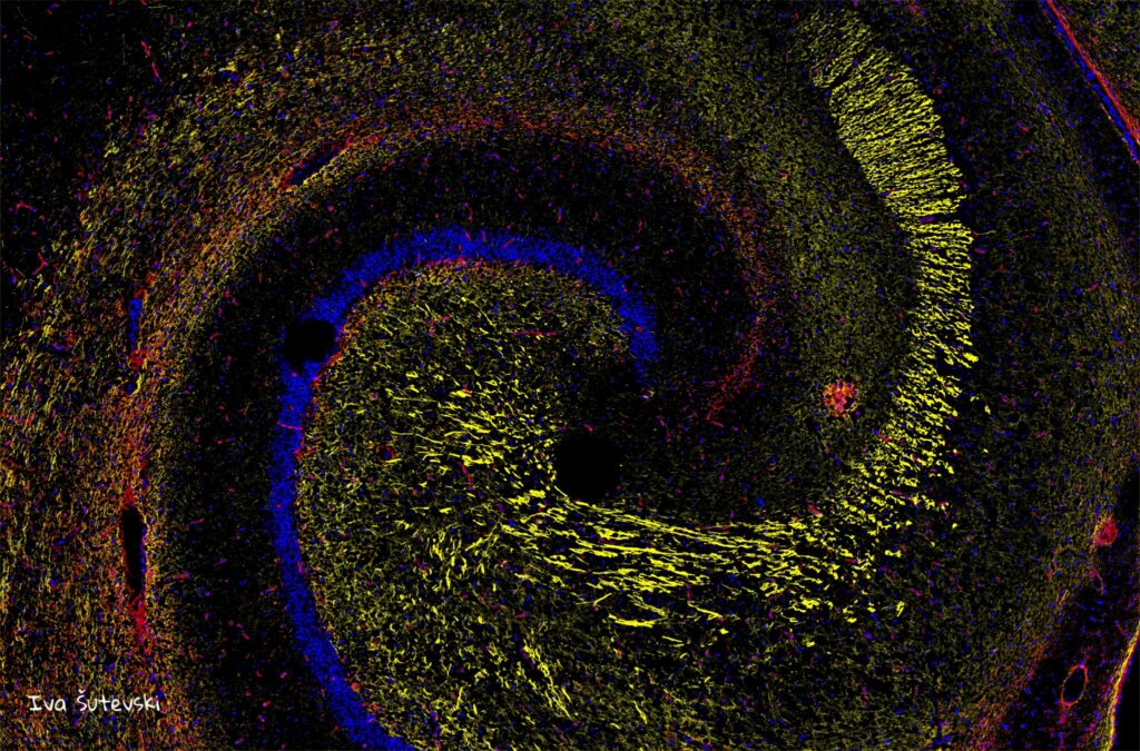
“Gastrula Nebula”
Confocal image of a murine embryonic organoid illustrating the collective migration of cells on a surface coated with laminin. Actin filaments, labelled with phalloidin, and nuclei, stained with Hoechst, reveal detailed cellular architecture and remarkably structured tissue organisation.
Zeiss LSM 880 Confocal Microscopy
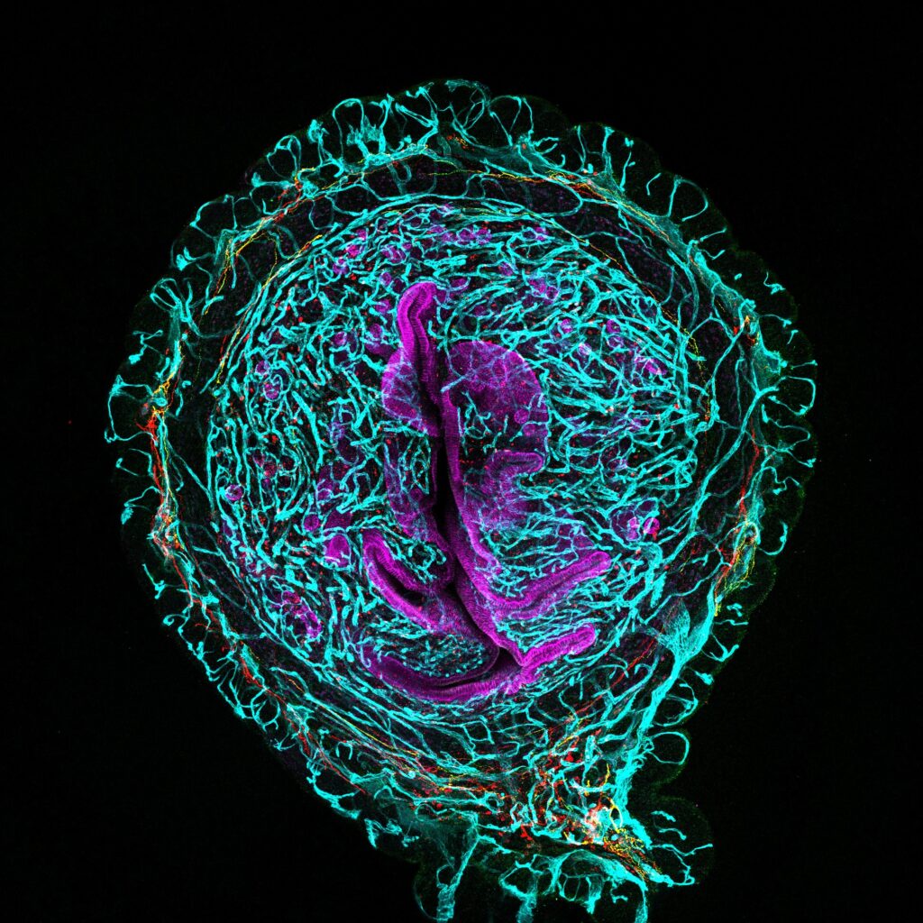
3rd Place:Frédéric FERCOQ, Parasites and Free-living Protists team, National Museum of Natural History
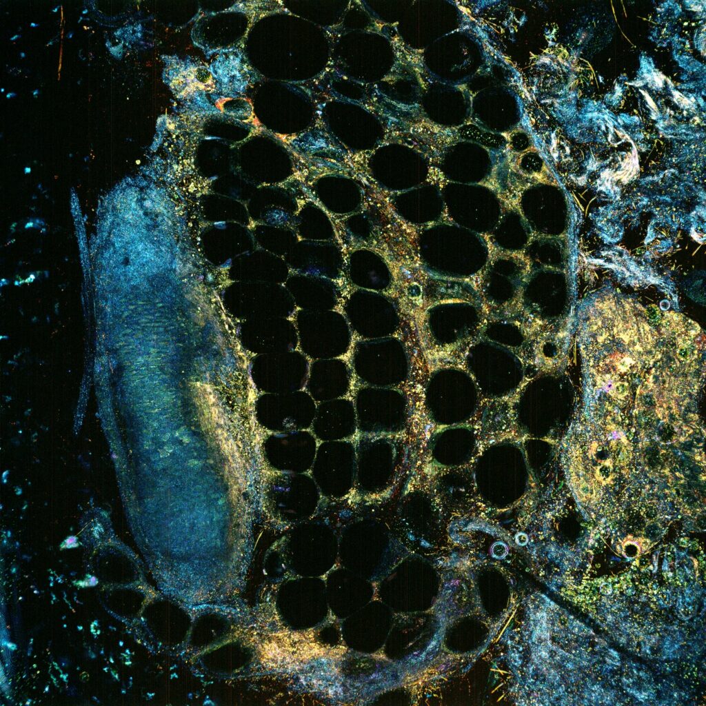
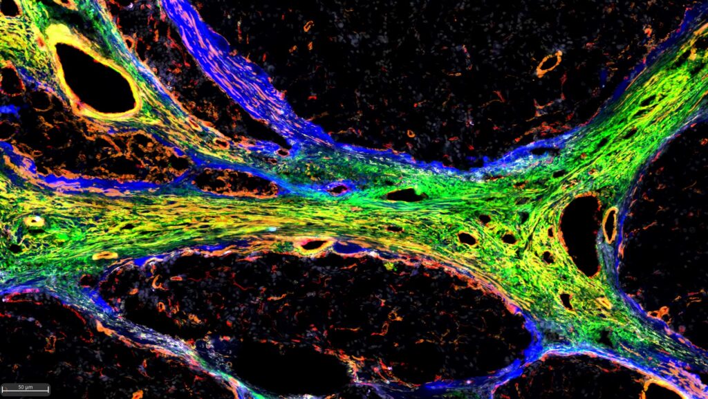
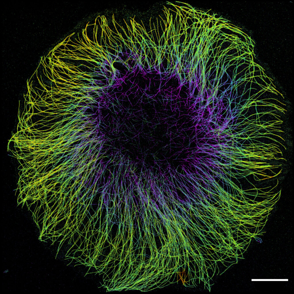
“Filarian explosion”
Internal architecture of Litomosoides sigmodontis, a parasitic nematode used as a model to better understand filarial infections. Subjected to strong internal pressure to maintain its structure, this nematode suffered a rupture of its cuticle during handling, resulting in the expulsion of certain organs, including the ovary and intestine. The cytoskeleton appears in orange, and the DNA in cyan.
Confocal Microscopy
Explore all the images submitted here
ImageJ=1.53t unit=inch
Advanced microscopy workshop in Bordeaux from November 4th to 7th, 2024.
This advanced training course aims to (1) present the theoretical foundations, (2) clarify and synthesize the various existing approaches to both sample and instrument preparation, and (3) provide an overview of solutions for handling and processing the data acquired. These objectives will be addressed through the prism of two important biological fields of application: Neuroscience and 3D Cell Cultures. Indeed, the versatility of light sheet methods means that questions in these two fields can be addressed at a wide range of scales, from the whole brain or organoid, to the study of the nervous system of small living organisms or brain slices, right down to the single molecule inside spheroids. To address these themes, we will draw on the expertise of the Bordeaux FBI site, whether in neuroscience or in the growth and imaging of 3D cell cultures.
The course will be structured around 4 main thematic tracks, addressing the issues of sample preparation and data analysis for given samples. Participants will have the choice of following one of these tracks, or navigating between them according to their interests. The tracks will be :
P1: Large sample imaging – Clearing & Expansion
P2: 3D cellular models Culture & Imaging
P3: Neuronal network imaging
P4: Image Analysis
The format of the course will include lectures and seminars in the morning, providing a theoretical grounding in the various areas covered (sample preparation, imaging, image processing) and presenting the latest developments in these fields, and practical workshop in the afternoon on the various sites of the Bordeaux node (IINS, BIC, VoxCell).
Ihssane Idrissi / Rémi Galland (Interdisciplinary Institute for Neurosciences, Bordeaux, France)
Vincent Studer (Interdisciplinary Institute for Neurosciences, Bordeaux France)
Gustavo de Medeiros (Friedrich Miescher Institute for Biomedical Research, Basel, Switzerland)
Georges Debrégeas (Jean Perrin Laboratory, Paris France)
Thai Truong (University of Southern California, Los Angeles USA)
Angela Getz / Mathieu Ducros (Bordeaux Imaging Center, Bordeaux France)
Alexandra Fragola (Institut des Sciences Moléculaires d’Orsay, Orsay France)
Emmanuel Faure (Laboratory of Computer Science, Robotics and Microelectronics, Montpellier France)
Johannes Roos (Johannes Kepler University, Linz Austria)
Philippe Girard (IJM, Paris, France)
Carole Siret (CIML, Marseille, France)
Guillaume Maucort (BIC, Bordeaux France)
Workshops on
Whole brains imaging by Ultramicroscopy
3D imaging of neuronal expanded samples by AxL (3i) microscope
3D entire small animal imaging
3D Cellular models culture and imaging using the soSPIM technology
Micro-niche creation for 3D cell culture and 3D imaging using the HS-ISM technique
Neurospheres culture and imaging using the MuViSPIM
Brain slices imaging using a Lattice Light Sheet Microscope
Single Cell electroporation for Brain slices labelling
Functional neuronal network imaging in ZebraFish
Orchestrating complex bioimage workflows using the Arkitekt solution
Napari for 3D data handling
How to segment a 3D dataset in just a few clicks?
Organizing committee
Coordinators: Mathieu Ducros & Rémi Galland
& FBI Work Group on « Multiscale Light-Sheet Microscopy »
Sponsors
Deadline: May 31th, 2024
The three national infrastructures ProFi, France-BioImaging and FRISBI along with the GIS IBiSA are pleased to announce a third call for a funded access to IBiSA-labelled facilities. Our aim is to promote IBiSA facilities networking through transdisciplinary research projects.
Applications should request access to at least two different IBiSA facilities from two disciplines (structural biology, Biological imaging and proteomics, see below a non-exhaustive list). The call is open to any academic laboratory.
Modalities for application are described in the attached document.
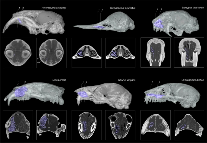
Most mammals can maintain a relatively constant and high body temperature. This is considered to be a key adaptation for theses species, enabling them to successfully colonize new habitats and survive harsher environments. Scientists from the Institut des Sciences de l’Evolution de Montpellier (ISEM) investigate the possible correlation between the maxilloturbinal in the anterior nasal cavity and the body temperature maintenance by using Micro Tomography (or MicroCT) at the MRI core facility (FBI Montpellier node). This technique was essential in this study as it rebuilt the hypothesis around body temperature maintenance. Here is what they found.
MicroCT: Image in a non-destructive way
First of all, what is Micro Tomography? Micro Tomography, or Micro-CT, is a 3D imaging technique using X-rays to see inside biological material, at a small animal or body part level. Slice by slice, this technology scans the object in a series of 2D images that are reconstructed in a 3D model. Micro CT is, thus, non-destructive. This means that it can be used to image a sample without having to cut it! Not only your material is still in one piece but you can use it for further experiments.
Phylogenetic studies as an example of application
In this study examining the correlation between skull structure and the stabilization of body temperature, MicroCT was the key. The presence and the relative size of the maxilloturbinal has been proposed as a hypothesis that reflects the endothermic conditions and basal metabolic rate in extinct vertebrates. Among bony structures, respiratory turbinals (e.g., maxilloturbinal) are interesting anatomical structures that may offer important insights to the origins of endothermy, in other words to the origin of warm-blooded animals. Indeed, respiratory turbinals are highly vascularized, which amplifies the surface area and offers an effective mechanism to avoid loss of internally-produced and costly heat.
You probably figured it out: scientists needed to compare the structure of the maxilloturbinal in order to take conclusion. This is when Micro Tomography was very useful. They scanned 424 individuals from 310 mammal species using high-resolution X-ray micro-computed tomography, with approximatively half of the samples imaged at MRI, part of our Montpellier node. Using the obtained comparative 3D µCT dataset, they explored the anatomical diversity of the maxilloturbinal based on relative surface area, morphology and complexity. They specifically tested the relationship between multiple parameters such as the size-corrected basal metabolic rate (cBMR), the relative surface area of the maxilloturbinal (Maxillo RSA) or body temperature.
And the results surprisingly showed that…
…there is no evidence to relate the origin of endothermy and the development of some turbinal bones! Even though scientists used a comprehensive dataset of Micro CT-derived maxilloturbinals spanning most mammalian orders, they demonstrate that neither corrected basal metabolic rate nor body temperature significantly correlate with the relative surface area of the maxilloturbinal. These results challenge the hypothesis of thermal regulation being linked to respiratory bone structure.
So, what could be linked with the thermoregulation of mammals? Researchers proposed 3 more hypothesis. First of all, environmental conditions could have a bigger role: “the maxilloturbinal function could have a more prominent heat/moisture exchange role in species that face harsh environmental conditions, thus helping to limit spurious heat and moisture loss”. Another major role of the maxilloturbinal is water conservation. As an example, the naked mole-rat avoid breathing through the mouth when performing energy intensive digging because the lips close behind the digging incisors and this species has the lowest value of predicted Maxillo RSA of the entire sample. But most of all, the factor could be a multifactorial physiological question. What is the relation of the maxilloturbinal with the overall nasal cavity? Do other functions play a role in the evolution of this body part, such as its protective role against toxic elements? Is it linked with brain cooling?
Well, imaging will certainly give them an answer in the future!
Detailed view of the maxilloturbinal in selected mammalian species with peculiar thermal and metabolic conditions or that undergo different forms of heterothermy (https://doi.org/10.1038/s41467-023-39994-1)
Get access to one of our services!
You need Micro-CT or another imaging technology or expertise that France-BioImaging provides? To get open access, please login via Euro-BioImaging website! You just have to choose the technology you want to use, then submit your proposal. All applications will be processed by the Euro-BioImaging Hub in close relation with France-BioImaging. And of course, all scientists regardless of their affiliation, area of expertise or field of activity can benefit from open access services! Users whose projects will be validated by Euro-BioImaging will benefit from a waiver for the access cost on France-BioImaging core facilities (https://france-bioimaging.org/access/).
Martinez, Q., Okrouhlík, J., Šumbera, R. et al. Mammalian maxilloturbinal evolution does not reflect thermal biology. Nat Commun14, 4425 (2023). https://doi.org/10.1038/s41467-023-39994-1
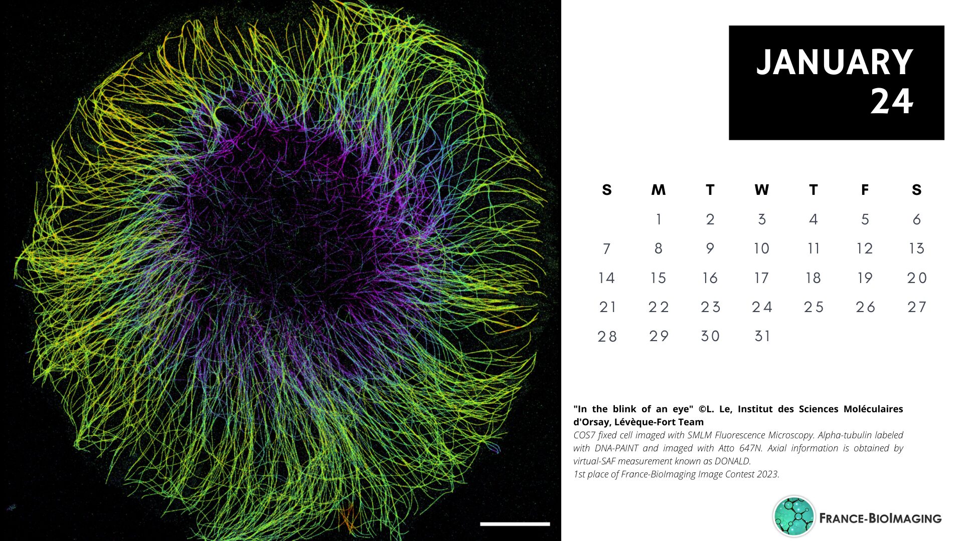
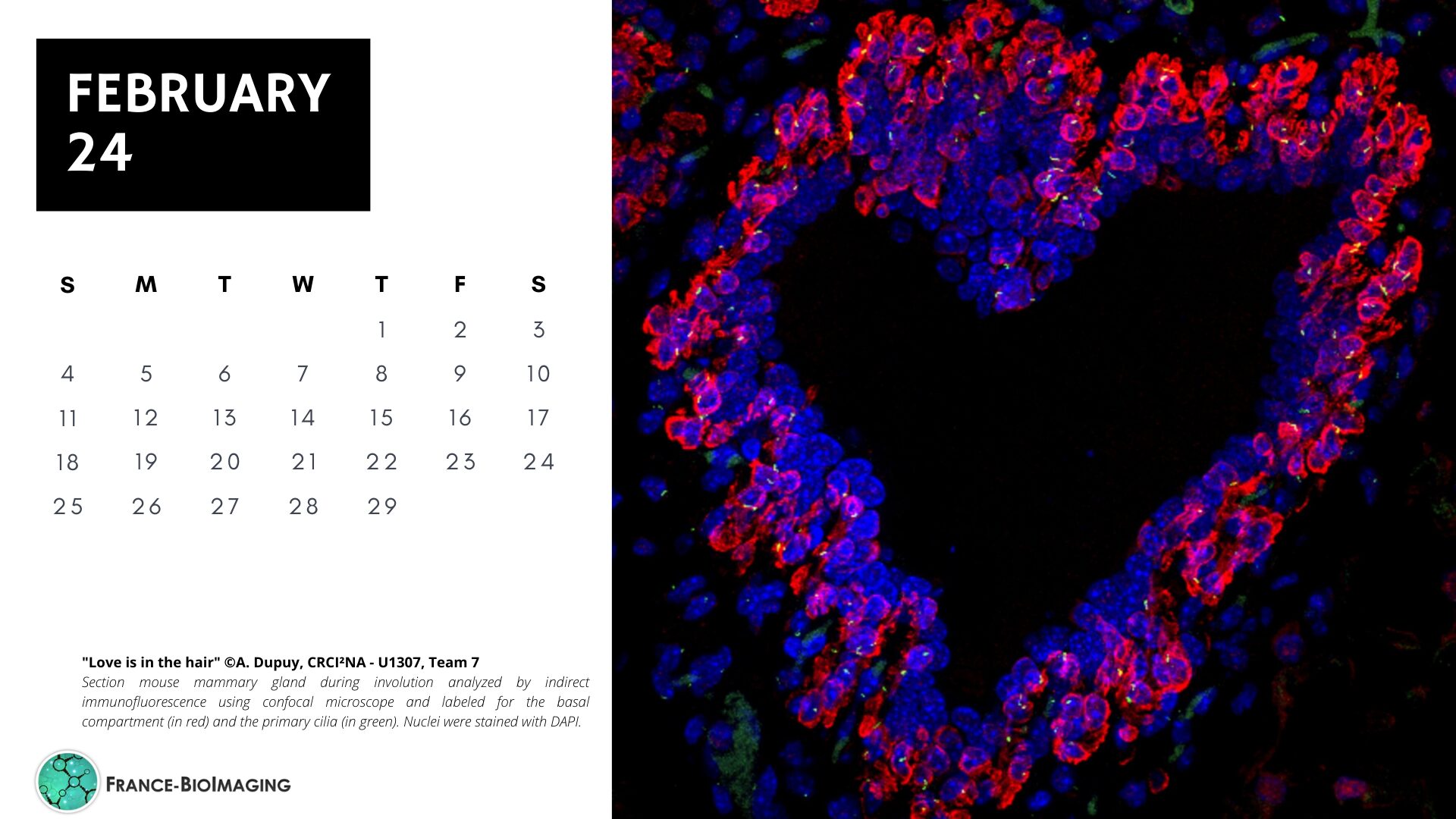
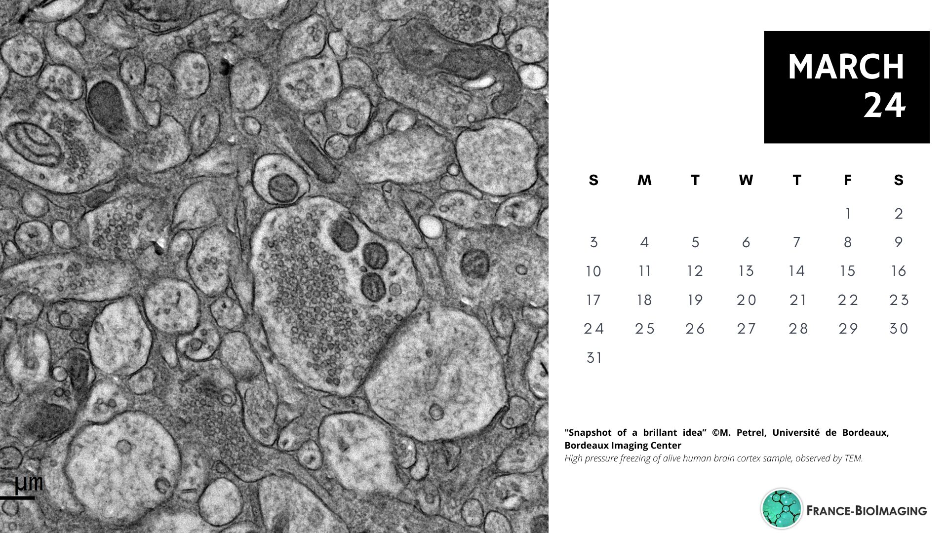
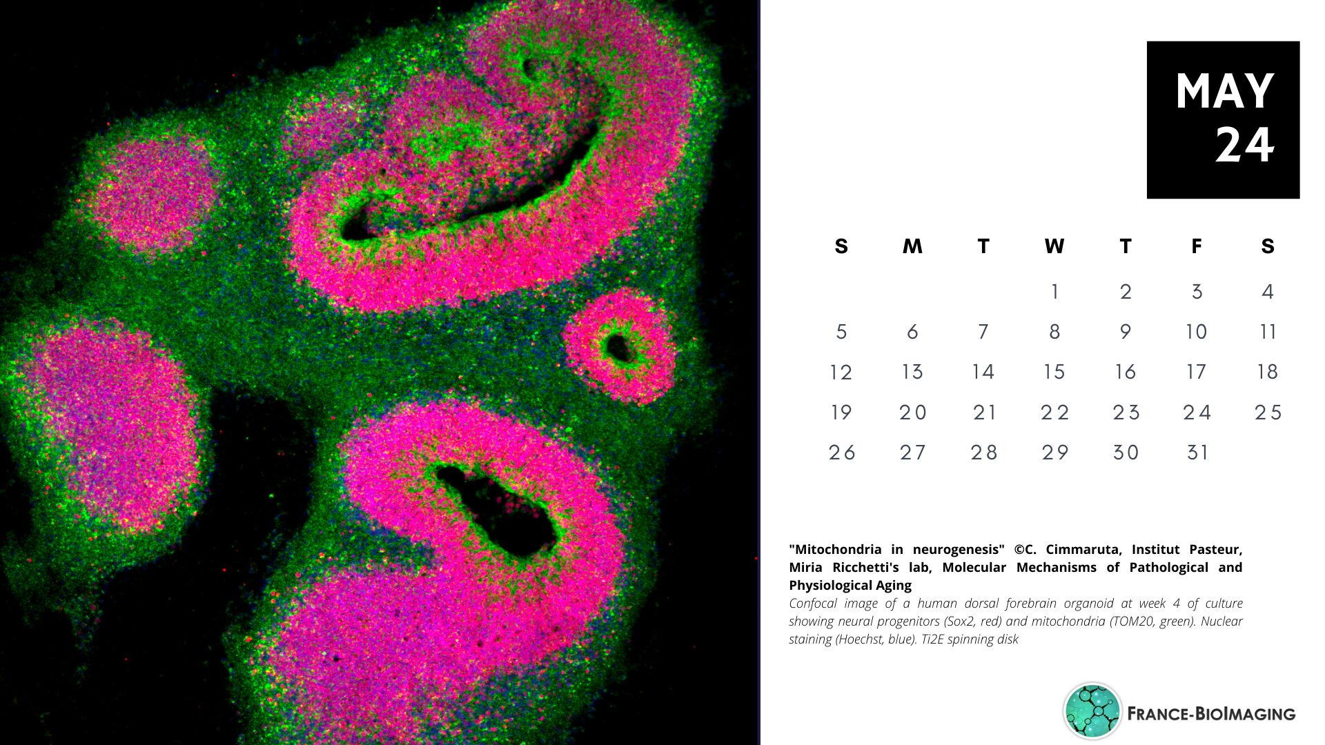
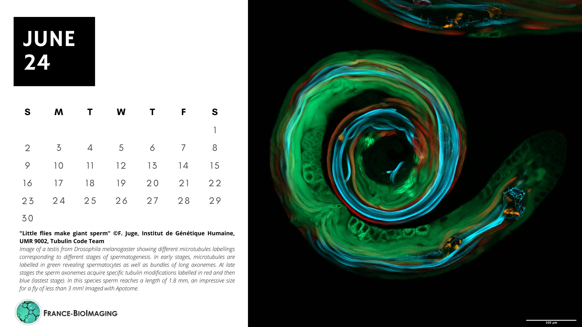
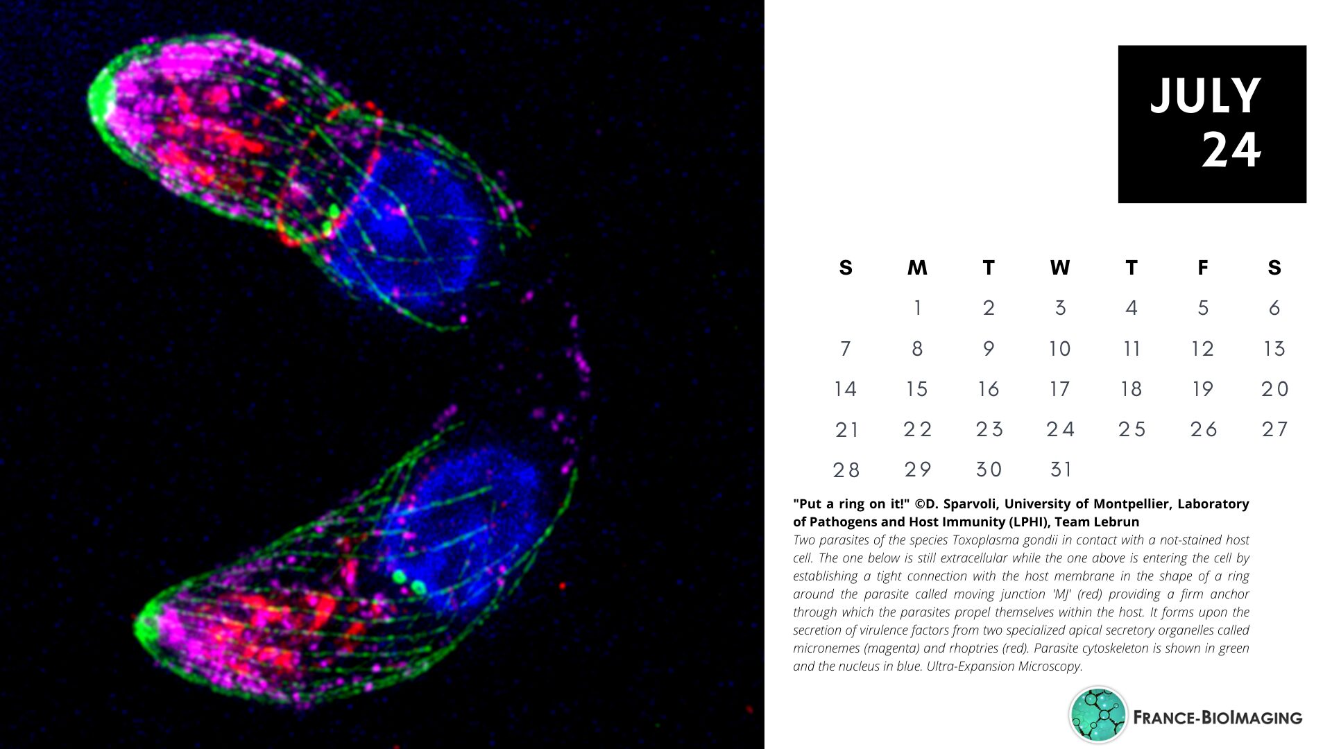
Explore the beauty of the invisible world through the 2024 FBI digital calendar!
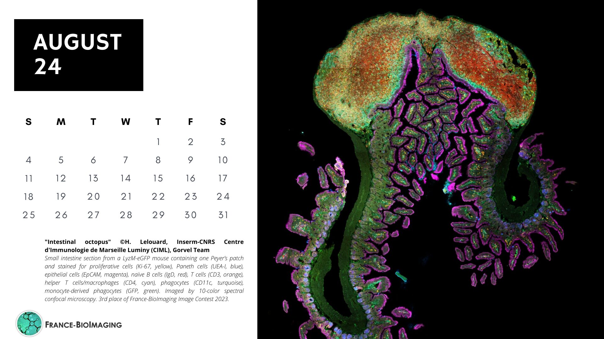
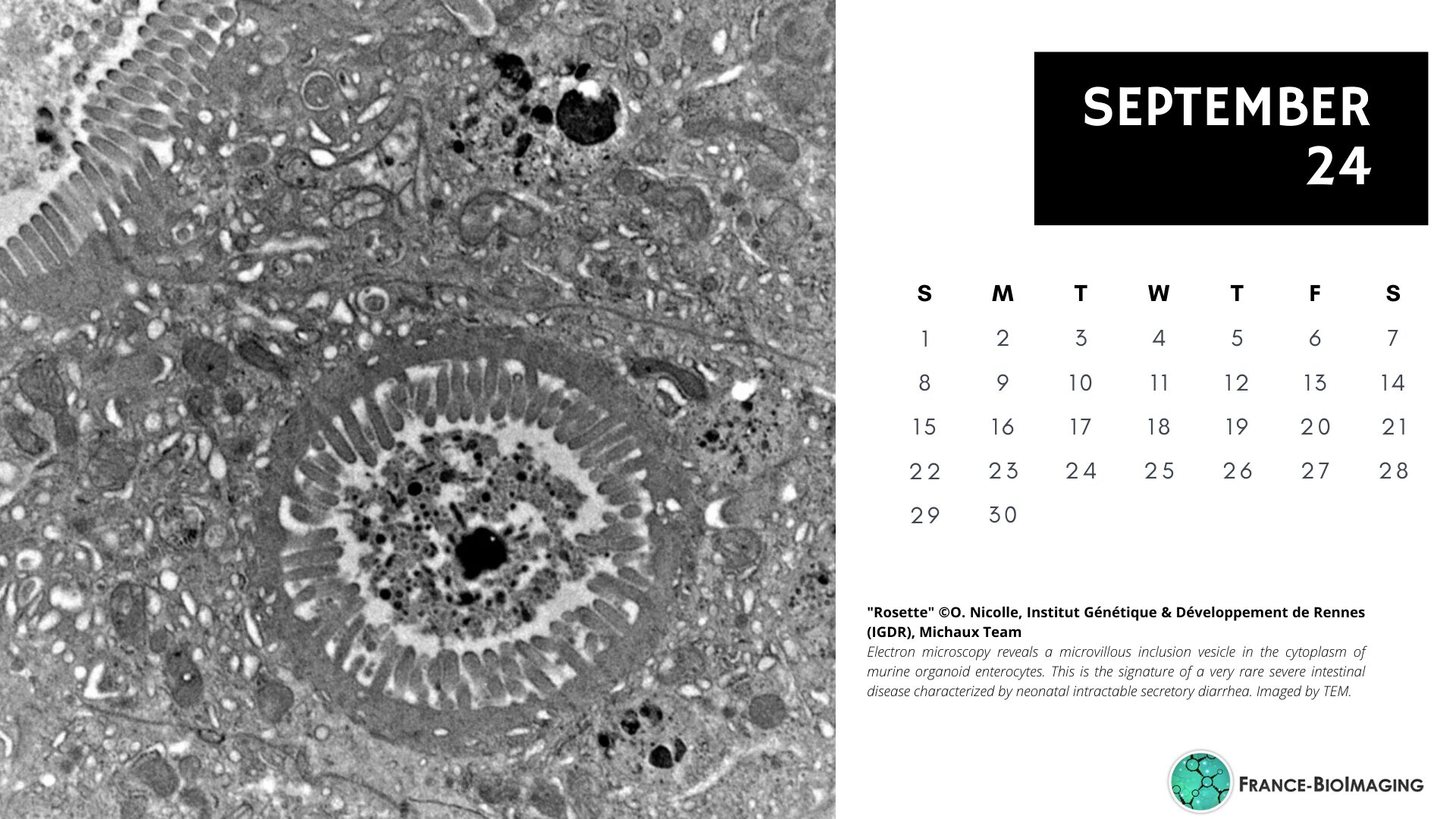
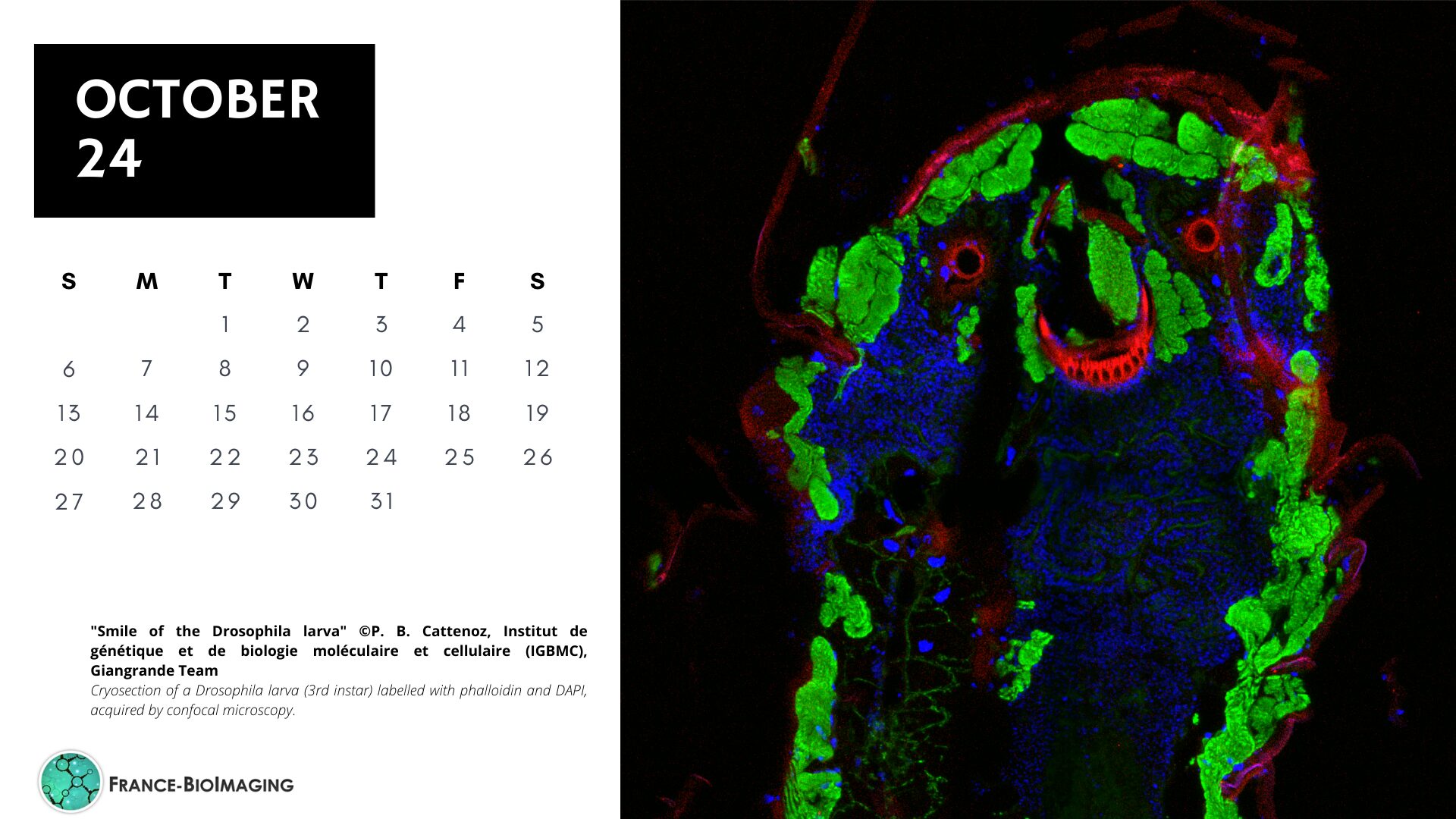
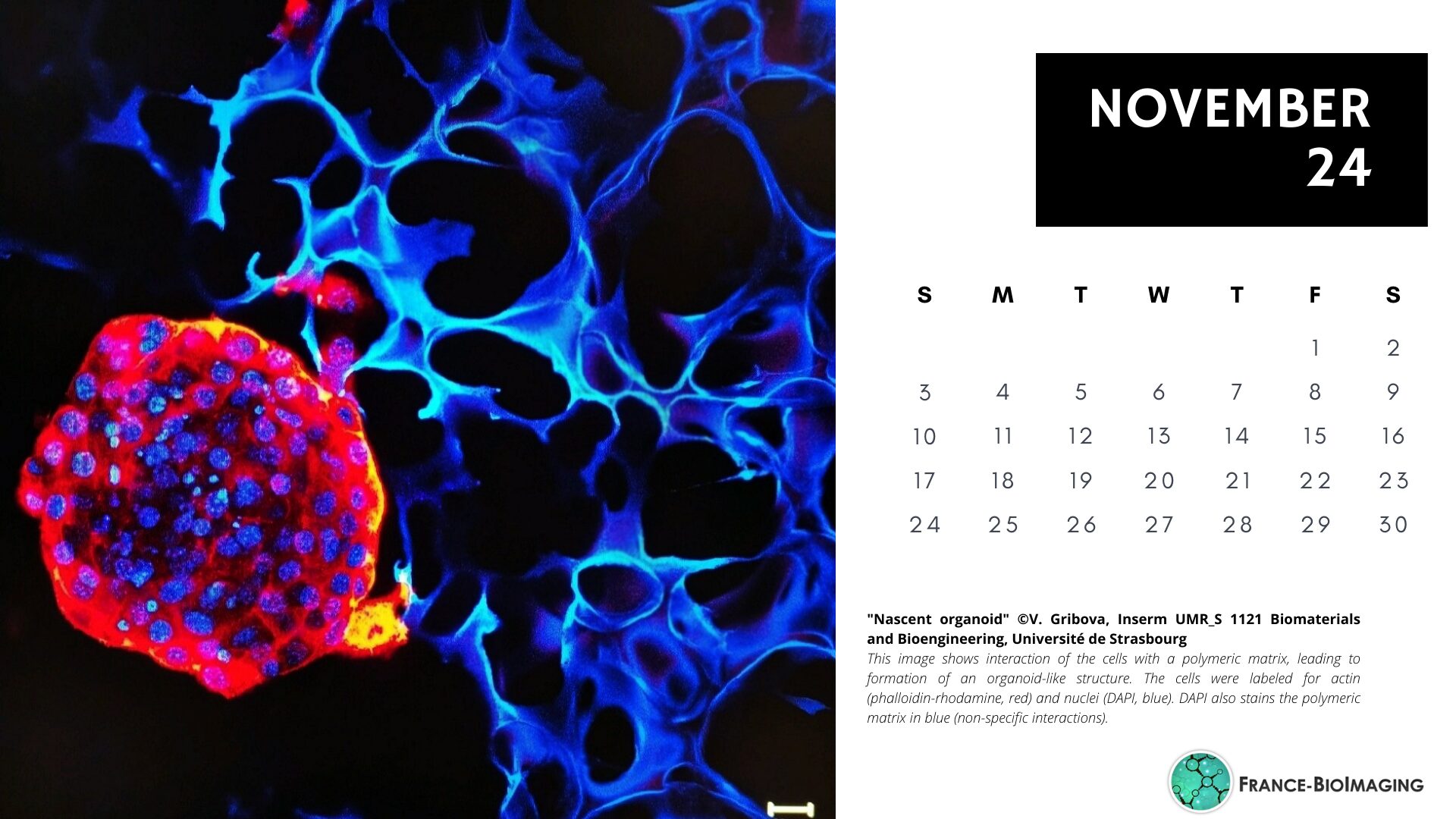
Enjoy the diversity of microscopy techniques, models and applications represented, one image at a time. All 12 images used for this calendar were submitted to France-BioImaging Image Contest 2023.
A big thank you again to all the participants!
You can download the A4 print version (one month per page) 2024 FBI digital calendar here:
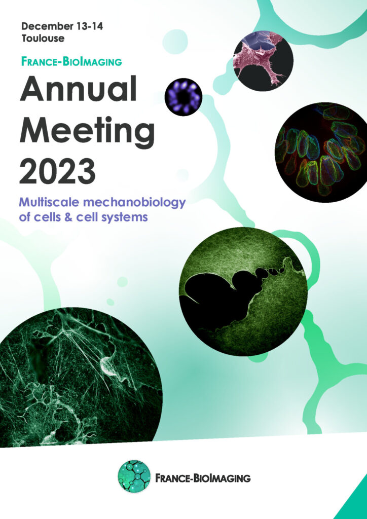
On December 13th and 14th 2023, we have the pleasure to invite you to our Annual Meeting, to be hosted by our brand new FBI Toulouse Node at the Centre de Biologie Intégrative of the Université Toulouse 3 – Paul Sabatier.
We will be happy to celebrate yet another year of achievements and developments in bioimaging with all the members of the community.
This year, the Annual Meeting will focus on “Multiscale mechanobiology of cells and cell systems“, a topic specially selected for being one of Toulouse node’s expertise.
Mechanobiology aims to apply biophysical approaches to measure and perturb forces in complex physiological systems, and to develop in vitro systems including different types of organoids, enabling the controlled manipulation of cells and tissues and the measurement of mechanical forces to study cell mechanics and morphogenesis.
This “mechanobiology” theme is including (i) how cells generate and transmit forces, (ii) the impact of forces on cell/tissue dynamics, (iii) the impact of extrinsic forces on cells and tissues, and (iv) the development of new tools for manipulating and measuring forces in cells and tissues in a controlled, non-invasive way.
We invite the France-BioImaging Community to present their mechanobiology-related projects during the second day of the Annual Meeting around a dedicated session with selected talks. We strongly encourage you to submit an abstract for a talk or a poster presentation during your registration!
The winner of the best talk and the best poster presentation will win their registration fees for one 2024 microscopy related event of their choice!
Core facilities will also have an opportunity to present a poster.
En venant de Bordeaux, contourner Toulouse par l’Est (direction Montpellier) ; avant le péage prendre la sortie no 23 direction Rangueil ou la no 19 direction Ramonville, puis suivre Université Paul Sabatier.
En venant de Montpellier, après le péage, prendre la sortie no 19 direction Ramonville, puis suivre Université Paul Sabatier.
Une navette et le tramway relient l’aéroport et le centre ville de Toulouse (Transports en communs)
De là, il est possible de prendre le métro (lignes A ou B) pour rejoindre la station « Ramonville St Agne » (ligne B, direction Ramonville) : environ 1h de trajet
Le CBI est à 500 mètres à l’Ouest de la station et accessible à pieds ou en bus.
Il est également possible de prendre un taxi depuis l’aéroport : compter au moins 1/2h de trajet hors heures de pointe
En train
Depuis la Gare SNCF Matabiau:
Prendre le métro LIGNE A direction Basso Cambo, changer à la station “Jean Jaurès” et prendre la Ligne B pour rejoindre la station « Ramonville St Agne » : comptez environ 40 minutes depuis la gare.
Le CBI est à 500 mètres à l’Ouest de la station et accessible à pieds ou en bus.
Hôtels conseillés :
Nous vous conseillons de privilégier des hôtels en centre-ville.
Prise en charge des missions:
Se rapprocher de votre noeud FBI (fonds mission), sauf pour les intervenants qui seront directement contactés pour la prise en charge de leur missions.
France-BioImaging and all the French community aims to develop and promote innovative imaging technologies and methods. But microscopy images can also take an artistic, creative look and make the invisible world beautiful, allowing people to see the visual appeal of the life sciences.
We enjoyed the diversity of the images submitted with many different microscopy techniques, models and applications represented. A big thank you to all the participants!
The National Coordination Team and the Executive Board are proud to announce the winners of the FBI Image Contest 2023:
1st Place: Laurent LE, Lévêque-Fort Team, Institut des Sciences Moléculaires d’Orsay
“In the blink of an eye”
COS7 fixed cell. Alpha-tubulin labeled with DNA-PAINT and imaged with Atto 647N. Axial information is obtained by virtual-SAF measurement known as DONALD.
SMLM Fluorescence Microscopy with DNA-PAINT with DONALD detection
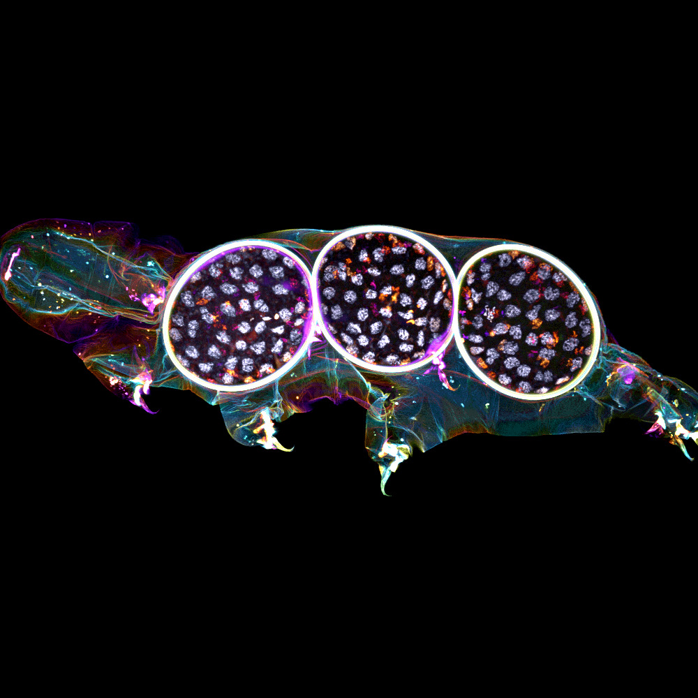
2nd Place:Gonzalo QUIROGA-ARTIGAS, Team Contrôle cytoplasmique de la stabilité du génome, Centre de recherche en Biologie Cellulaire de Montpellier
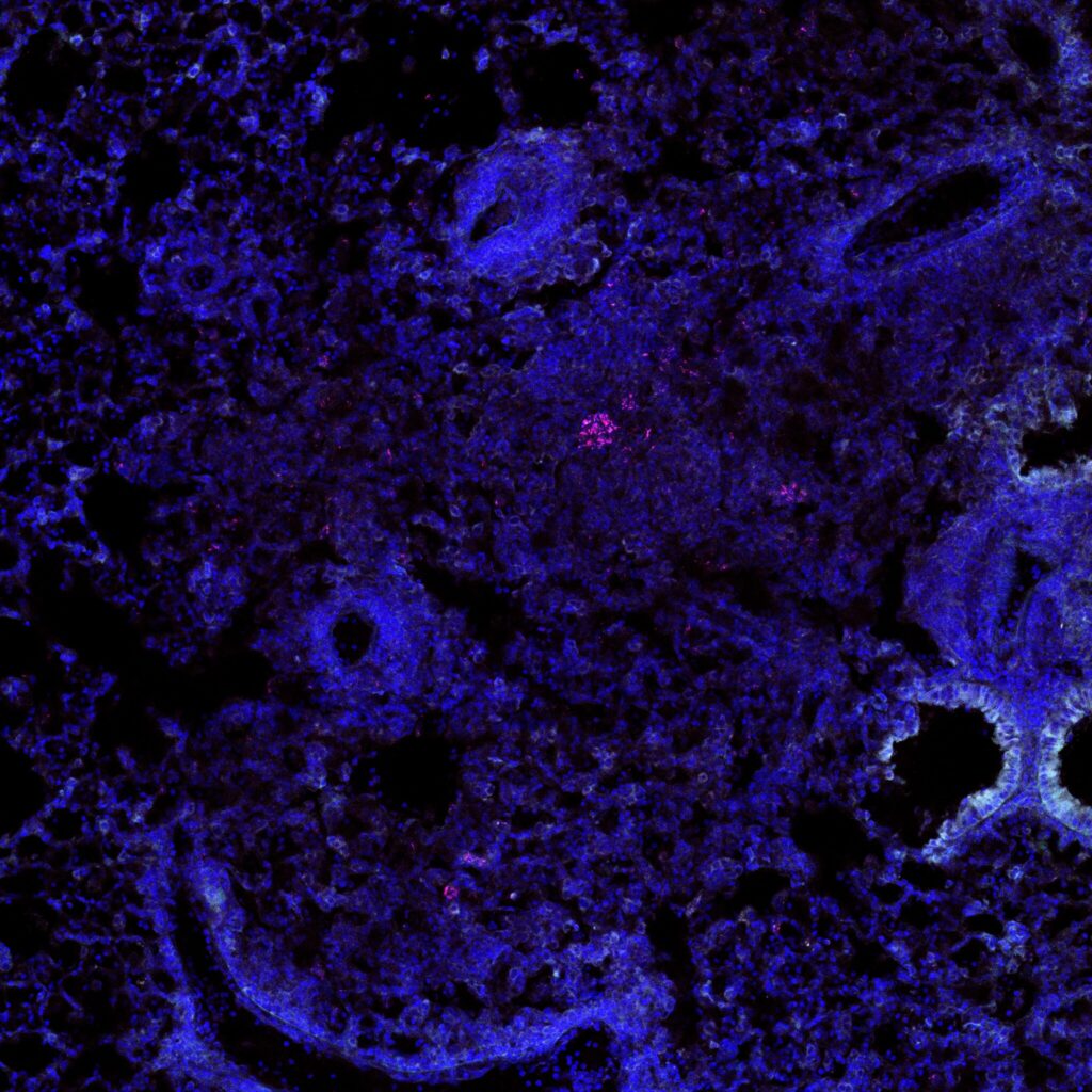
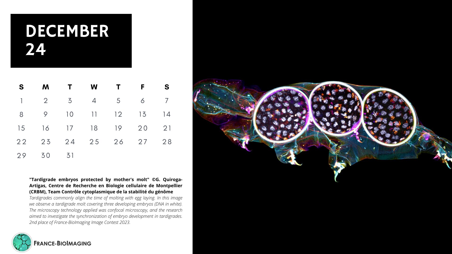
“Tardigrade embryos protected by mother’s molt”
Tardigrades commonly align the time of molting with egg laying. In this image we observe a tardigrade molt covering three developing embryos (DNA in white). The microscopy technology applied was confocal microscopy, and the research aimed to investigate the synchronization of embryo development in tardigrades.
Confocal microscopy
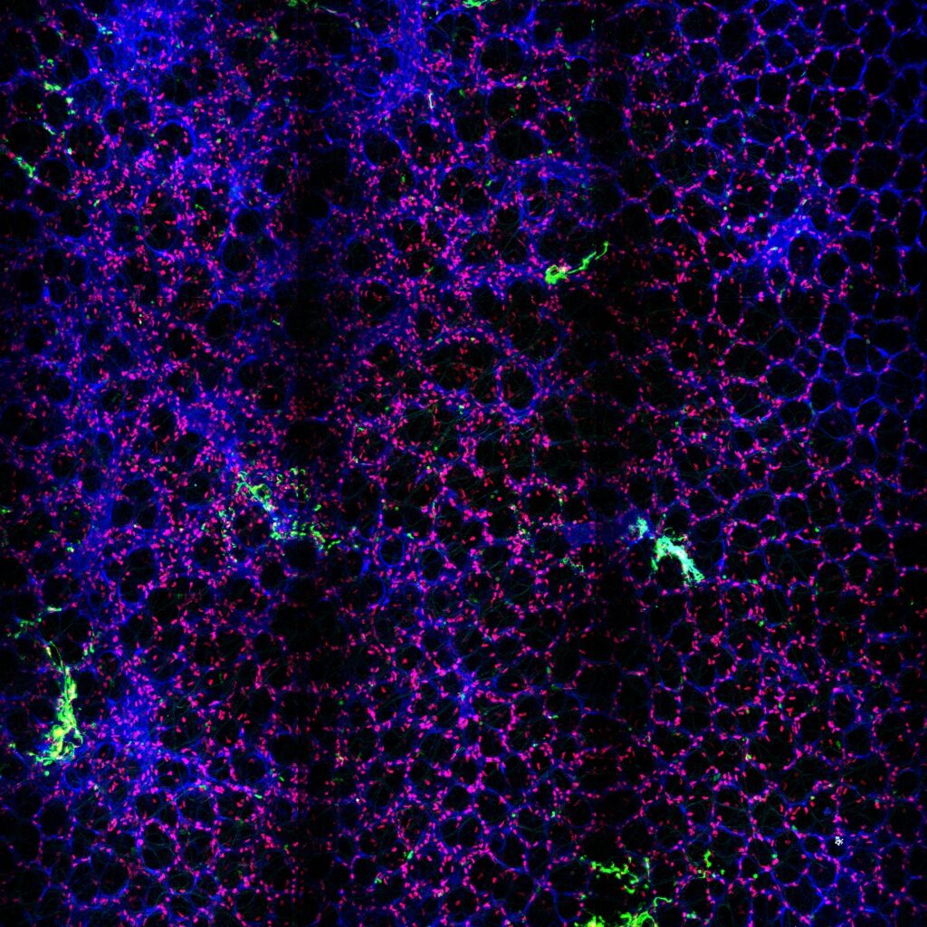
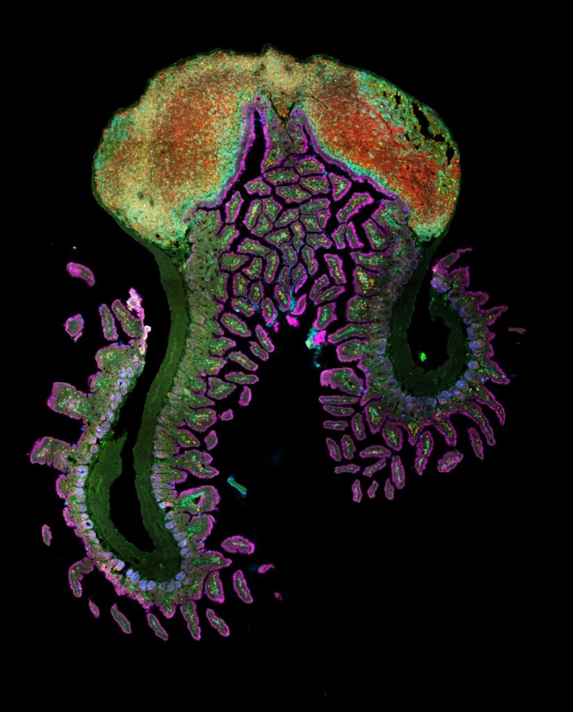
3rd Place:Hugues LELOUARD, Gorvel team, Centre d’Immunologie de Marseille Luminy
“Intestinal octopus”
Small intestine section from a LyzM-eGFP mouse containing one Peyer’s patch and stained for proliferative cells (Ki-67, yellow), Paneth cells (UEA-I, blue), epithelial cells (EpCAM, magenta), naive B cells (IgD, red), T cells (CD3, orange), helper T cells/macrophages (CD4, cyan), phagocytes (CD11c, turquoise), monocyte-derived phagocytes (GFP, green).
10-color spectral confocal microscopy
Congratulations to the winners!
Explore all the images submitted here:
As stated in the Terms & Conditions of the contest, foreign participants non-affiliated to a French institution are featured in the gallery, but were not evaluated as part of the contest.
As the 2023 edition of the France-BioImaging Image Contest admissions is still running, we wanted to highlight our previous winners and their projects. Here is a quick throwback to our 2022 winners.
Before getting to the heart of the matter, we want to remind you that you still have time (before November 10th) to submit your best images and try to win your registration fees for one 2024 microscopy-related event! Please make sure you upload your images on the following link:
Last year, we enjoyed the winning images submitted for their artistic take and their quality. Thanks to Carole SIRET, Magalie BENARD and Frédéric FERCOQ for their beautiful images!
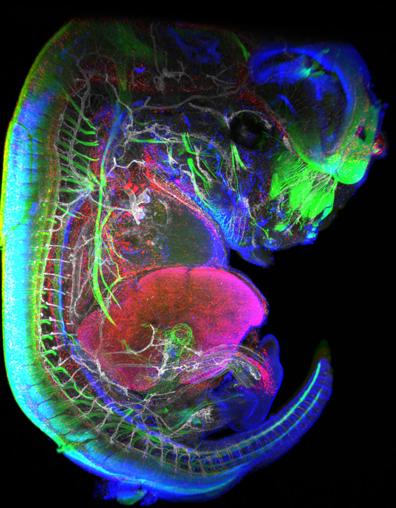
1st Place: Carole SIRET, Van de Pavert Team, Centre d'Immunologie de Marseille-Luminy
"Little Monster"
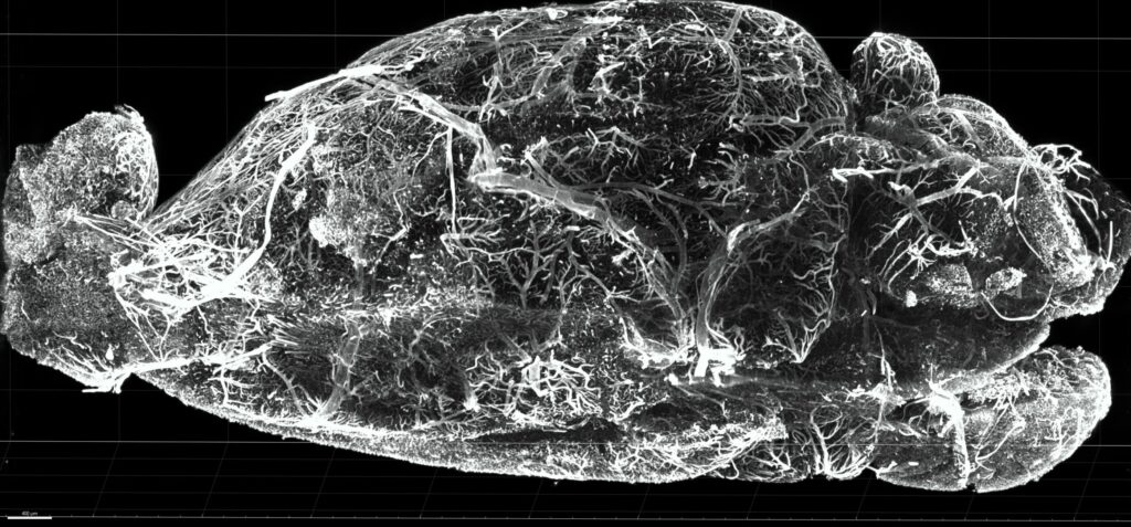
The embryonic formation of lymph nodes, small organs essential for the immune response, is now known. Using light sheet microscopy, scientists were able to determine the dynamics at work in this 13.5-day-old mouse embryo. In blue, the lymphoid cells (LTi), derived from the haematogenous endothelium, a specific tissue of the embryo. They pass into the liver where they proliferate before migrating through the body to give rise to lymph nodes. The 3D information obtained thus makes it possible to follow the interactions of lymph nodes with their environment, in particular with nerve cells, in green, and blood vessels, in white. The lymphatic endothelial cells and some macrophages are visible in red.
Lightsheet Microscopy
Carole Siret is a Research engineer, expert in Lightsheet microscopy, at the Centre d’Immunology Marseille Luminy (CIML) since 2018. She is working in Dr Serge van de Pavert team where they study immune system development. They are particularly interested in the lymph nodes (LN) formation during mouse embryogenesis.
The image she submitted is a projection from a lightsheet acquisition on the UMII (Miltenyi). This image illustrates an E13.5 mouse embryo stained for neurons, LTi (Tissue inducer cells which are the precursor cells for the lymph node), lymphatic and blood vessels. This acquisition was done in the context of the study of the role of Cxcl12 in embryonic LN formation. From previous work it is clear that Cxcl13 and Ccl21 are not expressed present near blood vessels, but it likely that some chemokines, possibly Cxcl12, could be expressed on the endothelial cells. We focus on Cxcl12 since this chemokine has shown to be important for the attraction of several hematopoietic cells. Although it was shown that the receptor for Cxcl12, Cxcr4, is expressed by the mature hematopoietic inducer cells, it is not clear whether it also expressed by the progenitor hematopoietic inducer cells. Next to the possible attraction of hematopoietic cells towards the lymph node anlagen, Cxcl12 is involved in the attraction of nerve fibers. Therefore, the possible role of Cxcl12 could be to both attract hematopoietic cells as well as nerve fibers to initiate a region which is permissive to form lymph nodes.
Thanks to the France-Bioimaging Image Contest, Carole participated to the SFI Congress, where, this year, it was a special joint conference both between the Société Française d’Immunologie (SFI) and the Deutsche Gesellschaft für Immunologie (DGfI). It was a great opportunity to exchange with people at the cutting edge of the immunology field.
2nd Place:Magalie BENARD, Plateforme de Recherche en IMAgerie CEllulaire de Normandie (PRIMACEN), Research infrastructure HeRacLeS, Inserm US 51, CNRS UAR 2026,
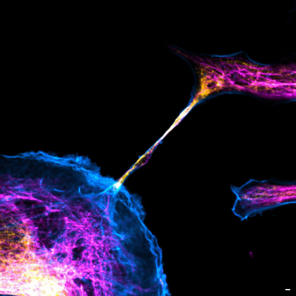
"The communication link with others"
Image of a cellular interconnection between two human tumor cells whose cytoskeleton has been labeled with anti-tubulin (ATTO-647N), anti-vimentin (AlexaFluor594) antibodies and with Phalloidin probe (AlexaFluor488). Scale bar 1µm.
Confocal microscopy
Magalie Bénard is a Research Engineer and the Technical Manager at the Cellular Imaging Facilty PRIMACEN (Plate-forme de Recherche en IMAgerie CEllulaire de Normandie).
The image she submitted is a confocal image representing a cellular interconnection tunneling nanotube (TNTs) between two human tumour cells. In a cancer case, some cells are able to express spontaneously TNTs with cytoskeleton protein composition corresponding to specific role of this communication mechanism. In the winning image, the TNT is composed of tubulin (magenta), actin (cyan) and vimentin (yellow) proteins. Called TNT1, this nanotube allows the transfer of intracellular elements such as RNA, proteins or organelles. Moreover, due to the thinness of TNTs, their photo-sensitivity and their fragility, live-cell imaging is technically challenging with regards to potentially damaging methods. Magalie and her team have developed an adapted method to observe TNTs in living cell with high resolution imaging (STED) enhanced by FLIM by using red and near infrared probes.
France-Bioimaging sponsored her participation to the ELMI (European Light Microscopy Initiative Meeting June 6-9, 2023) congress. During this event, she had the chance to present her project through a poster. This congress also offered a great opportunity to have an overview and the last updates on state-of-the-art imaging techniques.
3rd Place:Frédéric FERCOQ, Parasites et Protistes Libres (PPL), Museum National d'Histoire Naturelle
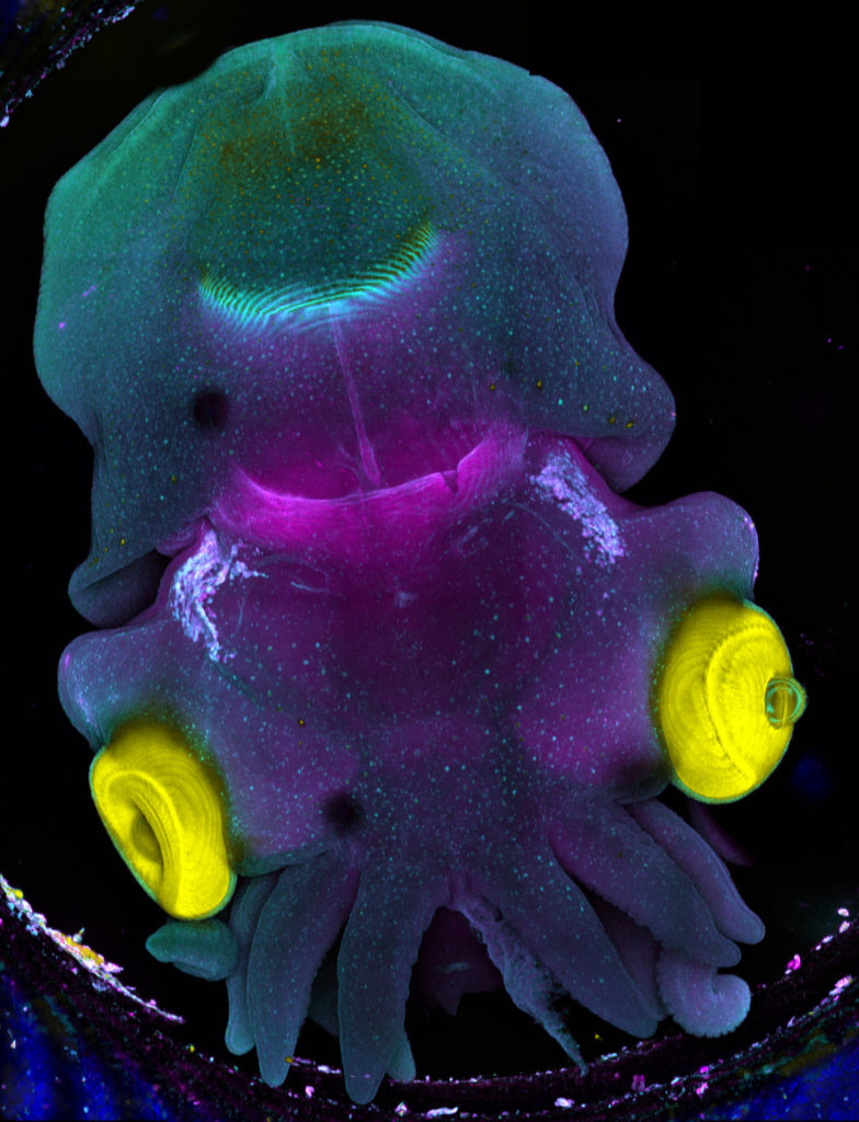
"Sepia"
Stage 25 cuttlefish embryo (Sepia officinalis) observed under a confocal microscope. The cuttlefish was cleared and the tissue autofluorescence was captured.
This image was produced in collaboration with Laure BONNAUD-PONTICELLI and Luis MOLINA from the BOREA laboratory.
Confocal microscopy
Frédéric Fercoq is a postdoc scientist in the Parasitology laboratory of the Muséum National d’Histoire Naturelle (MNHN) in Paris. My main interest is on how myeloid cells participate to the control of parasitic infections, but sometimes at the price of collateral tissue damage. This project involves a lot of microscopy of immune cells, parasites and host tissues to analyse the complex interactions taking place at the site of infection.
The image he submitted has nothing to do with his main project! As he has the chance to work on very different topics and models, this image was acquired as a proof of concept for imaging full embryos of the cuttlefish Sepia officinalis for Frédéric's colleagues Laure BONNAUD-PONTICELLI and Luis MOLINA (BOREA laboratory, MNHN). They work on the nervous system of cephalopod and on the influence of environmental factors during its development. They are now optimizing fluorescent staining for neuronal markers to test the effect of light on the nervous system in situ.
France-Bioimaging sponsored his participation to the FOM (Focus on Microscopy) 2023 congress in Porto. He had the chance to be granted the opportunity to both present his current project through a poster and to give an oral presentation. He was also amazed by the new avenues opened up by the cutting-edge imaging techniques presented throughout the conference.
The France-BioImaging Image Contest is back for its 5th edition!
This image contest is open to all within the imaging community: core facility staff and users, R&D labs teams and co-workers, students… Submit your best microscopy images for a chance to showcase your skills, research and creativity to the French bioimaging community and beyond, allowing people to see the visual appeal of the life sciences. Images from the contest will be featured on France-BioImaging communication tools, online and in print.
France-BioImaging and all the French community aims to develop and promote innovative imaging technologies and methods. But microscopy images can also take an artistic, creative look and make the invisible world beautiful.
We are all eager to see your work !
Prizes
1 to 3 images will be awarded depending on the quantity and quality of the entries submitted. France-BioImaging will cover the registration fees for one 2024 microscopy related event of the winners’ choice (FOM, ELMI, EMC, COMULIS conference, etc.).
Important: Only French or foreign participants affiliated to a French institution can enter the contest. Foreign participants non-affiliated to a French institution can submit images and will be featured in the gallery, but will not be evaluated as part of the contest.
Submission deadline: Friday, November 10th, 2023, 23h59 UTC+2.
Expansion microscopy (ExM) is a relatively new super-resolution method based in the isotropic dilatation of the biological sample in order to overcome the diffraction limit of conventional microscopy. Since its development in 2014, many laboratories have been implementing and adapting the technique to their needs, and the biological applications of ExM grow exponentially.
The ExM working group from the French fluorescence microscopy network (RTmfm), with the support of France-BioImaging, is organizing a workshop on ExM at Bordeaux on October 2, 2023. During the workshop, we will cover different aspects of ExM protocols with experts in biochemistry and biology, and we will present examples of biological applications.
Invited speakers:
Marine Laporte (Institut NeuroMyoGène, Université Claude Bernard, Lyon).
Sven Truckenbrodt (E11 Bio, Alameda, California, US).
Maxence Wisztorski (University of Lille, Inserm, CHU Lille, U1192).
We use cookies on our website to give you the most relevant experience by remembering your preferences and repeat visits. By clicking “Accept All”, you consent to the use of ALL the cookies. However, you may visit "Cookie Settings" to provide a controlled consent.
This website uses cookies to improve your experience while you navigate through the website. Out of these, the cookies that are categorized as necessary are stored on your browser as they are essential for the working of basic functionalities of the website. We also use third-party cookies that help us analyze and understand how you use this website. These cookies will be stored in your browser only with your consent. You also have the option to opt-out of these cookies. But opting out of some of these cookies may affect your browsing experience.
Necessary cookies are absolutely essential for the website to function properly. These cookies ensure basic functionalities and security features of the website, anonymously.
Cookie
Duration
Description
cookielawinfo-checkbox-analytics
11 months
This cookie is set by GDPR Cookie Consent plugin. The cookie is used to store the user consent for the cookies in the category "Analytics".
cookielawinfo-checkbox-functional
11 months
The cookie is set by GDPR cookie consent to record the user consent for the cookies in the category "Functional".
cookielawinfo-checkbox-necessary
11 months
This cookie is set by GDPR Cookie Consent plugin. The cookies is used to store the user consent for the cookies in the category "Necessary".
cookielawinfo-checkbox-others
11 months
This cookie is set by GDPR Cookie Consent plugin. The cookie is used to store the user consent for the cookies in the category "Other.
cookielawinfo-checkbox-performance
11 months
This cookie is set by GDPR Cookie Consent plugin. The cookie is used to store the user consent for the cookies in the category "Performance".
viewed_cookie_policy
11 months
The cookie is set by the GDPR Cookie Consent plugin and is used to store whether or not user has consented to the use of cookies. It does not store any personal data.
Functional cookies help to perform certain functionalities like sharing the content of the website on social media platforms, collect feedbacks, and other third-party features.
Performance cookies are used to understand and analyze the key performance indexes of the website which helps in delivering a better user experience for the visitors.
Analytical cookies are used to understand how visitors interact with the website. These cookies help provide information on metrics the number of visitors, bounce rate, traffic source, etc.
Advertisement cookies are used to provide visitors with relevant ads and marketing campaigns. These cookies track visitors across websites and collect information to provide customized ads.


