Fostering technology transfer from the R&D teams to the core facilities of FBI
▽ Scroll down
Tag: Microscopy
France BioImaging primary mission is to develop, promote, disseminate and provide access to innovative instruments and imaging technologies in the field of bioimaging to scientists. Fostering the technological transfers is at the heart of this mission, and for this France BioImaging relies on a strong association of leading R&D research teams with core facilities.
However, several bottlenecks exist and often hamper or prevent successful technology transfer:
A lack of human resource leads to difficulties in transferring and stabilizing the technology which is not enough user-friendly
A technology that is too specific, with not enough user base
A difficulty to contract with industry through institutional offices for industrial valuation
In the context of image analysis: the instability of open software economical model, inter-operability, large data handling and algorithm complexity
As a way to tackle these bottlenecks, France BioImaging launched in January 2021 its first “FBI Internal Call 2021: Technology transfer from the R&D teams to the core facilities” to promote the transfer of new technologies (instrumentation, probes, staining methods, software, data analysis or data visualization) from the R&D teams to the facilities of France BioImaging, for access and service to end-users. The outcome of the transfer project had to ensure for the prototype to be usable by the end-users until the interpretation of the data. The project had also to include a sustainability plan and a training plan to guide both facility staff and end-users toward autonomy.
The project selection was organized by the National Coordination of France-BioImaging and applications were assessed according to the following evaluation criteria:
Innovation and originality of the proposal
Scientific quality, implementation, timeline
Competitive positioning
Adequacy of resources with the proposed project
Economic impact and tech transfer potential and perspectives
Estimation of the user market and potential for user adoption
Plan for training and sustainability.
For the first edition of the “FBI Internal Call 2021: Technology transfer from the R&D teams to the core facilities”, 5 projects were selected:
Icy@FBI: Jean-Christophe Olivo-Marin (IPDM Node): Broadening the scope of applications of Icy (http://icy.bioimageanalysis.org/) by implementing several key new bioimage analysis components
BIC-HCS-SMLM: Jean-Baptiste Sibarita (Bordeaux Node), Technological transfer of a Single-Molecule-based High Content Screening platform to the Bordeaux Imaging Center
CloudFISH: Marcello Nollmann (Montpellier Node), A tool for the analysis of single-molecule RNA and DNA FISH images
MorphoNet: Emmanuel Faure (Montpellier Node), An interactive online morphological browser to explore complex multi-scale data
BioImageIT (https://bioimageit.github.io/#/about): Jean Salamero, Sylvain Prigent (IPDM Node), An open source framework for integration of image data management with analysis
Each selected project was awarded with a 80k€ grant for salary and/or equipment, and several positions are currently available: https://france-bioimaging.org/jobs/
This call will be renewed in 2023.
Don’t forget to register before June 15th for MiFoBio autumn school, 5-12th November 2021 (Presqu’île de Giens)!
Important information for the registered participants: an email with the links to access the training videos was sent on February 15th, 2020. Please check your inbox and SPAM folder!
If you registered after February 14th, 2021, you will receive an email with the links within two days.
France BioImaging (FBI) is organizing a remote training on Light-Sheet Fluorescence Microscopy (LSFM), which enable 3D imaging of biological samples with unprecedented spatio-temporal resolutions and low perturbing effects.
LSFM methods actually cover a large variety of implementations which allow imaging a wide range of sample types, from single cell to whole organs or organisms both live and fixed. These new imaging capabilities are revolutionizing the way we visualize our samples and address biological questions. However, imaging with a light-sheet microscope raises many questions about the choice of the set-up depending on the sample to image, the sample preparation and mounting protocols or the data management (storage, visualization, quantification). Thus, it can be difficult to find its way through the numerous microscope implementations, protocols and tools that have been extensively developed over the last 20 years. We therefore decided to review all those questions in a remote training.
Our goal is to help people who want to jump into the world of 3D imaging and are seeking the best solution for their samples and biological questions. In that perspective, we will provide a comprehensive picture including all the possibilities and challenges regarding LSFM.
Format:
The training will be divided in 3 parts:
Theoretical courses on LSFM
Practical demonstrations of several LSFM implementations available throughout the FBI infrastructure
Live online question-and-answer session
For the two first parts, videos will be available on a Youtube FBI channel. The participants will have 3 weeks, from the 15th of February to the 5th of March 2021, to watch those videos and will be invited to ask questions or comment.
FBI experts will then answer all questions during a live interactive video chat on the third week of the training (5th of March where participants will have the opportunity to directly interact with the experts.
Program:
1. Theoretical aspects of LSFM (15th to 26th of February 2021)
Here are the three main questions concerning the imaging with a light-sheet microscope: (1) what LSFM type should I use for my experiment, (2) How do I prepare and mount my sample, and (3) how to visualize and analyze my data sets. The first part of this training will address these three questions through three theoretical courses:
Course 1: Theoretical principles and numerous implementations overview of LSFM
P. Girard (Institut Jacques Monod, Paris-Centre)
Course 2: Sample preparation and mounting principles – highlight on clearing approaches
Carol Siret (CIML, Marseille)
Course 3: Reconstruction, Visualization and Analysis software overview.
Cesar Augusto Valades (Institut Curie, Paris-Centre),
2. Practical demonstrations of several LSFM implementation and experiments (15th of February to 5th of March 2021)
In the second part of the training we will propose several videos on various systems available in the FBI laboratories and imaging platforms covering diverse types of LSFM design and applications.
Each video will feature a specific set-up and experts will present how to run an experiment on them focusing on three main aspects: (1) sample preparation and mounting methods, (2) image acquisition processes, and (3) visualization of the data-sets.
Lattice Light Sheet Microscope – (Home-made and 3i versions)
Mathieu Ducros (BIC, Bordeaux)
Ludovic Lecomte, Jean Salamero and Cesar Valaldes-Cruz (Institut Curie, Paris-Centre)
Single-objective Single Plane Illumination Microscope(soSPIM) – Home-made
Rémi Galland (IINS, Bordeaux)
Dual inverted Single Plane Illumination Microscope(diSPIM) – 3i (Marianas)
Elric Esposito et Julien Fernandes (Institut Pasteur, Paris-Centre)
An online video session will conclude the training where FBI experts will answer all participants’ questions. You can ask questions either in advance in the comment box of the Youtube video, or during the Q&A session in a chat box. The Q&A session will be divided in sections, each related to a specific video.
To register:
In order to register to the Light-Sheet Fluorescence Microscopy remote training, please fill out the registration form available here.
Registration is free but mandatory in order to receive the links to the training videos.
Extended deadline: February 19th, 2021
We look forward to your participation !
The PICsL-FBI microscopy core facility is located on two sites: Centre d’Immunologie de Marseille Luminy (CIML) and Institut de Biologie du Développement de Marseille (IBDM).The PICsL-FBI facility of the CIML called ImagImm (Imaging Immunity) via its microscopy resources – from the molecule to whole organisms – is dedicated to help its users deciphering cellular mechanisms in the fields of immunology.
Major research implications of the ImagImm facility:
In collaboration with Tomasz Trombik (Faculty of Biotechnology, University of Wroclaw – Wroclaw, Poland), Sophie Brustlein (Institut de Convergences Centuri, AMU,CNRS – Marseille, France) and Nicolas Bertaux (Institut Fresnel, AMU, Centrale Marseille, CNRS – Marseille, France), Sébastien Mailfert and Didier Marguet published the procedure for implementing spot variation Fluorescence Correlation Spectroscopy (svFCS) measurements using a classical fluorescence microscope that has been customized1. This publication is following the technology transfer made in 2018: the svFCS developed by Didier Marguet’s lab was duplicated by Sébastien Mailfert and Sophie Brustlein and built from scratch in 7 days on site, in Poland.
Dynamic biological processes in living cells, including those associated with plasma membrane organization, occur on various spatial and temporal scales, ranging from nanometers to micrometers and microseconds to minutes, respectively. Such a broad range of biological processes challenges conventional microscopy approaches. The published protocol includes a specific performance check of the svFCS setup and the guidelines for molecular diffusion measurements by svFCS on the plasma membrane of living cells under physiological conditions. Additionally, a procedure for disrupting plasma membrane raft nanodomains by cholesterol oxidase treatment is provided and how these changes in the lateral organization of the plasma membrane might be revealed by svFCS analysis. This fluorescence-based method can provide unprecedented details on the lateral organization of the plasma membrane with the appropriate spatial and temporal resolution.
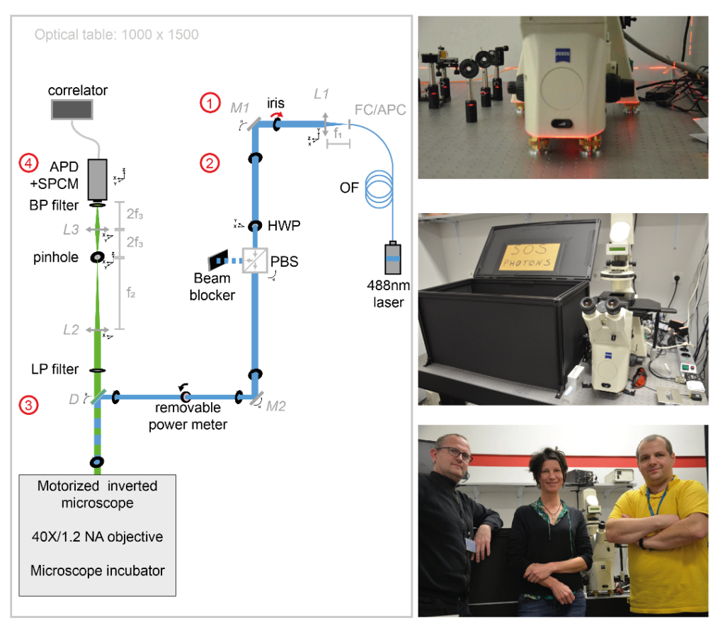
Figure 1:Schematic view of excitation and emission optical paths of the svFCS setup and pictures of the setup. The svFCS setup contains four modules: (1) the output of a fibered 488 nm laser is collimated, (2) a combination of a half-wave plate and polarizing beamsplitter sets the optical power, (3) the laser beam focused on the sample after traveling through a tube-lens free motorized microscope, and (4) the fluorescence is detected through a confocal-like detection path onto an avalanche photodiode coupled to a single photon counting module, which delivers a signal to a hardware correlator. Simplicity gives the system its sensitivity, robustness, and ease of use.
SAPHIR : a Shiny application to analyze tissue section images
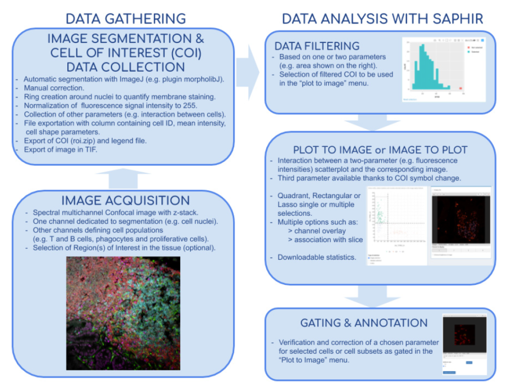
In collaboration with Hugues Lelouard (CIML, Inserm, CNRS, AMU) and Elodie Germani, Mathieu Fallet published a powerful method for both basic and medical research to study cell populations in tissues using immunofluorescence. Image acquisitions performed by confocal microscopy notably allow excellent lateral resolution and more than 10 parameter measurement when using spectral or multiplexed imaging. Analysis of such complex images can be very challenging and easily lead to bias and misinterpretation. They developed the Shiny Analytical Plot of Histological Images Results (SAPHIR), an R shiny application for histo-cytometry using scatterplot representation of data extracted by segmentation. It offers many features, such as filtering of spurious data points, selection of cell subsets on scatterplot, visualization of scatterplot selections back into the image, statistics of selected data and data annotation. This application allows to quickly characterize labeled cells, from their phenotype to their number and location in the tissue, as well as their interaction with other cells.
Figure 2: Flow chart of tissue image analysis from image acquisition and segmentation (left side) to extract data analysis with SAPHIR (right side)
Wound healing in C. elegans
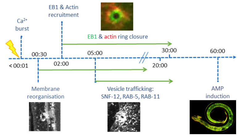
In collaboration with Nathalie Pujol and Jonathan Ewbank (CIML, Inserm, CNRS, AMU), Mathieu Fallet and Sébastien Mailfert participated in the project on the immune response by showing that wounding provokes a reorganization of plasma membrane subdomains3. The skin protects animals from infection and physical damage. In Caenorhabditis elegans, wounding the epidermis triggers an immune reaction and a repair response, but it is not clear how these are coordinated. Previous work implicated the microtubule cytoskeleton in the maintenance of epidermal integrity (Chuang et al., 2016). Taffoni et al. show the reorganization of the plasma membrane subdomains by a simple wounding system. This is followed by recruitment of the microtubule plus end-binding protein EB1/EBP-2 around the wound and actin ring formation, dependent on ARP2/3 branched actin polymerization. They show that microtubule dynamics are required for the recruitment and closure of the actin ring, and for the trafficking of the key signaling protein SLC6/SNF-12 toward the injury site. Without SNF-12 recruitment, there is an abrogation of the immune response. These results suggest that microtubule dynamics coordinate the cytoskeletal changes required for wound repair and the concomitant activation of innate immunity.
Figure 3: Time line of events
References:
Mailfert, S., Wojtowicz, K., Brustlein, S., Blaszczak, E., Bertaux, N., Łukaszewicz, M., Marguet, D., Trombik, T. Spot Variation Fluorescence Correlation Spectroscopy for Analysis of Molecular Diffusion at the Plasma Membrane of Living Cells, JoVE, 165, 1-19 (2020).
Germani, E., Lelouard, H., Fallet, M. SAPHIR: a Shiny application to analyze tissue section images, F1000Research, Faculty of 1000, 9, 1276-1285 (2020).
Taffoni, C., Omi, S., Huber, C., Mailfert, S., Fallet, M., Rupprecht, J-F,. Ewbank, J., Pujol., N. Microtubule plus-end dynamics link wound repair to the innate immune response, eLIFE, 9, e45047 (2020)
Registration is now open for the Virtual Early Career European Microscopy Congress 2020!
Following the cancellation of emc2020, this virtual meeting will provide an opportunity for Early Career Scientists who would have attended and presented at the congress, to still present their work at an International Meeting this year.
Registration Fees The registration fees can be found below.
Price
Early Career You are a student or an Early-Career Researcher (less than 3 years since the completion of your PhD by November 2020). Thank you to the European Microscopy Society for subsidising to enable free registration for Early Career Researchers.
FREE
EMS Member You are a member of the European Microscopy Society. Member fees are applicable only if your membership status is active at the time of registration. Your membership status will be verified, so you must be sure that your annual subscription is current.
For help and queries If you have any questions regarding this event, please do email katejermey@rms.org.uk or visit the website www.emc2020.eu for further information.
The France BioImaging Image Contest is back for its 3rd edition!
This image contest is open to all within the imaging community: core facility staff and users, R&D labs teams and co-workers, students… Submit your best microscopy images for a chance to showcase your skills, research and creativity to the French bioimaging community and beyond, allowing people to see the visual appeal of the life sciences. Images from the contest will be featured on France BioImaging communication tools, online and in print.
France BioImaging and all the French community aims to develop and promote innovative imaging technologies and methods. But microscopy images can also take an artistic, creative look and make the invisible world beautiful.
We are all eager to see your work !
Prizes
1 to 3 images will be awarded depending on the quantity and quality of the entries submitted. France BioImaging will cover the registration fees for one 2022 microscopy related event of the winners’ choice (FOM, ELMI, EMC, COMULIS conference, etc.).
Important: Only French or foreign participants affiliated to a French institution can enter the contest. Foreign participants non-affiliated to a French institution can submit images and will be featured in the gallery, but will not be evaluated as part of the contest.
Submission deadline: Friday, October 15th, 2021, 23h59 UTC+2.
The school is dedicated to teaching the basics and wider context necessary to understand recent advances and current challenges in biological and medical imaging. Cutting-edge techniques using a wide range of image-formation mechanisms — including magnetic resonance imaging, positron emission tomography, infrared and optical microscopy, electron microscopy and X-ray imaging — will be discussed, with a focus on multimodal and multiscale imaging methods, together with supporting technologies such as computer-aided image analysis and modelling.
The school will provide different tracks for participants with a background in life sciences and physical sciences, respectively. Furthermore, specialized lectures will address current topics in biological and medical imaging. The students will have lectures in the morning and practical sessions in the afternoon (either hands-on lab work or lab demonstrations, depending on the field). The program will be rounded off with a practical day and an industry day.
The school follows a challenging and demanding schedule. It addresses excellent MSc and PhD students as well as scientists from industry with background in biology, chemistry, computer science, engineering, mathematics, medical science or physics. We plan to admit about 60 participants (internally and from abroad).
Admission will be decided based on the applicant’s curriculum vitae, a statement of purpose and applicant’s references. Students who have not yet started a PhD program may apply for a stipend. Interested students are kindly asked to submit their application pack including their study grades and reference letter on our homepage (www.excite.ethz.ch)
The application deadline is Monday, 23 April 2018. The notification of acceptance will be given by 25 May 2018.
This conference is organized by the “Réseau d’Imagerie Cellulaire Paris-Saclay”.
The two most common techniques of vibrational micro-spectroscopy are infrared (IR) and Raman. These two sophisticated tools enable to visualize the inherent vibrational spectra of biochemical constituents of a cell or a tissue. Therefore, IR and Raman microscopy provide the specific and distinct “fingerprint” spectrum of each cell and offer the powerful possibility of high contrast images without any external labeling.
Recently, significant developments of these approaches provided a better access of these two techniques by the biologist community. Currently, IR and Raman microscopy are used for tissues, biopsies, animal and plant analyses in order to visualize proteins (C-H3 bonds), lipids (C-H2 bonds), water (O-H bonds), membranes, myelin, chromophores such as flavins, etc …
(This training session will be taught in French only).
Formation CNRS
Objectifs :
Apprendre les fondamentaux de la microscopie photonique
Acquérir les bonnes pratiques
Découvrir les techniques avancées
Acquérir de l’autonomie sur un ensemble de techniques de microscopie fréquemment rencontrées dans les laboratoires de biologie
After 14 years, the Multinational Congress on Microscopy will again be organized in Croatia on September 24-29, 2017. In its 13th issue, the traditional conference series is returning to Istria, this time to the beautiful coastal town of Rovinj.
MCM2017 is jointly organized by 8 societies: Austrian Society for Electron Microscopy (ASEM), Croatian Microscopy Society (CMS), Czechoslovak Microscopy Society (CSMS), Hungarian Society for Microscopy (HSM), Italian Society of Microscopical Sciences (SISM), Serbian Society for Microscopy (SSM), Slovenian Society for Microscopy (SDM) and Turkish Society for Electron Microscopy (TEMD).
MCM2017 will bring together leading experts and young researchers that develop microscopy methods and apply them in the fields of life and material sciences. It will also include a trade exhibition in order to encourage exchange between the producers of microscopy-related equipment and researchers.
MCM conferences have always been an excellent opportunity for microscopists to exchange ideas and experience and to establish new cooperations and joint projects. Our aim is to provide an optimal balance between talks given by world-renowned scientists and a possibility for talented young scientists to introduce themselves to an international audience.
We believe this conference will be a highly rewarding professional and networking experience for all. Additionally, we encourage you to take this opportunity to explore the highlights of coastal town Rovinj with its beautiful surroundings and to experience the unique local blend of nature, culture and gastronomy.
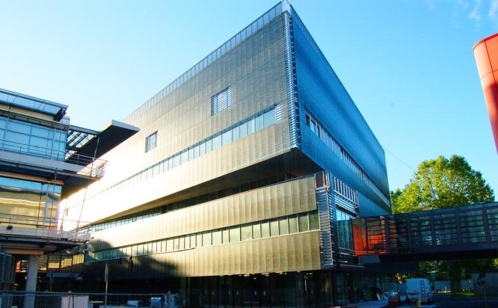
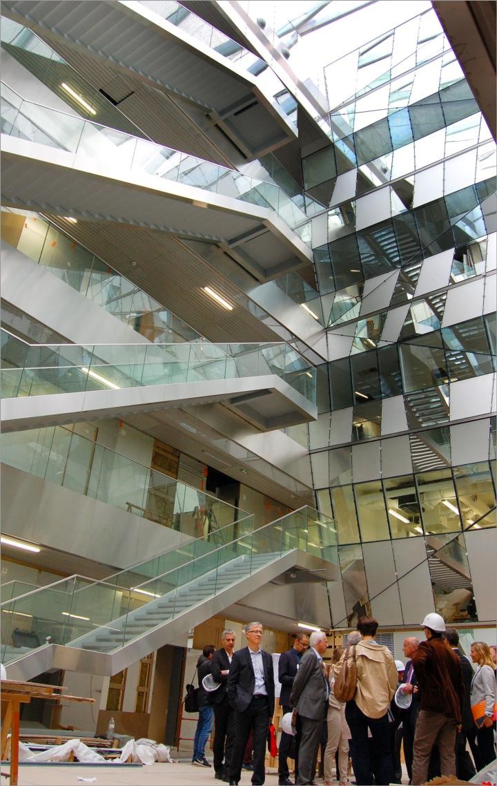
The BIC is setting up in a brand new space
In the last weeks of October 2016, the BIC has settled in a brand new building, constructed by the Regional Council of Aquitaine as part of the Neurocampus project. This building, of around 13 000 m2, is shared with the Interdisciplinary Institute for Neuroscience (IINS) and the Institute for Neurodegenerative Disorders (IMN). This building, constructed in two years, cost 47 M€ and is part of a large project to develop Neuroscience and imaging in Aquitaine. The new building is conveniently located and connected by footbridges between the Magendie Neuroscience center and the Center for functional genomics (CGFB) that hosts several core facilities.
In total, the BIC will occupy 1000 m2, split between the CGFB and the new building. The major part in the new building is dedicated to photonic microscopy. Electron microscopy instruments, including two brand new ones coming in 2017, will be dispatched between the CGFB and Neurocampus building. In these new spaces, users have access to a culture room and also a room with analysis stations. Other rooms are dedicated to each kind of microscopy (one room for live cells imaging, one room for multiphoton, one room for confocal, one room for new scanning electron microscope etc…). Special rooms are dedicated to host R&D projects as well as confidential collaborations with industry.
Development of training capacities at the BIC – joint projects with the Cajal School of Neuroscience
The BIC has engaged for many years in active training programs for imaging at all levels (beginners to advanced training) for local, national and transnational users. The BIC personnel also participates extensively to various theoretical and hands on training/showcase activities in France and abroad (MifoBio, NeuBias, etc…). Within the strategy to develop the BIC-FBI training, we are engaging a partnership with the Cajal Advanced Neuroscience Training Program to develop special ima ging training for Neuroscience. The Cajal school is a European FENS and IBRO initiative in partnership with Bordeaux Neurocampus and the Champalimaud Foundation, which offers state-of-the-art hands-on training courses in neuroscience.
Construction of a light sheet microscope for super resolution imaging inside living samples
Fast and non-damaging imaging of single molecules inside live organisms is essential to study physiologically relevant biochemical mechanisms occurring at the subcellular level. For example, the dynamic organization of transmitter receptors at the membrane of excitatory neurons should, ideally, be studied in vivo in the brain of animal models. Unfortunately super resolution techniques such as PALM1, STORM23 and uPAINT4 are mostly restricted to the sample external surfaces and are unable to image inside live samples.
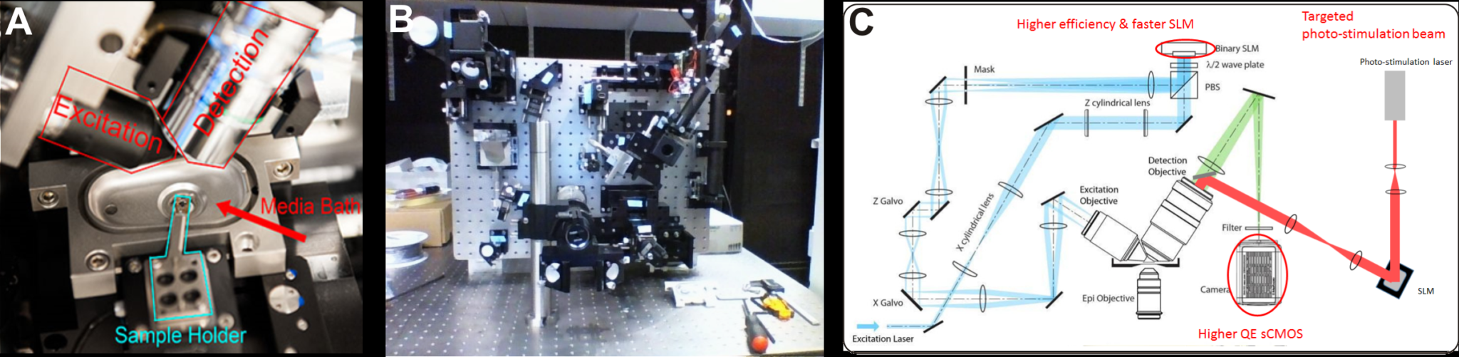
For these reasons the Bordeaux Imaging Center is developing a new light sheet microscope specially dedicated to image single molecules into live samples. Light sheet fluorescence microscopy (LSFM) is recognized as the method of choice to image thick live samples. Compared to other fluorescence imaging modalities such as wide field, confocal, structured illumination, two-photon or STED, LSFM strongly reduces out of focus fluorescence, decreases photobleaching and phototoxicity, and improves temporal resolution. Among the numerous technical implementations of LSFM 5, we decided to build a lattice light sheet microscope (LLS) because it has been specifically designed to perform super resolution imaging in thick live samples 6. Indeed In LLS the illumination beam is shaped by a spatial light modulator (SLM) to produce a < 1 µm thick excitation plane over a length of > 50 µm at the sample. A 1.1 NA detection objective ensures efficient light collection required for high localization precision. Illumination and detection objectives are both long working distance and water immersion, thus allowing observation of live samples up to 5 mm in diameter. (Fig 1 A)
Our LLS microscope is mostly based on the documentation freely and kindly shared by Eric Betzig’ group (HHMI Janelia Farms, USA).
Photo Credits: Mathieu Ducros
Fig 1. (A) The sample is placed at the intersection of the excitation and detection objective optic axes in a temperature controlled perfusion chamber. It is held at the tip of motorized arm on a 5 mm diameter cover slip (from 6). (B) The LLS microscope under construction in June 2016. (C) In blue and green the optical path of the excitation and detection beams respectively (from 6). A higher efficiency SLM, higher QE camera should improve the light budget compared to the original specifications. In addition, a targeted laser beam (red) will allow precise photo-conversion of light sensitive molecules.
We made a few modifications compared to the original specifications of the LLS as described in 6 : our microscope will be equipped with a laser combiner including 4 high power lasers at 405 nm (300mW), 488 nm (1 W), 560 nm (2 W), 642 nm (2W), a higher efficiency SLM (Fourth Dimension DD QXGA) and a sCMOS camera with improved quantum efficiency (Hamamatsu ORCA Flash V2). These improvements should mitigate the weak throughput of the LLS beam path, and, in turn, improve molecule localization precision and/or time resolution. In addition, a targeted photostimulation beam will be coupled through the detection objective to photo stimulate or photoconvert with a high spatial and temporal resolution photosensitive molecules.
STORM, PALM and PAINT imaging modalities will be fully compatible with the constructed LLS.
The microscope construction by Mathieu Ducros, INSERM research Engineer on the BIC, started in April (Fig 1B). First images are expected by the end of 2016. Once our LLS is fully operational and running, it will be accessible to all BIC users under the supervision of a local engineer.
For this project we are supported financially by the GIS IBiSA, LABEX brain and FBI.
References
Betzig, E. et al. Imaging intracellular fluorescent proteins at nanometer resolution. Science 313, 1642–1645 (2006).
Rust, M. J., Bates, M. & Zhuang, X. Sub-diffraction-limit imaging by stochastic optical reconstruction microscopy (STORM). Nat. Methods 3, 793–795 (2006).
van de Linde, S. et al. Direct stochastic optical reconstruction microscopy with standard fluorescent probes. Nat. Protoc. 6, 991–1009 (2011).
Giannone, G. et al. Dynamic superresolution imaging of endogenous proteins on living cells at ultra-high density. Biophys. J. 99, 1303–1310 (2010).
Santi, P. a. Light sheet fluorescence microscopy: a review. J. Histochem. Cytochem. 59, 129–138 (2011).
Chen, B.-C. et al. Lattice light-sheet microscopy: Imaging molecules to embryos at high spatiotemporal resolution. Science (80-. ). (2014). doi:10.1126/science.1257998
We use cookies on our website to give you the most relevant experience by remembering your preferences and repeat visits. By clicking “Accept All”, you consent to the use of ALL the cookies. However, you may visit "Cookie Settings" to provide a controlled consent.
This website uses cookies to improve your experience while you navigate through the website. Out of these, the cookies that are categorized as necessary are stored on your browser as they are essential for the working of basic functionalities of the website. We also use third-party cookies that help us analyze and understand how you use this website. These cookies will be stored in your browser only with your consent. You also have the option to opt-out of these cookies. But opting out of some of these cookies may affect your browsing experience.
Necessary cookies are absolutely essential for the website to function properly. These cookies ensure basic functionalities and security features of the website, anonymously.
Cookie
Duration
Description
cookielawinfo-checkbox-analytics
11 months
This cookie is set by GDPR Cookie Consent plugin. The cookie is used to store the user consent for the cookies in the category "Analytics".
cookielawinfo-checkbox-functional
11 months
The cookie is set by GDPR cookie consent to record the user consent for the cookies in the category "Functional".
cookielawinfo-checkbox-necessary
11 months
This cookie is set by GDPR Cookie Consent plugin. The cookies is used to store the user consent for the cookies in the category "Necessary".
cookielawinfo-checkbox-others
11 months
This cookie is set by GDPR Cookie Consent plugin. The cookie is used to store the user consent for the cookies in the category "Other.
cookielawinfo-checkbox-performance
11 months
This cookie is set by GDPR Cookie Consent plugin. The cookie is used to store the user consent for the cookies in the category "Performance".
viewed_cookie_policy
11 months
The cookie is set by the GDPR Cookie Consent plugin and is used to store whether or not user has consented to the use of cookies. It does not store any personal data.
Functional cookies help to perform certain functionalities like sharing the content of the website on social media platforms, collect feedbacks, and other third-party features.
Performance cookies are used to understand and analyze the key performance indexes of the website which helps in delivering a better user experience for the visitors.
Analytical cookies are used to understand how visitors interact with the website. These cookies help provide information on metrics the number of visitors, bounce rate, traffic source, etc.
Advertisement cookies are used to provide visitors with relevant ads and marketing campaigns. These cookies track visitors across websites and collect information to provide customized ads.