An image from the Bordeaux Imaging Center published in Science et vie
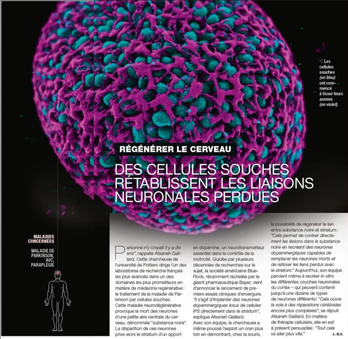
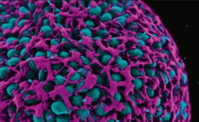
The image depicts a spheroid of human stem cells (green) and its actin cytoskeleton (purple), produced by Philippe Cohen during its PhD at Treefrog. This nice picture serves as an illustration for an article covering the use of stem cells for regenerative medicine.
Acquisition was made by Philippe Cohen on a scanning confocal microscope and 3D rendering was done by Jérémie Teillon using Agave software.
Agave is a free 3D visualization software, using light path-trace light rendering.
The Bordeaux Imaging Center team offers training and support on 3D commercial softwares such as Imaris and Arivis as well as other freeware such as Agave. Don’t hesitate to contact them (bic@u-bordeaux.fr) if you are interested in 3D rendering and visualization of your microscopy data!
Agave software:
https://www.allencell.org/software-and-code.html
https://www.allencell.org/pathtrace-rendering.html
Article (in French):
https://www.science-et-vie.com/corps-et-sante/regenerer-le-cerveau-des-cellules-souches-retablissent-les-liaisons-neuronales-p-58266#dossier-58457