A powerful high speed, low phototoxicity microscopy method to achieve super-resolved images
An innovative technology to look at thick samples at high resolution? Marc Tramier, a group leader at the Institute of Genetics & Development of the University of Rennes/INSERM/CNRS, and scientific director of MRic (Microscopy Rennes Imaging Centre), is currently working with his team on Random Illumination Microscopy (RIM), a fast and easy to use microscopy technique with low phototoxicity. His facility, which is part of the Bretagne-Loire Node of France-BioImaging, offers RIM as a Euro-BioImaging Proof-of-Concept study, and is now accepting applications for projects. He explains the ideas behind RIM in the article below.
The idea of Random Illumination Microscopy is to use the speckle of the illumination laser in wide field to create a structured illumination pattern at the diffraction limit. By varying the pattern from image to image using a diffracting element (in our case a SLM), scientists are able to acquire a stack of images (around 100 images) on a camera which corresponds to a cumulative homogeneous illumination. By resolving the inverse problem, a super-resolved image is, then, reconstructed, at the focal plane with unprecedented optical sectioning. In comparison to conventional SIM, RIM is able to work in depth inside diffusive samples as the speckle is insensitive to diffusion.
A transfer full of advantages
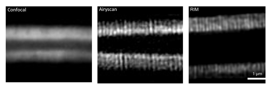
The method was first implemented by Thomas Mangeat – that we are happy to welcome in our new Toulouse node! – and collaborators in Toulouse (Mangeat et al., 2021. doi: 10.1016/j.crmeth.2021.100009). In the MRic, after the transfer of the prototype, the facility was able to image microvilli of intestine in c-elegans (depth > 50µm) having a spatial resolution of around 100 nm. This structure is impossible to be revealed by conventional confocal microscopy. Before the use of RIM, only the airyscan approach allowed us to resolve the microvilli but with higher illumination power (photobleaching of the sample) and longer acquisition time (around 10 times more). Now with RIM, we are able to follow microvilli in the living C–elegans at the second time-scale during several minutes.
RIM is one of the powerful methods to achieve super-resolved images in depth at high speed with very low phototoxicity. This makes a very nice compromise of z-sectioning and super-resolution with wide field illumination particularly adapted to thick live samples. Beside reconstruction and data analysis, MRic is offering a user-friendly system, with a complete set of microscopy methods for live sample investigation, from wide-field to light sheet including spinning disk, confocal and airyscan. And of course, this technology is available in open access through France-BioImaging and Euro-BioImaging!

How to apply to use RIM:
Random Illumination Microscopy is part of the Euro-BioImaging Proof-of-Concept study, in collaboration with our Nodes. The Proof-of-Concept study makes it possible to introduce exciting, new imaging technologies to our portfolio that were previously unavailable via our network. We are currently accepting applications to use these technologies as part of the Proof-of-Concept study. Be part of this study – and contribute to community-wide continuous technological innovation!
All scientists, regardless of their affiliation, area of expertise or field of activity can benefit from Euro-BioImaging’s pan-European open access services. Potential users of these new technologies are encouraged to submit project proposals via our website. To do so, you can Login to access our application platform, choose the technology you want to use and the facility you wish to visit, then submit your proposal. All applications will be processed by the Euro-BioImaging Hub. As usual, users will benefit from advice and guidance by technical experts working at the Nodes, training opportunities, and data management services.
For more information: info@eurobioimaging.eu
Thank you Marc Tramier and Marianna Childress, communication officer of Euro-BioImaging, for the original article.