A yeast responsible for horse sweat in wine: the Brettanomyces bruxellensis
Brettanomyces bruxellensis is one of the most damaging spoilage yeasts in the wine industry because of its impact on the beverage’s flavor. Lysiane Brocard, research engineer specialised in plant biology at the Bordeaux Imaging Center (FBI Bordeaux node), recently co-published an article on this yeast cell surface and bioadhesion properties.
Fruits are transformed into beverages through fermentation processes carried out by microorganisms naturally present in the environment. In wine, yeasts and bacteria play this role and contribute to the development of volatile compounds. Scientists targeted Brettanomyces bruxellensis in this study, a yeast famous for the production of volatile phenols, characterized by horse sweat odors which is – usually – not very enjoyable for the consumer.
A yeast characterized by bioadhesion abilities
This specific odor comes from volatile compounds, the 4-ethylgaïacol (4EG) and 4-ethylphenol (4 EP), that winemakers try to avoid. Beside adding an unpleasable flavor to the beverage, the issue is that the spoilage yeast is persistent in cellars over several years, resulting in recurrent wine contamination. This suggests a bioadhesion process that helps the microorganism to survive in its environment. To put it simply, bioadhesion is the ability of an organism to adhere on a surface to, then, participate in the formation of a biofilm (which is defined as “a structured community of microorganisms adhered to a surface and producing an extracellular matrix”).
Here, 54 strains of B. bruxellensis were characterized for their cell surface physico-chemical and bioadhesion properties. And all of them have shown bioadhesion abilities (after only three hours on stainless steel) both on synthetic medium and wine. Enough to highlight the persistence of our favorite horse sweat flavored yeast.
How did bioimaging help in this project?
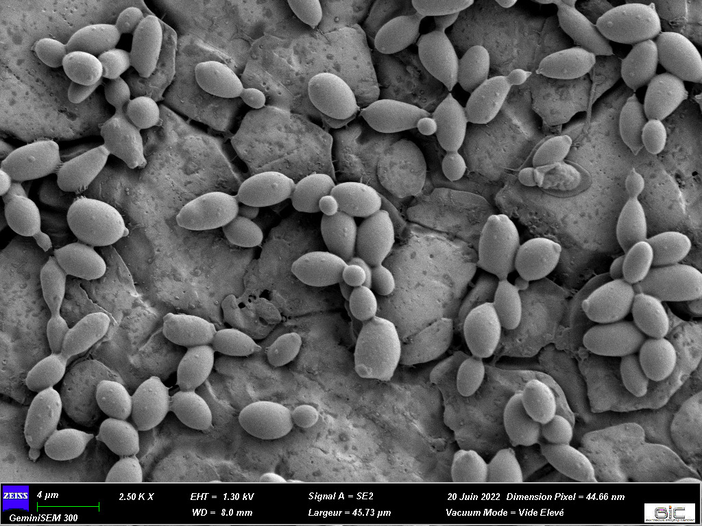
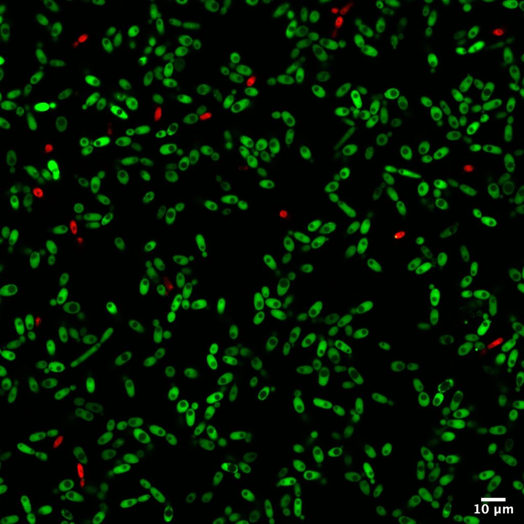
Among all the analytical methods used in this study, microscopy helped identify the structure of the biofilms formed with B. bruxellensis. Two imaging techniques were used: confocal microscopy and scanning electron microscopy. The first one offers the advantage of realizing live imaging without being too time-consuming. With fluorescent dyes, the status of cells can be easily determined at the same time as the cell repartitions and concentrations.
Moreover, comparisons have been made thanks to confocal microscopy to determine if some strains of B. bruxellensis could form biofilm with only one cell layer or if they proliferate in three dimensions. To complete these observations, scanning electron microscopy was performed at the Bordeaux Imaging Center (FBI Bordeaux node) with the help of Isabelle Svahn, expert of this type of microscopy. It was a great addition to this study as these observations validated the morphological variability among Brettanomyces strains.
Bioimaging helped a broader project about Brettanomyces led by Isabelle Masneuf-Pomarède who works in the Institut des Sciences de la Vigne et du Vin of Bordeaux. Isabelle studies the persistence and proliferation of Brettanomyces over the years. Isabelle’s PhD student, Paul Le Montagner, carried out most of the experiments published in this paper. Thanks to them for this amazing paper!

Original article: Paul Le Montagner, Morgan Guilbaud, Cécile Miot-Sertier, Lysiane Brocard, Warren Albertin, Patricia Ballestra, Marguerite Dols-Lafargue, Vincent Renouf, Virginie Moine, Marie-Noëlle Bellon-Fontaine, Isabelle Masneuf-Pomarède, High intraspecific variation of the cell surface physico-chemical and bioadhesion properties in Brettanomyces bruxellensis, Food Microbiology, Volume 112, 2023, 104217, ISSN 0740-0020, https://doi.org/10.1016/j.fm.2023.104217
You are interested in our bioimaging services, including technologies and expertise?
Please contact us at: contact@france-bioimaging.org