Dear colleague,
Dear FBI community,
Following the decision of the Executive Board of June 30, 2016, the National Coordination is proud to announce that the winners of the FBI Image Contest 2016 are:
1. Sébastien Mailfert – Centre d’immunologie de Marseille Luminy – Aix Marseille Université with “Dalton”
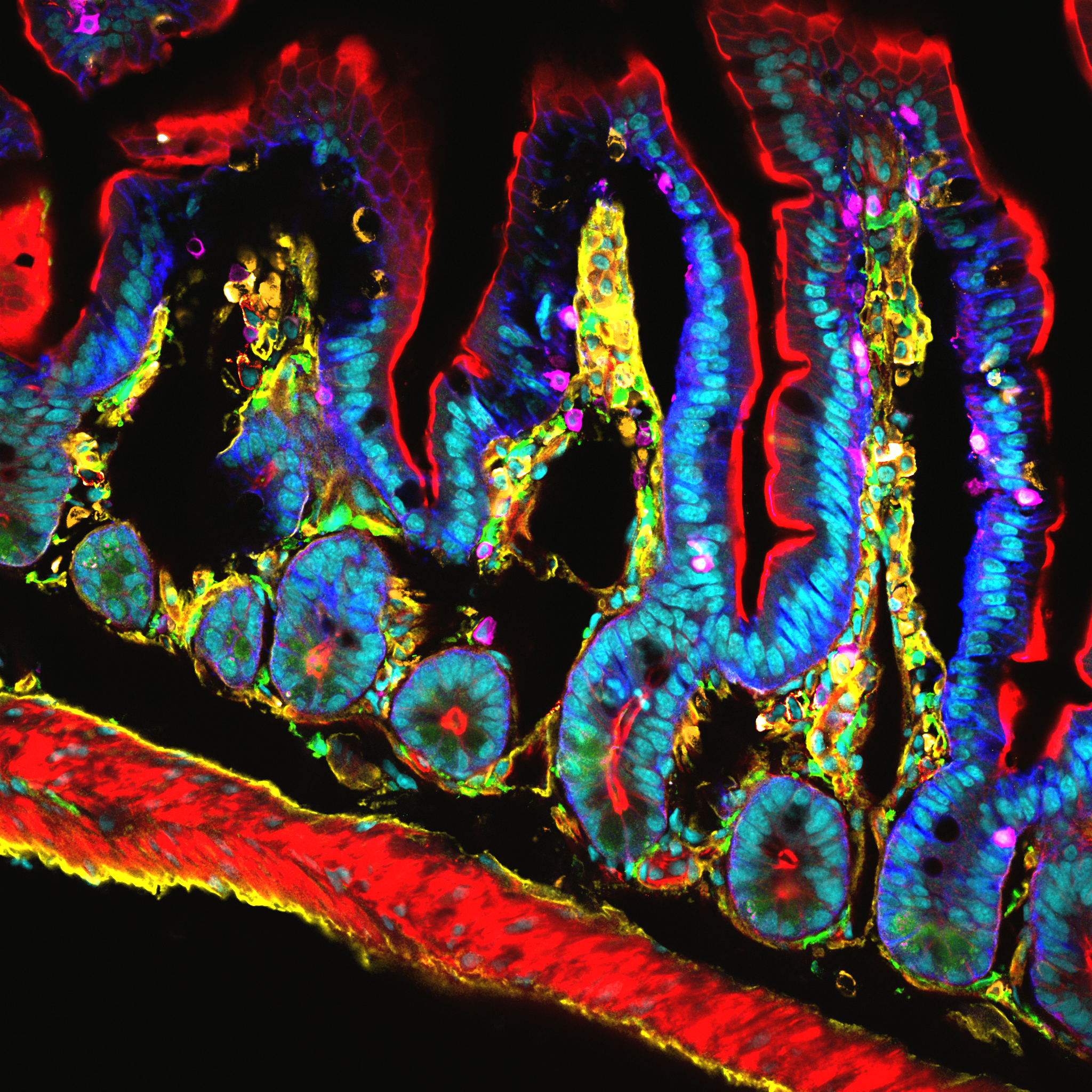
Dalton © Hugues Lelouard, Mailfert Sébastien & Mathieu Fallet CIML CNRS-INSERM-AMU
Confocal microscopy
Revealing sub-population of immune cells on small intestine by 10 colors spectral imaging
Confocal microscopy
Revealing sub-population of immune cells on small intestine by 10 colors spectral imaging
AND with “Le Saint Pierre Méditerranéen”
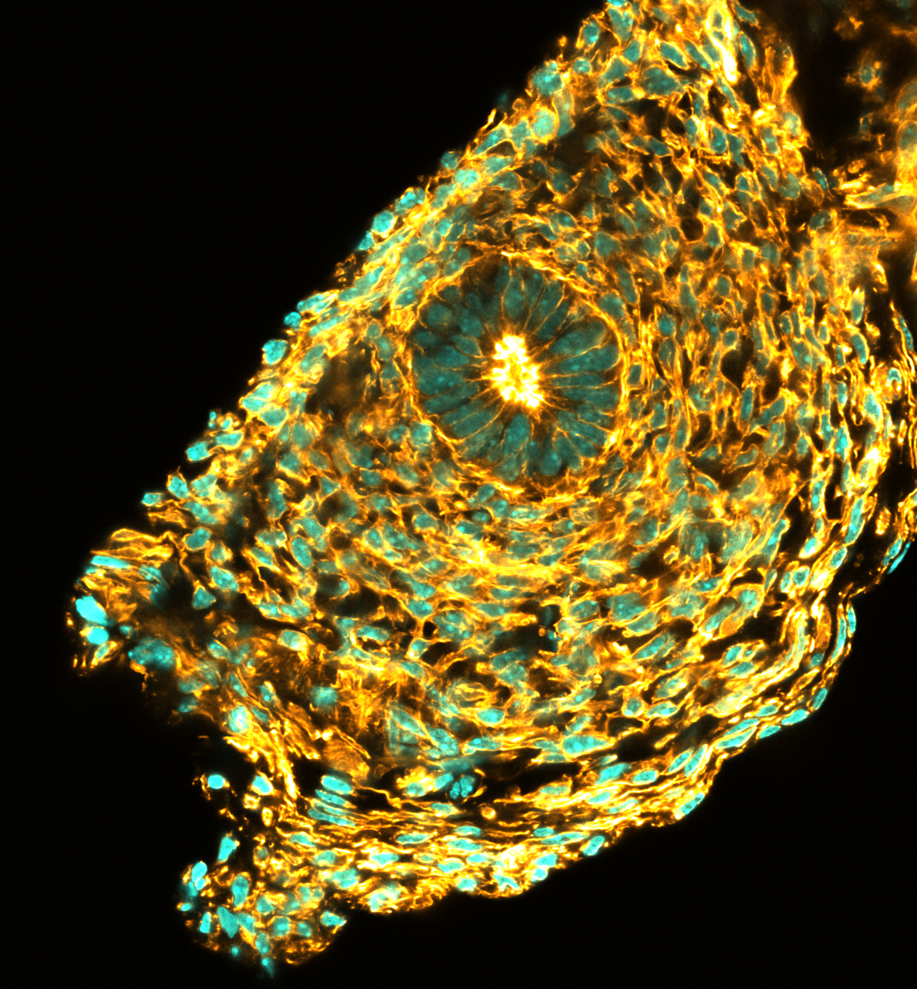
Le St Pierre Mediterraneen © Noushin Mossadegh & Mailfert Sébastien – CIML, CNRS-INSERM-AMU
A l’aise comme un poisson dans l’eau, les spermatozoïdes se reposent dans leur habitat, qui ressemble à un œil, avant leur grande migration. Coupe d’un testicule de nouveau-né de souris. Marquage en immunofluorescence des noyaux cellulaires (DAPI) représentés ici en cyan. L’actine représentée ici en jaune, révèle le «squelette de la cellule» (phalloïdine marquée avec le fluorochrome Alexa-647). L’image représente 256×256µm sur 4096×4096 pixels. La coupe est d’une épaisseur de 20µm. Image de microscopie confocale sur Leica SP5 ; laser 405nm et laser blanc à 633nm ; objectif 40X, O.N. 1.25, immersion à huile.
A l’aise comme un poisson dans l’eau, les spermatozoïdes se reposent dans leur habitat, qui ressemble à un œil, avant leur grande migration. Coupe d’un testicule de nouveau-né de souris. Marquage en immunofluorescence des noyaux cellulaires (DAPI) représentés ici en cyan. L’actine représentée ici en jaune, révèle le «squelette de la cellule» (phalloïdine marquée avec le fluorochrome Alexa-647). L’image représente 256×256µm sur 4096×4096 pixels. La coupe est d’une épaisseur de 20µm. Image de microscopie confocale sur Leica SP5 ; laser 405nm et laser blanc à 633nm ; objectif 40X, O.N. 1.25, immersion à huile.
2. Michael Lang – Institut Jacques Monod – ImagoSeine with “Fly Monster”
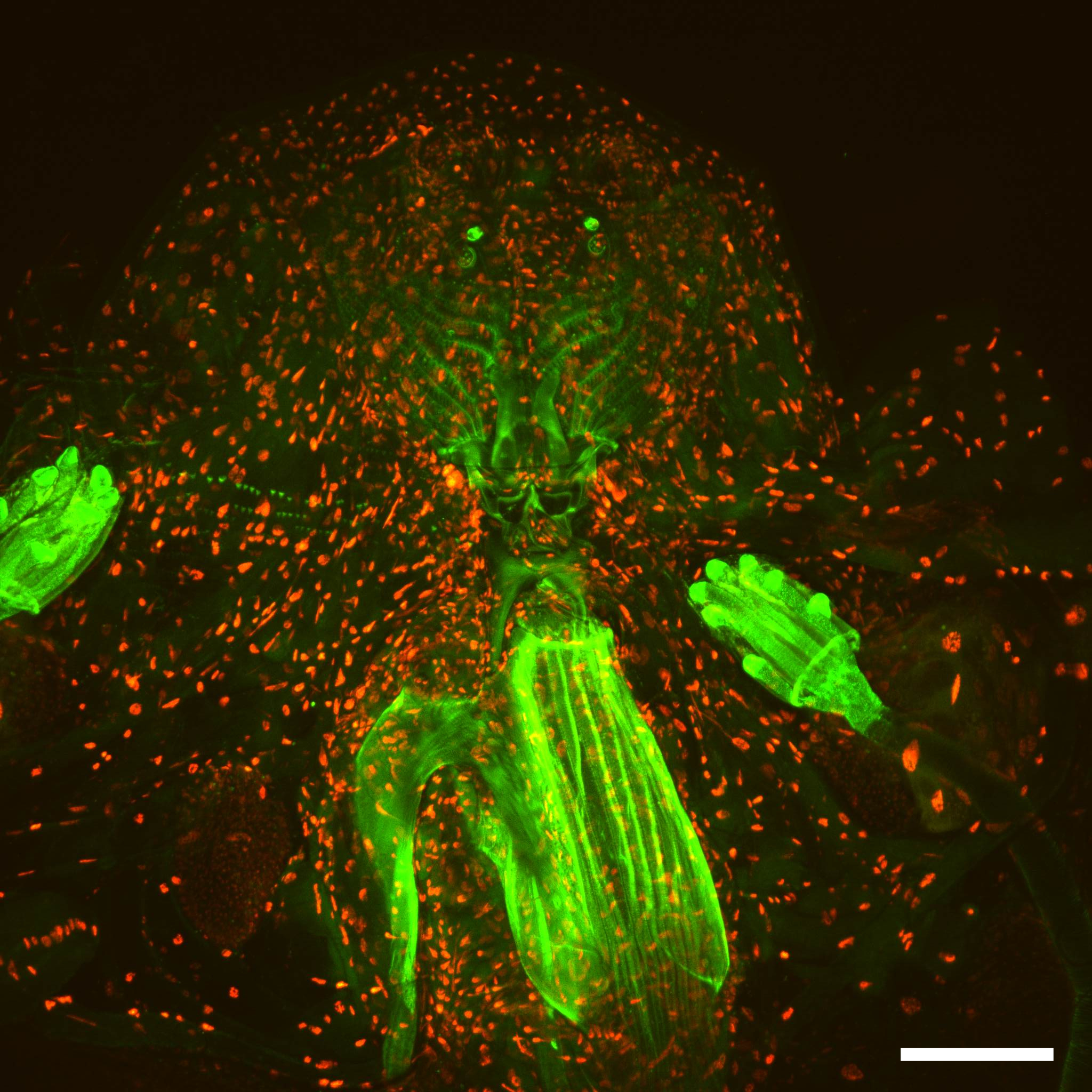
Fly monster © Orestis Faklaris & Michael Lang – Institut Jacques Monod, CNRS UMR7592 – ImagoSeine
Multifocal confocal microscopy (spinning disk, Spinning CSU W1)
Drosophila third instar larval head, nuclear-RFP, neronal-GFP and green autofluorescence, 25x magnification, scale bar is 100 μm.
Multifocal confocal microscopy (spinning disk, Spinning CSU W1)
Drosophila third instar larval head, nuclear-RFP, neronal-GFP and green autofluorescence, 25x magnification, scale bar is 100 μm.
Thank you to the participants for their great contribution:
- Sébastien Mailfert – Centre d’immunologie de Marseille Luminy – Aix Marseille Université
- Ariane Peyret – Laboratoire de Chimie des Polymères Organiques – Bordeaux Imaging Center
- Melina Petrel – Bordeaux Imaging Center – Université Bordeaux Segalen
- Patrice Mascalchi – Interdisciplinary Institut of Neuroscience – University of Bordeaux – Bordeaux Imaging Center
- Michael Lang – Institut Jacques Monod – ImagoSeine
- Olga Nagy – Institut Jacques Monod, Drosophila Evolution Group – ImagoSeine
- Théophile Déjardin – Institut Jacques Monod – ImagoSeine
- Liu Zeng Zhen – Institut Jacques Monod – ImagoSeine
- Melina Heuze – Institut Jacques Monod – ImagoSeine
- Orestis Faklaris – Institut Jacques Monod – ImagoSeine
- David Pereira – Laboratoire Matière et Systèmes Complexes – ImagoSeine
Thank you also to the core facilities staff and heads for having forwarded the contest to their users and for providing them state of the art Bioimaging.
The next edition of our Image Contest will open early 2017. Get ready!
The National Coordination