Les inscriptions pour la seconde édition du TED ImaBio lundi 2 mai à Montpellier sont ouvertes.

Le GDR ImaBio vous propose de venir decouvrir les dernières innovations des partenaires industriels du GDR , et pour les etudiants un focus sur les métiers du domaine.

Programme

The 7th edition of OPTOGEN will bring developers and end-users in the field of Optogenetics and Neurophotonics together. It will be a great occasion to meet in person for stimulating discussions and to reignite collaborations.

Registration and abstract submission are open.

Early registration will close on April 15th.

Thanks to the support of the IHU Foresight and of the sponsors, inscription is free for students registered before April 15th and some travel grants will be available.

Euro-BioImaging is organizing a third online User Forum on Tuesday, April 5, 2022 from 14:00-17:00 CEST. The topic is “At the Forefront of Neuroscience.”

This event will highlight the importance of cutting-edge imaging technologies in support of brain research and showcase the specific expertise available at our Nodes across Europe through case studies presented in tandem with the research community. 

In addition, keynote presentations from Hervé Boutin, University of Manchester, and Selma Ding, Max Planck Institute for Animal Behavior, will further reveal the potential of biological and biomedical imaging technologies to boost brain research. 


Register here: https://us02web.zoom.us/meeting/register/tZ0ucuytpj8iE92A-JjvPbvpNnmTF4Z7iRaC

More information: https://www.eurobioimaging.eu/news/join-us-for-the-third-euro-bioimaging-user-forum-at-the-forefront-of-neuroscience/

Programme

Deadline: April 15th, 2022

The three national infrastructures ProFi, France-BioImaging and FRISBI along with the GIS IBiSA are pleased to announce a call for a funded access to IBiSA-labelled facilities.

Our aim is to promote IBiSA facilities networking through interdisciplinary research projects.

Applications should request access to at least two different IBiSA facilities from two disciplines (structural biology, Biological imaging and proteomics). The call is open to any academic laboratory.

The amount of the financial support will be up to 5000 € per application to cover facility costs.

Applications should be submitted to: Call-IBISA-FBI-FRISBI-PROFI@i2bc.paris-saclay.fr
using the template document https://mycore.core-cloud.net/index.php/s/VNLU2jgfcP4o0NN


The deadline for this inter-infrastructure access call is 15 April 2022.

All submitted proposals will be peer-reviewed by independent experts and the final funding will be approved by a committee comprising 2 representatives from each infrastructure as well as representatives
from GIS IBISA. We advise the project PI to contact the chosen facilities in order to set-up the optimal experimental design.

Call description

The France-BioImaging LSFM workgroup is pleased to announce an INSERM workshop on Light-Sheet Fluorescence Microscopy imaging technics. 

This workshop will be divided in two parts:

  • A first theoretical part in Bordeaux from the 16th to the 18th of May 2022 which will cover basic principles, applications and challenges of LSFM imaging through seminars,
  • A second practical part along June 2022 where you will have the possibility to choose the set-up on which you want to be trained amongst many systems available within the France-BioImaging community.

If you want to discover, learn and/or deepen your knowledge about this vast and powerful family of 3D imaging technics, this training is for you!


You will find all the information (program, registration details, venue and accommodations, …) about this event on the following poster and website (Atelier 268): https://evenium-site.com/site/atelier-de-l-inserm-268

REGISTRATION DEADLINE: March 4, 2022

Programme

Poster

Understanding how development is coordinated in multiple tissues and gives rise to fully functional organs or whole organisms necessitates microscopy tools. Over the last decade numerous advances have been made in live-imaging, enabling high resolution imaging of whole organisms at cellular resolution. Yet, these advances mainly rely on mounting the specimen in agarose or aqueous solutions, precluding imaging of organisms whose oxygen uptake depends on ventilation.

Engineers from the Institut Curie and Institut Jacques Monod implemented a multi-view multi-scale microscopy strategy based on confocal spinning disk microscopy, called Multi-View confocal microScopy (MuViScopy).

MuViScopy enables live-imaging of multiple organs with cellular resolution using sample rotation and confocal imaging without the need of sample embedding. They illustrated the capacity of MuViScopy by live-imaging Drosophila melanogaster pupal development throughout metamorphosis, highlighting how internal organs are formed and multiple organ development is coordinated. They foresee that MuViScopy will open the path to better understand developmental processes at the whole organism scale in living systems that require gas exchange by ventilation.

3D fusion reconstruction of eight different angles at 10x magnification. Animation of a 3D reconstruction after fusion of images acquired from eight angles by the MuViScope of an Ecad:3xGFP Drosophila pupa at 28 hAPF with a 10x objective. The animation starts with a 180° rotation along the A-P axis and then sequentially shows the eight different angles from a top view: 0° (red), 45° (orange), 90° (yellow), 135° (green), 180° (light blue), 225° (dark blue), 270° (purple) and 315° (pink). Fusion was performed using Huygens Fuser (SVI) and 3D visualization with Imaris software. The acquisition parameters are detailed in Table S1. Scale bar: 200 μm.

Olivier Leroy, Eric van Leen, Philippe Girard, Aurélien Villedieu, Christian Hubert, Floris Bosveld, Yohanns Bellaïche, Olivier Renaud; Multi-view confocal microscopy enables multiple organ and whole organism live-imagingDevelopment 15 February 2022; 149 (4): dev199760. doi: https://doi-org.insb.bib.cnrs.fr/10.1242/dev.199760

The MuViScope was co-funded by France-BioImaging.

Le Réseau d’Imagerie Cellulaire Paris-Saclay organise en avril prochain une visioconférence de 2 heures portant sur le thème de l’imagerie des organoïdes.

Titre : Pipeline Organoïdes de A à Z : Fabrication, Transparisation, Acquisition et Analyse d’images.

Thématique : Le RIC Paris-Saclay vous propose une sorte de pipeline sur l’imagerie des modèles organoïdes qui vous permettra de mieux appréhender l’ensemble du processus de leurs fabrications, leurs modes de transparisation, l’analyse des images 3D associées à ce type d’échantillon épais, et pour finir, l’importance de l’étude de ces modèles chez l’homme pour développer de nouveaux outils thérapeutiques.

Date : 14 avril 2022
Horaire : 14h à 16h

Programme : Auto-organisation cellulaire en systèmes 3D par imageries optiques — Tissue clearing : Adapted protocols for small samples — Analyses d’images et reconstruction 3D — Applications en Recherche translationnelle (Organoïdes hépatiques humains).

Inscription gratuite et obligatoire sur www.ric-paris-saclay.fr

Programme

Affiche

The thematic institute of molecular and structural basis of life sciences of the French National Alliance for Life Sciences and Health (Aviesan) has identified cellular (cryo) electron microscopy as a priority area to be developed in the future. To better understand the functional behaviour of living organism, it becomes increasingly important to describe its structural organization from atomic scales to integrated, multi-component biological systems. Recent developments in imaging have drastically transformed our perception of the molecular organization and of the dynamic interactions of molecules in cellular systems.

That is why, they are organizing a 2-days meeting entitled “Symposium on recent developments in cellular electron microscopy”, and dedicated to the latest progresses in the field of cellular electron microscopy at the interface with fluorescent microscopy and structural biology. The meeting is built around 4 scientific sessions, namely volumetric imaging, cryo-electron tomography, correlative light and electron microscopy, and new technological developments. The meeting is for molecular and cellular biologists to have an overview of the latest results and concepts that develop at this interface.

Due to the sanitary situation, vaccination pass and face masks are mandatory to attend the meeting.

Download the provisional program

ORGANIZING COMMITTEE

Yves Gaudin, I2BC

Carine Giovannangeli, AVIESAN

Bruno Robert, CEA

Patrick Schultz, IGBMC

France-BioImaging is launching a call to have up to two new Nodes joining the infrastructure.

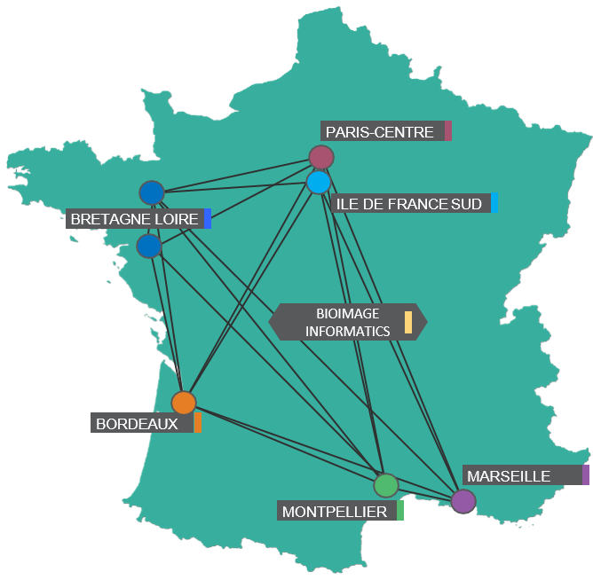
In 2022, France-BioImaging gathers 21 biological imaging facilities and 54 R&D teams specialized in biological imaging (see https://france-bioimaging.org/). It is structured in 6 local Nodes (Bordeaux, Bretagne-Loire, Ile-de-France-Sud, Marseille, Montpellier, Paris) and one transversal Node dedicated to bioimage informatics (IPDM). France-BioImaging is also the French node of Euro-BioImaging ERIC (see https://www.eurobioimaging.eu/).

The objectives of France-BioImaging are to promote the dissemination of the latest advances in all technologies and methods related to biological imaging, and their adoption by the users of the facilities that constitute the infrastructure.

In order to expand its technology and expertise portfolio and to provide the best possible geographical coverage to life scientists across France, France-BioImaging invites node candidates to submit their letter of intent here before May 31st, 2022.

Node candidates should:

  • Be made up of imaging facilities and R&D teams specialized in biological imaging, not necessarily grouped on the same campus but with a territorial logic.
  • Demonstrate readiness to provide open user access. The facilities must be open to the outside world beyond the regional level, i.e. to the national and international levels. Each Facility must have a valid IBiSA label, or must be in the process to have their integration in an already existing IBiSA facility validated in 2023, or must obtain this label in 2023. Each facility should have a structured organization and cost accounting. The facilities must have a reservation website for access to the equipment and a user training program. The facilities will be required to have or commit to a data management plan.
  • Gather R&D teams with a high degree of innovation and demonstrate a willingness to collaborate with the facilities. They must explain and demonstrate their technology and/or know-how transfer methods, particularly towards the local platforms, but not exclusively.
  • Bring real added value in relation to the current status of France-BioImaging, whether in terms of technologies, methodologies, scientific themes, novel user communities,… (for more information, visit the FBI website and consult the service offer). This is a key criteria.
  • Have an integrated and effective governance model.

More information on the selection criteria, expected commitments and documentation that needs to be provided can be found here.

Node selection will be done via a two-step process:

  • a letter of intent (deadline May 31st, 2022): the form to be completed is available here
  • a full application (deadline September 30th, 2022).

Applications will be reviewed at both steps by an International Board and decisions will be communicated at the end of 2022.

If at any time of the application process you have questions, please feel free to contact the France-BioImaging coordination Team by sending an email to contact@france-bioimaging.org.

We are looking forward to receiving your letter of intent!

Explore the beauty of the invisible world through the 2022 FBI digital calendar!

Enjoy the diversity of microscopy techniques, models and applications represented, one image at a time.

All 12 images used for this calendar were submitted to France-BioImaging image contest 2021. A big thank you again to all the participants!

You can download the A4 print version (one month per page) 2022 FBI digital calendar here:

If you wish to use it as your computer desktop, you can download a PNG version of each month here:

The France BioImaging Team wishes you a happy new year and all the best for 2022!

May 2022 bring new microscopy breakthroughs that will take biological research to new depths!

To start this new year, here is a beautiful & festive fluorescence microscopy image illustrating skeletal muscle fibers (Desmin, green) in co-culture with motor neurons (SMI-32) forming active Neuromuscular Junction in a cell culture method using murin primary cells in vitro. Nuclei are stained by DAPI (blue).

This image was submitted by A. Ghasemizadeh, from the Institut NeuroMyoGene, to France-BioImaging Image Contest 2021.