
Winning images – FBI Image Contest 2021
France BioImaging and all the French community aims to develop and promote innovative imaging technologies and methods. But microscopy images can also take an artistic, creative look and make the invisible world beautiful, allowing people to see the visual appeal of the life sciences.
We enjoyed the diversity of the images submitted with many different microscopy techniques, models and applications represented. A big thank you to all the participants!
The National Coordination Team and the Executive Board are proud to announce the winners of the FBI Image Contest 2021:
- 1st Place: Léna Meneux, Eye Team, Institut des Neurosciences de Montpellier
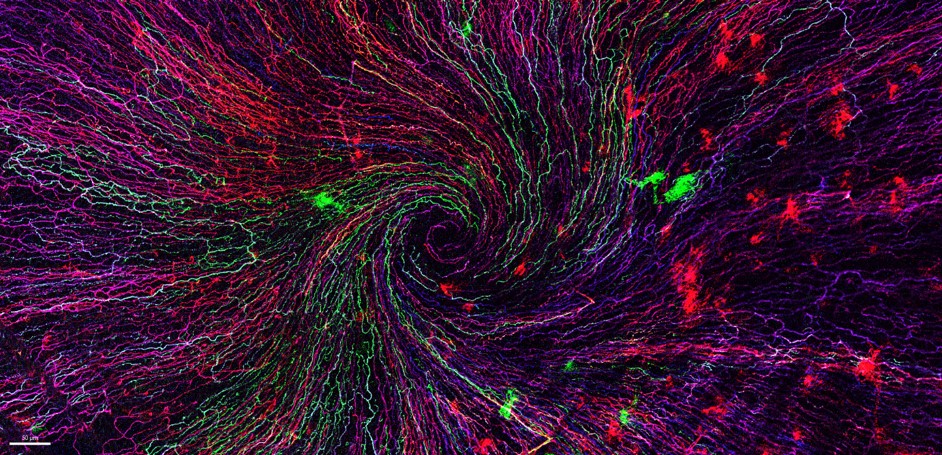
“The eye of the storm”
Sensory fibers of a mouse cornea imaged with a confocal microscope. The corneal nerves converge toward the centre forming a vortex. This particular transgenic mouse model allows stochastic expression of fluorescent proteins, unravelling the heterogeneity of the fiber origines inside the corneal epithelium.
Acknowledgements to Karine Loulier for the mouse model and Laetitia Hudececk for her help during the acquisition.
Confocal microscopy
- 2nd Place: Eunice HoYee Chan, Muscle Dynamics Team, Developmental Biology Institute of Marseille (IBDM)
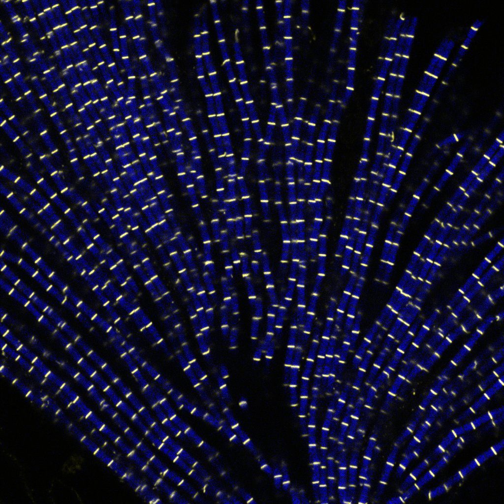
“Sarcomeric bouquet”
Myofibrils isolated from Drosophila indirect flight muscle labelled with titin (yellow) and actin (blue). Image captured from confocal microscope. We are studying the role of titin protein in muscle mechanics and organisation during development.
| Confocal LSM880 |
- 3rd Place: Camille Boutin, Biology of multiciliated cells Team, Developmental Biology Institute of Marseille (IBDM) & Nicolas Brouilly, PICsL Imaging facility, Electron Microscopy department
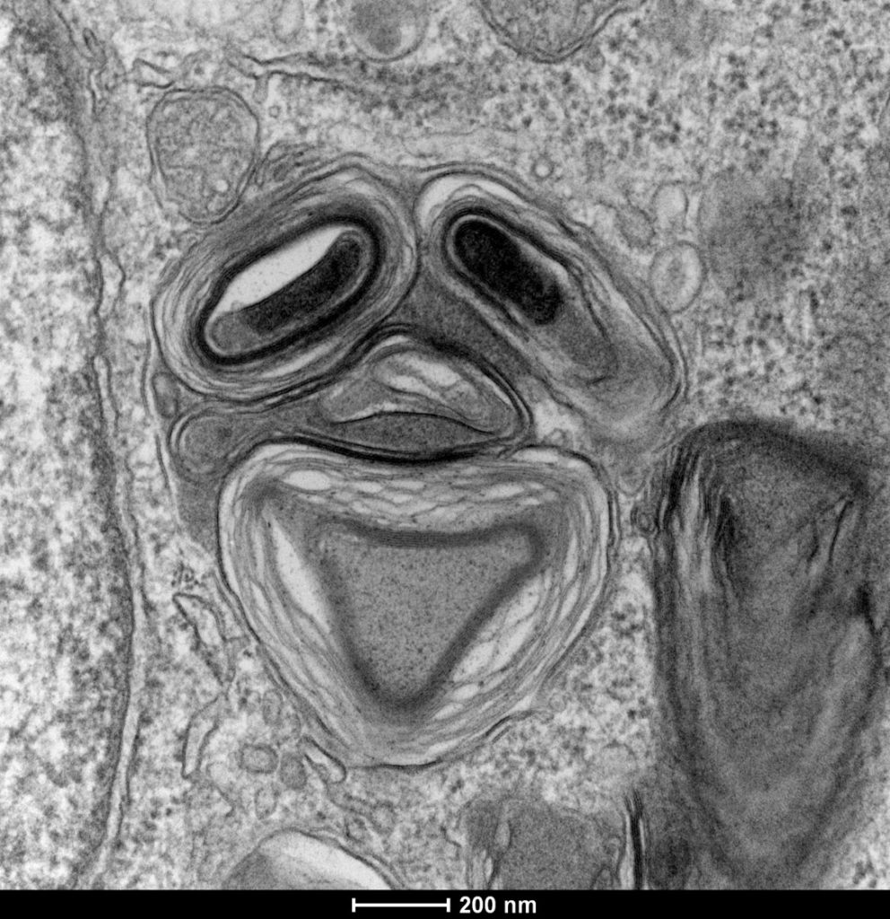
“Clown”
Lamellar structure in a differentiating multiciliated cell observed by transmission electron microscopy with a Tecnai G2 200kV FEI.
Transmission Electron Microscopy, Tecnai G2 200kV FEI
Congratulations to the winners!
Explore all the images submitted here:


As stated in the Terms & Conditions of the contest, foreign participants non-affiliated to a French institution are featured in the gallery, but were not evaluated as part of the contest.