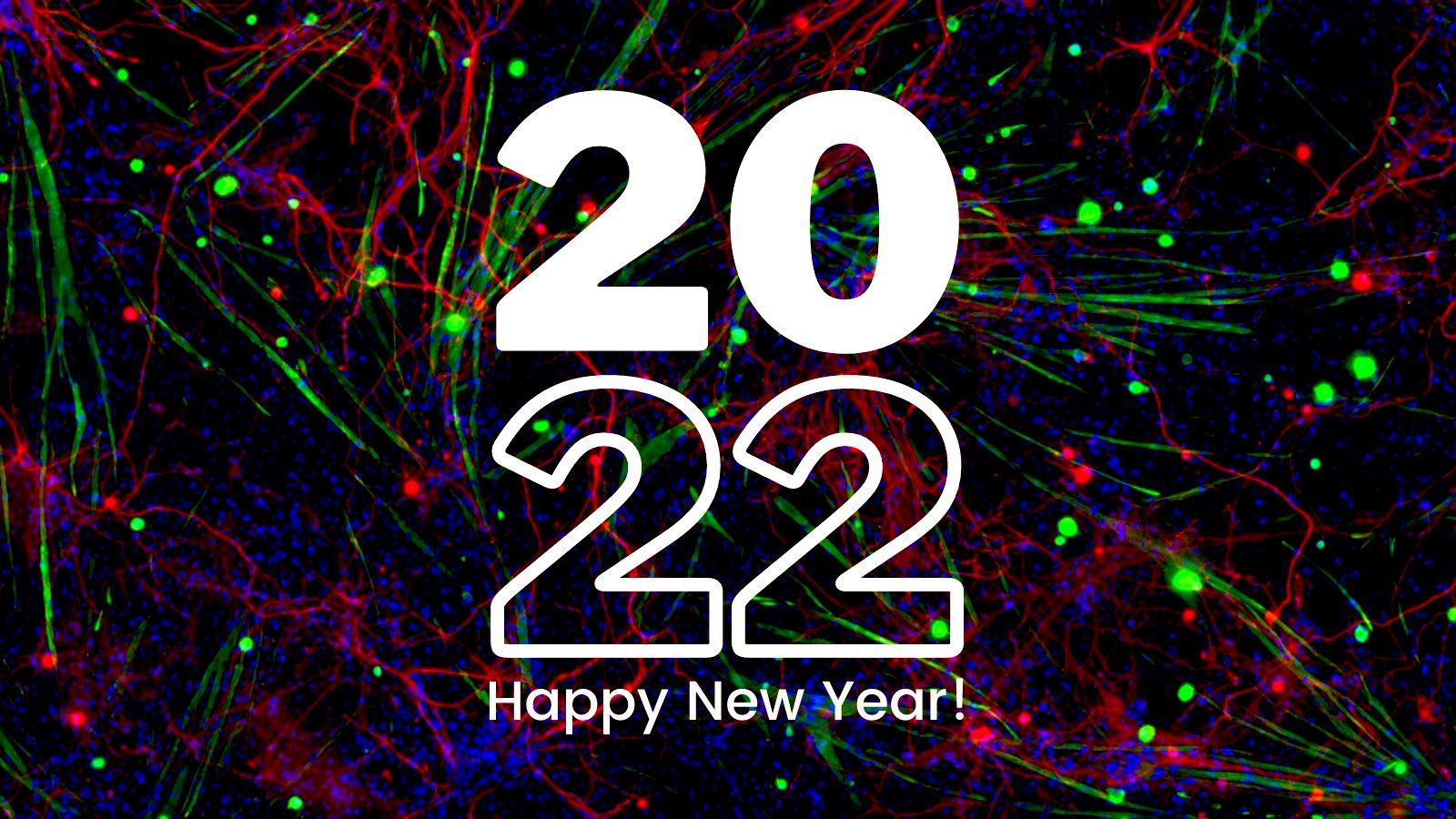
The France BioImaging Team wishes you a happy new year and all the best for 2022!
May 2022 bring new microscopy breakthroughs that will take biological research to new depths!
To start this new year, here is a beautiful & festive fluorescence microscopy image illustrating skeletal muscle fibers (Desmin, green) in co-culture with motor neurons (SMI-32) forming active Neuromuscular Junction in a cell culture method using murin primary cells in vitro. Nuclei are stained by DAPI (blue).
France BioImaging and all the French community aims to develop and promote innovative imaging technologies and methods. But microscopy images can also take an artistic, creative look and make the invisible world beautiful, allowing people to see the visual appeal of the life sciences.
We enjoyed the diversity of the images submitted with many different microscopy techniques, models and applications represented. A big thank you to all the participants!
The National Coordination Team and the Executive Board are proud to announce the winners of the FBI Image Contest 2021:
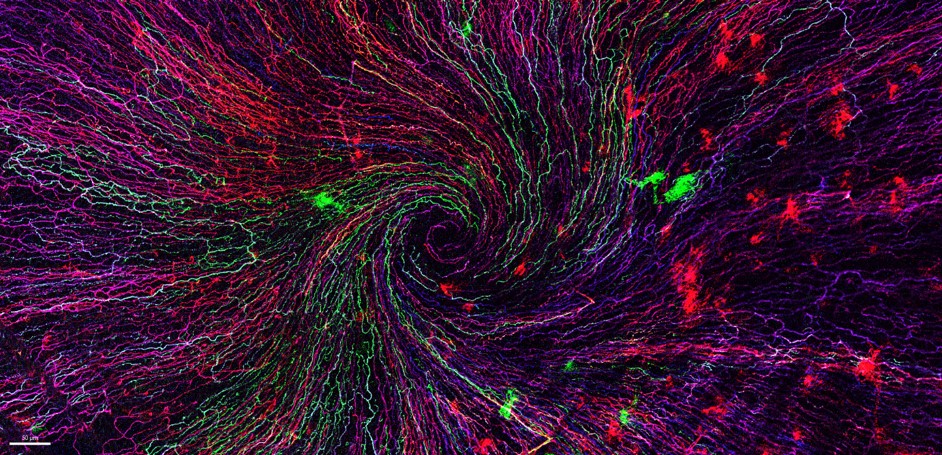
1st Place: Léna Meneux, Eye Team, Institut des Neurosciences de Montpellier
“The eye of the storm”
Sensory fibers of a mouse cornea imaged with a confocal microscope. The corneal nervesconverge toward the centre forming a vortex. This particular transgenic mouse model allows stochastic expression of fluorescent proteins, unravelling the heterogeneity of the fiber origines inside the corneal epithelium. Acknowledgements to Karine Loulier for the mouse model and Laetitia Hudececk for her help during the acquisition.
Confocal microscopy
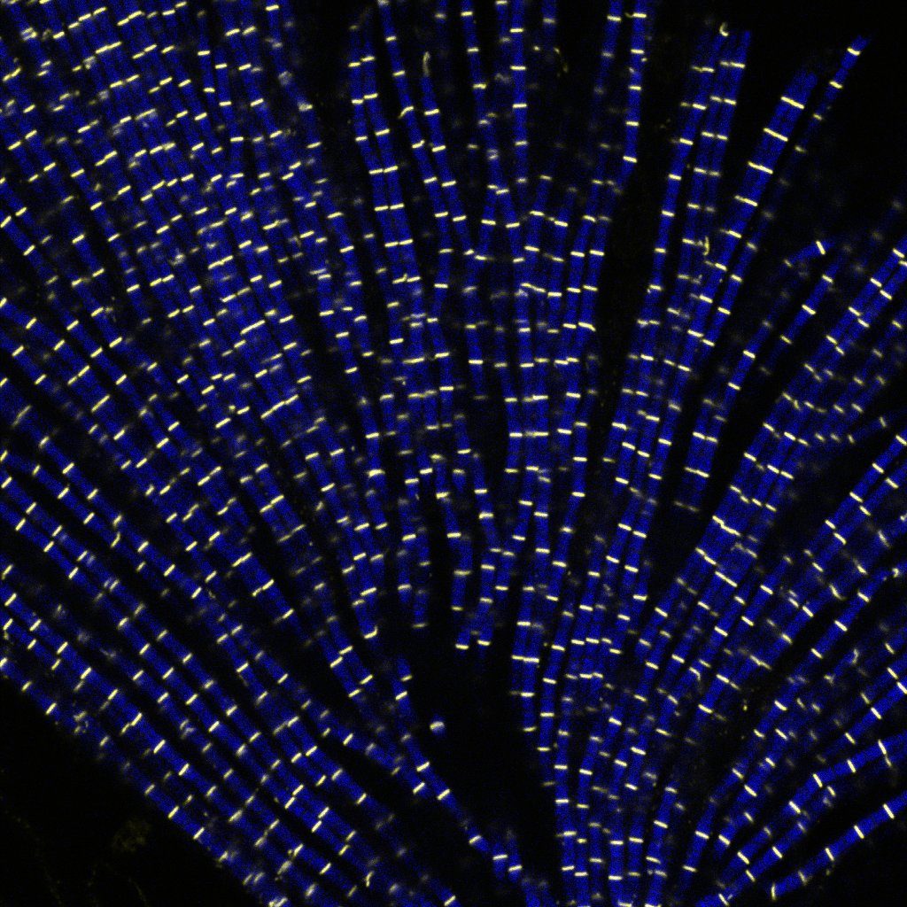
2nd Place:Eunice HoYee Chan, Muscle Dynamics Team, Developmental Biology Institute of Marseille (IBDM)
“Sarcomeric bouquet”
Myofibrils isolated from Drosophila indirect flight muscle labelled with titin (yellow) and actin (blue). Image captured from confocal microscope. We are studying the role of titin protein in muscle mechanics and organisation during development.
Confocal LSM880
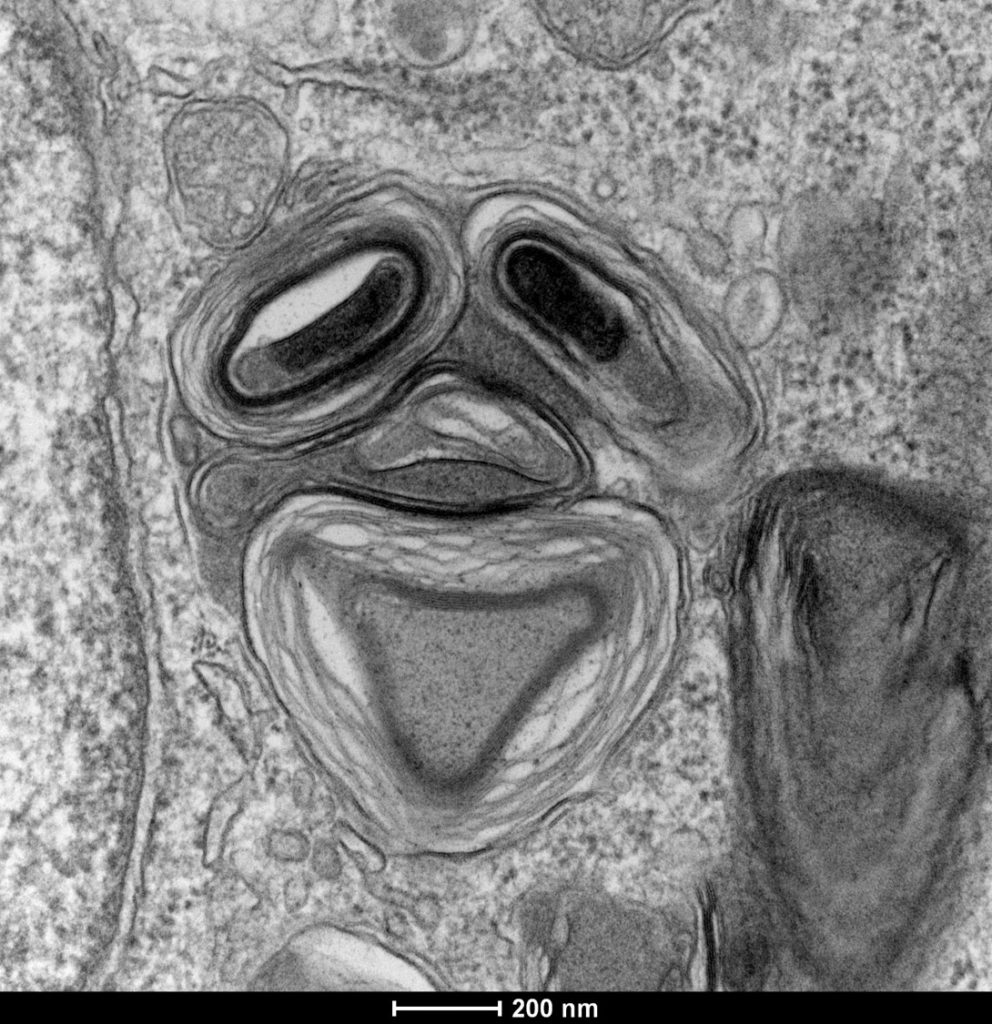
3rd Place:Camille Boutin, Biology of multiciliated cells Team, Developmental Biology Institute of Marseille (IBDM) &Nicolas Brouilly, PICsL Imaging facility, Electron Microscopy department
“Clown”
Lamellar structure in a differentiating multiciliated cell observed by transmission electron microscopy with a Tecnai G2 200kV FEI.
Transmission Electron Microscopy, Tecnai G2 200kV FEI
Congratulations to the winners!
Explore all the images submitted here:
As stated in the Terms & Conditions of the contest, foreign participants non-affiliated to a French institution are featured in the gallery, but were not evaluated as part of the contest.
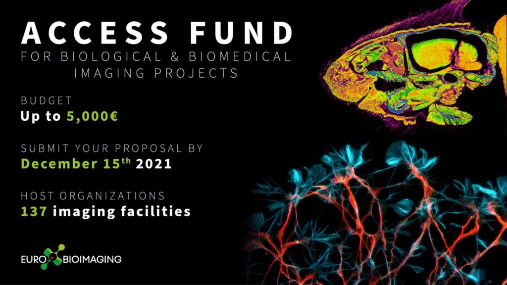
Euro-BioImaging first open call for user projects is open! If you have an idea for a biological or biomedical imaging project that you, your student or your close colleague could carry out in one of Euro-BioImaging Nodes, including France-BioImaging, now is the time to make this project come true with financial support from the Euro-BioImaging Access Fund.
How it works:
Submit your project proposal through the Euro-BioImaging web portal between October 20 and December 15, 2021, and indicate that you want to apply for the Euro-BioImaging Access Fund in order to be considered for a grant of up to 5.000 Euros to access the imaging services at a Euro-BioImaging Node. Projects will be evaluated by a committee of independent reviewers. Successful applicants will be notified by late January 2022 and successful projects should be started before July 2022.
What the funding covers:
The Euro-BioImaging Access Fund covers the user’s travel and accommodation costs as well as access and consumable costs at the imaging facilities that are part of Euro-BioImaging Nodes. For remote access projects shipment costs are also covered. Each successful applicant is eligible for up to 5.000 Euros of support.
Who is eligible:
All academic scientists, regardless of gender, nationality, home institution, career phase, or field of interest, are eligible to apply. We strongly encourage early career researchers to apply for this grant.
Projects should include transnational access to a Euro-BioImaging Node, i.e. the applicant’s home institution is in a different country than the Node where the project is to be performed. Due to the current sanitary situation, projects with non transnational access are elligible but transnational access will have priority.
All the external users/collaborators of France BioImaging facilities/labs are eligible:
any users from outside the institutional perimeter of France BioImaging nodes (i.e. from outside the following institutions: Aix-Marseille Université, Université de Montpellier, Université de Bordeaux, Université de Nantes, Université de Rennes 1, Université Paris-Saclay, Université de Paris, Université PSL -Paris Sciences & Lettres-, Généthon, Ecole Polytechnique, Institut Pasteur) who would like to use imaging technologies in one of FBI nodes: Paris Centre, Paris Ile-de-France-Sud, Marseille, Montpellier, Bordeaux, Bretagne-Loire. They can be French or international users – EU and non-EU
or users from a France BioImaging regional Node who want to access an equipment available in another FBI regional node.
Evaluation:
All applications will be evaluated for scientific excellence by a committee of independent reviewers. Selected projects will be assessed for technical feasibility and if needed receive technical advice from the Node providing the service.
How to apply:
Applicants are invited to visit our website to discover the range of technologies provided by Euro-BioImaging Nodes. Applicants will then follow the user access process described here: https://www.eurobioimaging.eu/about-us/how-to-access and indicate that they wish to apply for the Euro-BioImaging Access Fund in the application form.
Le club des Infrastructures Nationales en Biologie et Santé (INBS) organise son premier symposium autour d’une thématique qui les concernent toutes, la vie des données qui sont générées en leur sein.
Information pour les personnes inscrites:
Si vous avez opté pour une participation en présentiel:
En ce qui concerne les restrictions sanitaires, la présentation du pass sanitaire est requise et celui-ci sera contrôlé. Il peut être présenté sous forme numérique (sur téléphone) ou papier et consiste en soit un schéma vaccinal complet, soit la preuve d’un test négatif de moins de 48 heures. Aucune donnée vous concernant ne sera enregistrée.
Le renforcement des mesures sanitaires sur le campus ne permettra malheureusement pas d’assurer de pauses café ou de restauration sur place.
Si vous avez opté pour une participation en distanciel :
Ce symposium sur deux demi-journées aura de multiples objectifs:
Présentations des pratiques et développements en cours ou mis en place par les INBS ou des Infrastructures de Recherche d’autres disciplines autour des modalités de gestion de nos données (PGD de structure),
Pistes d’amélioration et de cohésion pour le futur, en ce qui concerne l’accès aux structures de stockage et de calcul scientifique,
Responsabilité morale et éthique vis-à-vis de ces données (FAIRisation, OpenScience éthique…) et leur valorisation,
Enfin, nous axerons aussi ce premier meeting sur les échanges d’expériences entre les INBS avec nos tutelles respectives.
Ce symposium est ouvert à tous. Il est cependant dédié en priorité à tous les personnels de nos communautés, c’est-à-dire à tous ceux qui participent à l’élaboration, la mise en œuvre des infrastructures nationales en Biologie-Santé et à l’accès aux prestations et services sur les différents sites qui les constituent.
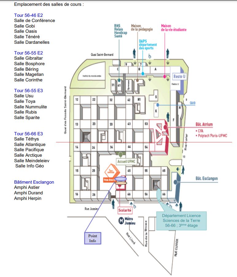
La situation sanitaire évolue mais reste complexe. Le symposium se déroulera en mode hybride dans l’Amphithéâtre Durand, Espace Esclangon sur le campus de Jussieu (Sorbonne Université), et en fonction des jauges imposées (inscriptions d’un nombre limite de participants en présentiel (60 personnes) + inscriptions pour le distanciel).
Going to Rendez-Vous Carnot 2021? Drop by our booth and say hello!
In one week from today, we will be travelling to Lyon to attend the Rendez-Vous Carnot 2021! This is the third time we’ll attend the Rendez-Vous Carnot as an exhibitor, in the Research Infrastructures Village. We are going to present France-BioImaging R&D ecosystem and the multiple advanced biological imaging technology developments taking place on FBI imaging platforms and R&D teams.
If you’re in Lyon between November 17 and November 18 attending the Rendez-Vous Carnot as well, be sure to drop by our booth and meet some of our colleagues at the venue:
Etienne Henry, France-BioImaging R&D and Tech-Transfer mission Officer
Jean Salamero, France-BioImaging Inter-Infrastructures Activities mission Officer
Besides getting to see in person what services France-BioImaging can offer in terms of R&D and tech transfer in the field of biological imaging, we also invite you to attend the Research Infrastructures special events:
On Nov. 17th 15h00 – 15h40: Flash presentations of successful academia-industry collaborations. France-BioImaging will present the success story of the “Nikon Imaging Center@Institut Curie-CNRS: 15 years of collaboration and integration in France-BioImaging”.
On Nov. 18th 14h00 – 17h00: Research Infrastructures and Health special event, in partnership with the Ministère de l’Enseignement Supérieur et de la Recherche.
What’s the RIs impact on Covid19 research and cancer research?
How can RIs work more closely with the France Health Innovation 2030?
What is France’s strategic investment plan in health & disease?
The club of INBSs is an informal consortium of the coordinations from 16 Research Infrastructures. It was initiated one year ago.
Why a CLUB?
Past institutional initiatives (DGRI, ANR…) stopped in 2019
Specificities and problems common to INBS (mostly distributed with complex perimeters and parent institutions). 21 Infrastructures in the 2018 roadmap of the Ministry of Research (16 members of the Club in October 2021) out of 70, for all disciplines.
Sharing of information on: MESRI WGs, ESFRI, Calls (from competition, to coordinated actions)
Overlapping service offer instead of benefiting from complementary technologies and skills between INBSs
New roadmaps (national, European). Régine (FLI) and Isabelle (IDMIT) propose themselves as facilitators. Changed to “Feedback from ESR/equipex+ for INBS RIs”.
February 23rd 2021
Meso-Centers and Data Center / Data management and openness. Perrine (FBI) and Jean François (IFB) as facilitators with invited participants.
April 6th 2021
Full costs and pricing “upcoming exercises”. Members of the MESRI WG on pricing, Myriam Ferro (Profi) and Yann Herault (Cellphedia) as facilitators
May 31st 2021
First propositions for the organization of a DATA-INBS symposium in December 2021
July 7, 2021
Indicators and Impact (Facilitated by MetaBoHUB, FBI and Cellphedia with invited participants): Automated Indicator Harvesting; Hierarchizing Relevant Indicators for INBS; KPIs and Impacts ; Sustainable Development Goals, are we concerned?
September 7th, 2021
Focus on year-end meetings:
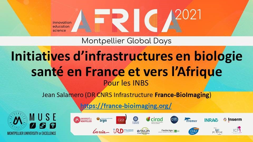
The NSAF (New Africa-France Summit in Montpellier. October) presentation of INBSs initiatives
Organization of the 1st open Meeting of the CLUB, 16 and 17 of December: « Les données des Infrastructures en Biologie et Santé: enjeux et perspectives ».
More information and free but mandatory registration at:
Deadline for face to face registration: 15th of November
The National Infrastructures in Biology and Health (INBS) club is organizing its first symposium on a theme that concerns them all: the life of the data generated within them.
This two half-day symposium will be in French and will have multiple objectives:
Practices and developments in progress or implemented by INBS or Research Infrastructures of other disciplines around the management of our data (PGD structure),
Ideas for improvement and cohesion for the future, regarding access to storage and scientific computing structures,
Moral and ethical responsibility towards these data (FAIRisation, ethical OpenScience…) and their valorisation,
Finally, we will also focus this first meeting on the exchange of experiences between the INBS and with our respective Institutions.
Cryogenic electron tomography (cryo-ET) visualizes the 3D spatial distribution of macromolecules at nanometer resolution inside native cells. However, automated identification of macromolecules inside cellular tomograms is challenged by noise and reconstruction artifacts, as well as the presence of many molecular species in the crowded volumes.
To overcome these obstacles, an international team of scientists from France, Spain and Germany, under the leadership of Charles Kervrann, from France BioImaging BioImage Informatics Node, developed a deep learning-based framework to quickly identify multiple classes of macromolecules in cryo-ET volumes. This DeepFinder program, now published in Nature Methods, builds upon convolutional neural networks that have already proven highly valuable in the microscopy field.
Overview of DeepFinder (from Moebel, E., et al., Nat Methods18, 1386–1394 (2021).
a) The DeepFinder workflow consists of a training stage (stage I) and an analysis (or inference) stage (stage II). These two stages correspond to five steps (represented by blue boxes) to locate macromolecular complexes within crowded cells.
b) Ribosome localization with DeepFinder in a cryo-electron tomogram of a C. reinhardtii cell. Tomographic slice with superimposed segmented cell membrane (gray) and ribosomes classified with respect to their binding states: membrane-bound (blue) and cytosolic (yellow).
c) Tomographic slices showing coordinates of detected ribosomes (colors correspond to b). The positions and classes were determined by analyzing the segmentation map shown in b. This analysis used 48 tomograms for training, one for validation and eight for testing. Scale bar, 60 nm.
Once trained, the inference stage of DeepFinder is faster than template matching and performs better than other competitive deep learning methods at identifying macromolecules of various sizes in both synthetic and experimental datasets. On cellular cryo-ET data, DeepFinder localized membrane-bound and cytosolic ribosomes (roughly 3.2 MDa), ribulose 1,5-bisphosphate carboxylase–oxygenase (roughly 560 kDa soluble complex) and photosystem II (roughly 550 kDa membrane complex) with an accuracy comparable to expert-supervised ground truth annotations. DeepFinder is therefore a promising algorithm for the semiautomated analysis of a wide range of molecular targets in cellular tomograms. It also serves as a prime example illustrating the importance of developing efficient, customized AI tools to accelerate knowledge generation in the biomedical life sciences.
DeepFinder has been implemented as a free, open-source program with an accessible graphical user interface.
The team is currently working on adapting it to fluorescence microscopy.
Moebel, E., Martinez-Sanchez, A., Lamm, L. et al. Deep learning improves macromolecule identification in 3D cellular cryo-electron tomograms. Nat Methods18, 1386–1394 (2021). https://doi.org/10.1038/s41592-021-01275-4
BioImage Informatics 2021 is an annual meeting in the processing, analysis, and extraction of information and knowledge from biological images. This conference will be held in virtual from November 29 to December 1, 2021, and is organised by Jean-Christophe Olivo-Marin (Institut Pasteur), Charles Kervrann (Inria), Jean Salamero (Institut Curie) and Jean-Yves Tinevez (Institut Pasteur).
This year’s edition of the BioImage Informatics conference will happen fully online, and rely on a very nice website built specially for the conference. There will be a dedicated space for poster presentations where presenters will be able to interact with the audience, leave a video or materials when they are not here, etc…
There will be a space for job fair and general announcements as well.
BioImage Informatics 2021 meeting will include, but not be limited to, the following topics:
Advanced analytical solutions for bioimage processing and analysis
Statistical spatial analysis of cellular or molecular distributions
Applications of machine and deep learning to analysis of cellular structure and related functions
Quantification of dynamic images and transport phenomena Automation of data acquisition and analysis
Dynamic cell imaging and biological processes
Reconstruction and analysis of structure and function of biological networks
Registration, correlation and fusion of multimodality data
BioImage Informatics will feature a variety of types of presentations: invited talks (45’), selected talks from abstracts (20’) and posters.
Invited Speakers
Yonina Eldar, Weissmann Institute, Israel
Michael Liebling, EPFL/IDIAP, Switzerland
Emma Lundberg, KTH Royal Institute of Technology, Sweden
Jong Chul Ye, KAIST, Korea
Abstract submission and Registration for BioImage Informatics 2021 are now open!
All abstracts for selected talk and poster consideration must be submitted by October 15, 2021 and should not exceed 350 words (excluding authors and affiliations).
You may submit as many abstracts as you like.
At least one author of each paper or poster must register and attend the conference in order to be listed in the conference programme as a presenter. Authors will have the opportunity to edit an originally-submitted abstract before it is published in conference proceedings.
Only accepted abstracts and fully paid registration will be printed in the PDF programme book.
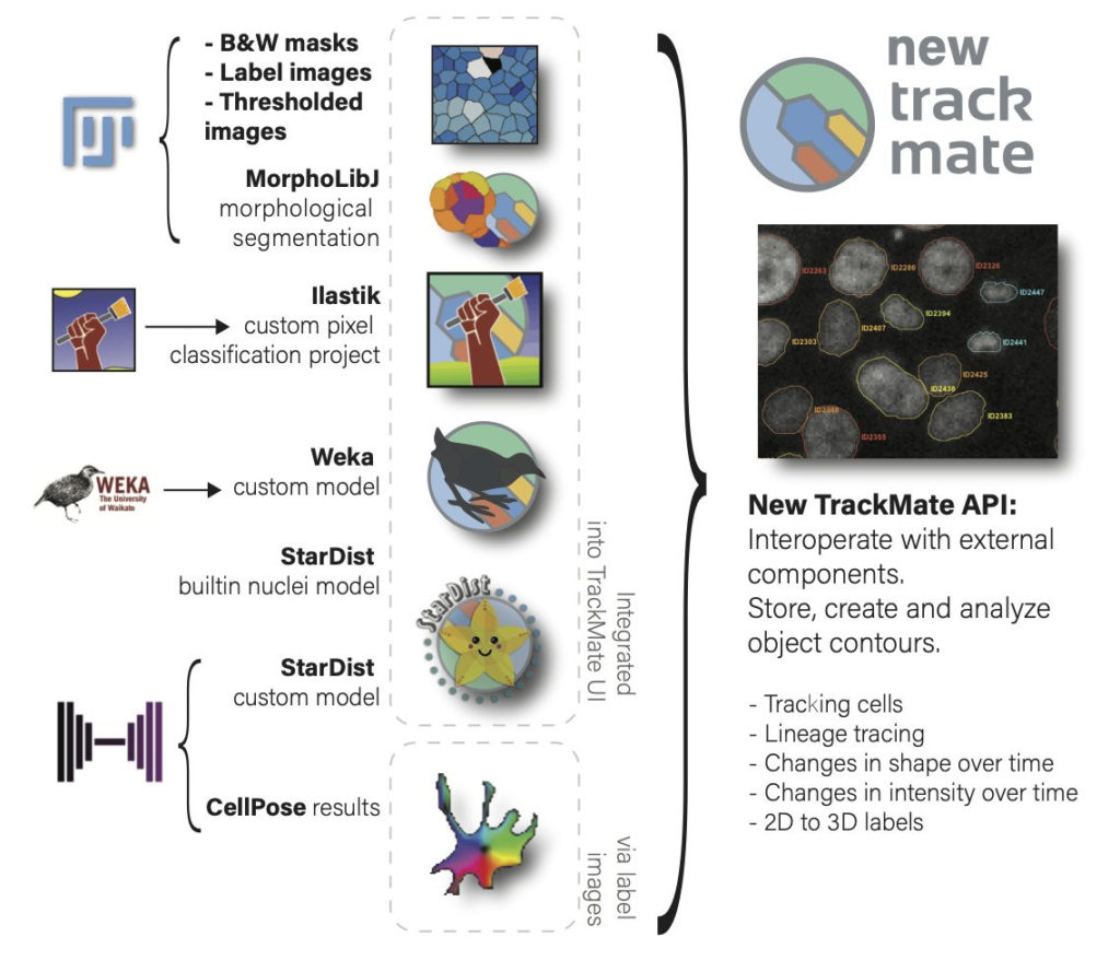
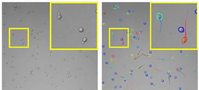
A new version of TrackMate is available now, with major changes that improve its versatility. TrackMate now integrates state-of-the-art segmentation algorithms from machine-learning and deep-learning such as StarDist, Ilastik and Weka.
TrackMate[1] is a Fiji plugin that address cell or organelle tracking in Life-Science microscopy images. Its main goals are to be user-friendly, interoperable and to serve as a platform to accelerate the development of novel tracking algorithms and analysis pipelines.
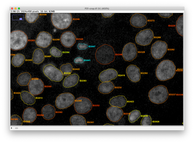
With this new version we rewrote almost entirely TrackMate so that it can integrate state-of-the-art segmentation algorithms and benefit from their output. For instance, TrackMate can now store, display, save, load and exploit object contours in 2D.
We also made a new application programming interface that will facilitate and accelerate reusing TrackMate in other analysis pipelines and allow 3rd party contributors to add new segmentation algorithms in TrackMate in an easy way. We used this API ourselves to add 7 new segmentation algorithms to TrackMate:
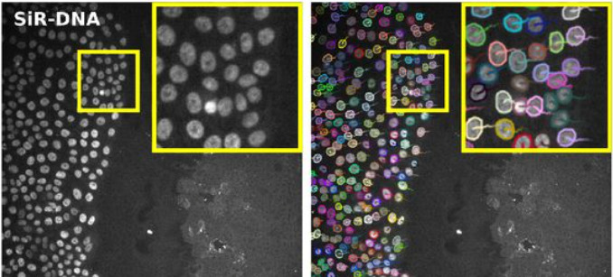
For instance, the StarDist[2] algorithm is integrated as two different detectors. The first one uses the built-in deep-learning model that can segment cell nuclei in fluorescence image in a wide range of situation. The robustness of the StarDist algorithm in turn positively impacts the robustness of tracking and allows for better detection of cell divisions with TrackMate tracking algorithms. This will facilitate cell migration studies.
The TrackMate StarDist integration also allows for specifying and using a custom deep-learning model. For instance, we trained a specific model to detect T-cells imaged in bright-field microscopy and track them over time. Before the emergence of such detection algorithms, the tracking of label-free cells was difficult.
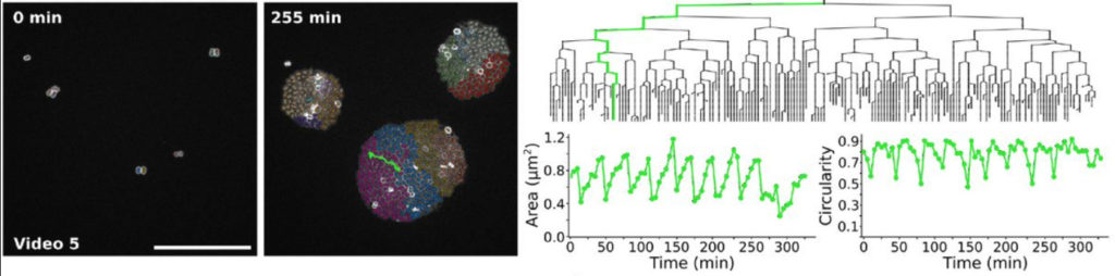
We also integrated the ilastik[3] segmentation software. A TrackMate user can input an ilastik classifier to detect objects then track them. We used them to study the bacterial growth of Neisseria meningitidis clones. The output of this analysis pipeline offers the lineage of each single cell along with its morphology and how it evolves across cell divisions.
The new capabilities of TrackMate can be used to address applications beyond tracking. For instance, it is now possible to use TrackMate to perform the segmentation of 3D objects using a slice-by-slice approach. This approach consists in segmenting objects in each 2D section of a 3D stack, then merging the segmentation results along Z in a subsequent step. This can be done in TrackMate, using the tracking step for merging. We implemented a novel tracking algorithm to foster this application, the overlap tracker. We could use this approach combining the cellpose[4] algorithm in 2D to segment 3D images of Arabidopsis thaliana floral meristem.
There are several other algorithms that are now offered to the TrackMate user, within a user-friendly software meant to interoperate with the key software of bioimage analysis. More importantly, TrackMate is an open-source academic software, and its new API will foster the development of new analysis pipeline with TrackMate and the integration of new algorithms by other developers, increasing the breadth of applications it can address for Life-Science researchers.
Bringing TrackMate in the era of machine-learning and deep-learningDmitry Ershov, Minh-Son Phan, Joanna W. Pylvänäinen, Stéphane U. Rigaud, Laure Le Blanc, Arthur Charles-Orszag, James R. W. Conway, Romain F. Laine, Nathan H. Roy, Daria Bonazzi, Guillaume Duménil, Guillaume Jacquemet, Jean-Yves TinevezbioRxiv 2021.09.03.458852; doi: https://doi.org/10.1101/2021.09.03.458852
[1] https://imagej.net/plugins/trackmate/
[2] Alejandro F. Frangi, Julia A. Schnabel, Christos Davatzikos, Carlos Alberola-López, and Gabor Fichtinger Uwe Schmidt, Martin Weigert, Coleman Broaddus, and Gene Myers. Cell detection with star-convex polygons. In Alejandro F. Frangi, Julia A. Schnabel, Christos Davatzikos, Carlos Alberola-López, and Gabor Fichtinger, editors, Medical Image Computing and Computer Assisted Intervention – MICCAI 2018, pages 265–273, Cham, 2018. Springer International Publishing. doi:10.1007/978-3-030-00934-2_30.
[3] Stuart Berg, Dominik Kutra, Thorben Kroeger, Christoph N Straehle, Bernhard X Kausler, Carsten Haubold, Martin Schiegg, Janez Ales, Thorsten Beier, Markus Rudy, Kemal Eren, Jaime I Cervantes, Buote Xu, Fynn Beuttenmueller, Adrian Wolny, Chong Zhang, Ullrich Koethe, Fred A Hamprecht, and Anna Kreshuk. ilastik: interactive machine learning for (bio)image analysis. Nature Methods, 16(12):1226–1232, 2019. ISSN 1548-7105. doi:10.1038/s41592-019-0582-9.
[4] Carsen Stringer, Tim Wang, Michalis Michaelos, and Marius Pachitariu. Cellpose: a generalist algorithm for cellular segmentation. Nature Methods, 18(1):100–106, jan 2021. doi:10.1038/s41592-020-01018-x.
Abstract submission is open for the Neurophotonics II Conference that will take place at Photonics Europe on April 3-7, 2022, in Strasbourg, France.
Please note that the submissions made to your conference can be viewed via your SPIE account. Details on how to access this information are listed at the end of this e-mail.
This conference focuses on cutting edge research and techniques used to investigate the brain and nervous system. Multiscale imaging and manipulating the living and intact brain are becoming important topics in neurophotonics. In this context, it’s mandatory to provide new strategies for optical measurements of neural function and develop tools such as optogenetics to enables the control of cellular function with light. Also, in terms of imaging, furthermore it’s often important to image the samples from nanoscale to whole organism scales, bridging the gap between technologies.
The conference aims to bring together engineers, optical and medical scientists, biologists, chemists, neuroscientists and physicians, bringing together researchers working in all aspects of neurophotonics. It will also serve as a forum to discuss existing and emerging techniques.
Topics include but are not limited to:
hybrid and multimodal approaches to neuroimaging
optical hemodynamic imaging and neuro-vascular interactions
mesoscopic, microscopic, and endoscopic imaging of neural structure and function
tissue scattering, clearing and de-scattering
superresolution microscopy and nanoscopy of the nervous system
novel reporters and actuators, optogenetics, bioluminescence
data analysis, machine learning, and image processing
analyzing circuitry, network function, and information processing
optics and brain disease
light shaping in the brain, holography
dissemination and commercialization of BRAIN technologies
France BioImaging primary mission is to develop, promote, disseminate and provide access to innovative instruments and imaging technologies in the field of bioimaging to scientists. Fostering the technological transfers is at the heart of this mission, and for this France BioImaging relies on a strong association of leading R&D research teams with core facilities.
However, several bottlenecks exist and often hamper or prevent successful technology transfer:
A lack of human resource leads to difficulties in transferring and stabilizing the technology which is not enough user-friendly
A technology that is too specific, with not enough user base
A difficulty to contract with industry through institutional offices for industrial valuation
In the context of image analysis: the instability of open software economical model, inter-operability, large data handling and algorithm complexity
As a way to tackle these bottlenecks, France BioImaging launched in January 2021 its first “FBI Internal Call 2021: Technology transfer from the R&D teams to the core facilities” to promote the transfer of new technologies (instrumentation, probes, staining methods, software, data analysis or data visualization) from the R&D teams to the facilities of France BioImaging, for access and service to end-users. The outcome of the transfer project had to ensure for the prototype to be usable by the end-users until the interpretation of the data. The project had also to include a sustainability plan and a training plan to guide both facility staff and end-users toward autonomy.
The project selection was organized by the National Coordination of France-BioImaging and applications were assessed according to the following evaluation criteria:
Innovation and originality of the proposal
Scientific quality, implementation, timeline
Competitive positioning
Adequacy of resources with the proposed project
Economic impact and tech transfer potential and perspectives
Estimation of the user market and potential for user adoption
Plan for training and sustainability.
For the first edition of the “FBI Internal Call 2021: Technology transfer from the R&D teams to the core facilities”, 5 projects were selected:
Icy@FBI: Jean-Christophe Olivo-Marin (IPDM Node): Broadening the scope of applications of Icy (http://icy.bioimageanalysis.org/) by implementing several key new bioimage analysis components
BIC-HCS-SMLM: Jean-Baptiste Sibarita (Bordeaux Node), Technological transfer of a Single-Molecule-based High Content Screening platform to the Bordeaux Imaging Center
CloudFISH: Marcello Nollmann (Montpellier Node), A tool for the analysis of single-molecule RNA and DNA FISH images
MorphoNet: Emmanuel Faure (Montpellier Node), An interactive online morphological browser to explore complex multi-scale data
BioImageIT (https://bioimageit.github.io/#/about): Jean Salamero, Sylvain Prigent (IPDM Node), An open source framework for integration of image data management with analysis
Each selected project was awarded with a 80k€ grant for salary and/or equipment, and several positions are currently available: https://france-bioimaging.org/jobs/
This call will be renewed in 2023.
The 6th edition of Global BioImaging annual gathering will have the theme “Imaging Research Infrastructures in a time of change” and will take place on the 8th and 9th September 2021 as an online event.
We use cookies on our website to give you the most relevant experience by remembering your preferences and repeat visits. By clicking “Accept All”, you consent to the use of ALL the cookies. However, you may visit "Cookie Settings" to provide a controlled consent.
This website uses cookies to improve your experience while you navigate through the website. Out of these, the cookies that are categorized as necessary are stored on your browser as they are essential for the working of basic functionalities of the website. We also use third-party cookies that help us analyze and understand how you use this website. These cookies will be stored in your browser only with your consent. You also have the option to opt-out of these cookies. But opting out of some of these cookies may affect your browsing experience.
Necessary cookies are absolutely essential for the website to function properly. These cookies ensure basic functionalities and security features of the website, anonymously.
Cookie
Duration
Description
cookielawinfo-checkbox-analytics
11 months
This cookie is set by GDPR Cookie Consent plugin. The cookie is used to store the user consent for the cookies in the category "Analytics".
cookielawinfo-checkbox-functional
11 months
The cookie is set by GDPR cookie consent to record the user consent for the cookies in the category "Functional".
cookielawinfo-checkbox-necessary
11 months
This cookie is set by GDPR Cookie Consent plugin. The cookies is used to store the user consent for the cookies in the category "Necessary".
cookielawinfo-checkbox-others
11 months
This cookie is set by GDPR Cookie Consent plugin. The cookie is used to store the user consent for the cookies in the category "Other.
cookielawinfo-checkbox-performance
11 months
This cookie is set by GDPR Cookie Consent plugin. The cookie is used to store the user consent for the cookies in the category "Performance".
viewed_cookie_policy
11 months
The cookie is set by the GDPR Cookie Consent plugin and is used to store whether or not user has consented to the use of cookies. It does not store any personal data.
Functional cookies help to perform certain functionalities like sharing the content of the website on social media platforms, collect feedbacks, and other third-party features.
Performance cookies are used to understand and analyze the key performance indexes of the website which helps in delivering a better user experience for the visitors.
Analytical cookies are used to understand how visitors interact with the website. These cookies help provide information on metrics the number of visitors, bounce rate, traffic source, etc.
Advertisement cookies are used to provide visitors with relevant ads and marketing campaigns. These cookies track visitors across websites and collect information to provide customized ads.