The France BioImaging Image Contest is back for its 3rd edition!

This image contest is open to all within the imaging community: core facility staff and users, R&D labs teams and co-workers, students… Submit your best microscopy images for a chance to showcase your skills, research and creativity to the French bioimaging community and beyond, allowing people to see the visual appeal of the life sciences. Images from the contest will be featured on France BioImaging communication tools, online and in print.

France BioImaging and all the French community aims to develop and promote innovative imaging technologies and methods. But microscopy images can also take an artistic, creative look and make the invisible world beautiful.

We are all eager to see your work !

Prizes

1 to 3 images will be awarded depending on the quantity and quality of the entries submitted. France BioImaging will cover the registration fees for one 2022 microscopy related event of the winners’ choice (FOM, ELMI, EMC, COMULIS conference, etc.).

Important: Only French or foreign participants affiliated to a French institution can enter the contest. Foreign participants non-affiliated to a French institution can submit images and will be featured in the gallery, but will not be evaluated as part of the contest.

Submission deadline: Friday, October 15th, 2021, 23h59 UTC+2. 

Click here to consult the terms and conditions of the contest. When you are ready, submit your entry by filling the form below. You can check out last editions’s entries for inspiration. One participant can submit several entries (up to 3).


This form is currently closed for submissions.

Important information for the registered participants: an email with the links to access the training videos was sent on February 15th, 2020. Please check your inbox and SPAM folder!

If you did not receive it, please send us an email to contact@france-bioimaging.org

If you registered after February 14th, 2021, you will receive an email with the links within two days.


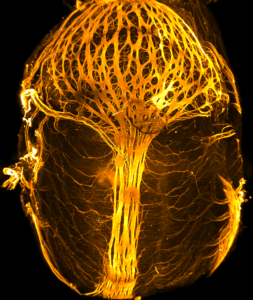
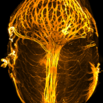
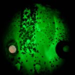
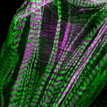
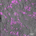
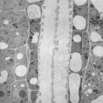
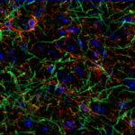
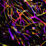
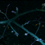
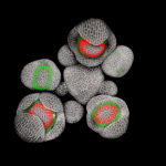
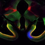
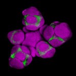
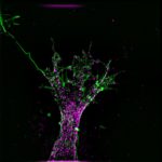
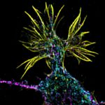
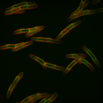
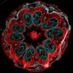
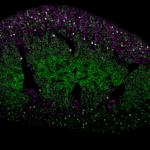
France BioImaging (FBI) is organizing a remote training on Light-Sheet Fluorescence Microscopy (LSFM), which enable 3D imaging of biological samples with unprecedented spatio-temporal resolutions and low perturbing effects.

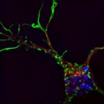
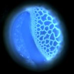
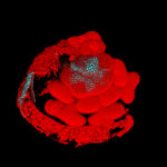
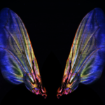
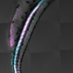
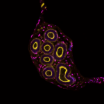
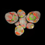
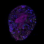
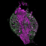
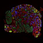
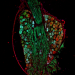
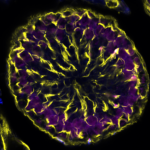
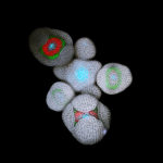
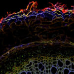
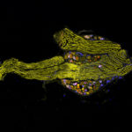
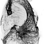
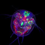
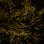
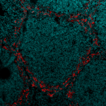
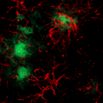
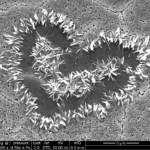
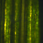
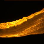
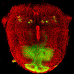
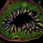
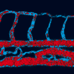
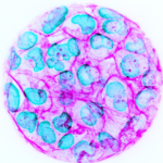
LSFM methods actually cover a large variety of implementations which allow imaging a wide range of sample types, from single cell to whole organs or organisms both live and fixed. These new imaging capabilities are revolutionizing the way we visualize our samples and address biological questions. However, imaging with a light-sheet microscope raises many questions about the choice of the set-up depending on the sample to image, the sample preparation and mounting protocols or the data management (storage, visualization, quantification). Thus, it can be difficult to find its way through the numerous microscope implementations, protocols and tools that have been extensively developed over the last 20 years. We therefore decided to review all those questions in a remote training.

Our goal is to help people who want to jump into the world of 3D imaging and are seeking the best solution for their samples and biological questions. In that perspective, we will provide a comprehensive picture including all the possibilities and challenges regarding LSFM.

Format:

The training will be divided in 3 parts:

  1. Theoretical courses on LSFM
  2. Practical demonstrations of several LSFM implementations available throughout the FBI infrastructure
  3. Live online question-and-answer session

For the two first parts, videos will be available on a Youtube FBI channel. The participants will have 3 weeks, from the 15th of February to the 5th of March 2021, to watch those videos and will be invited to ask questions or comment.

FBI experts will then answer all questions during a live interactive video chat on the third week of the training (5th of March where participants will have the opportunity to directly interact with the experts.
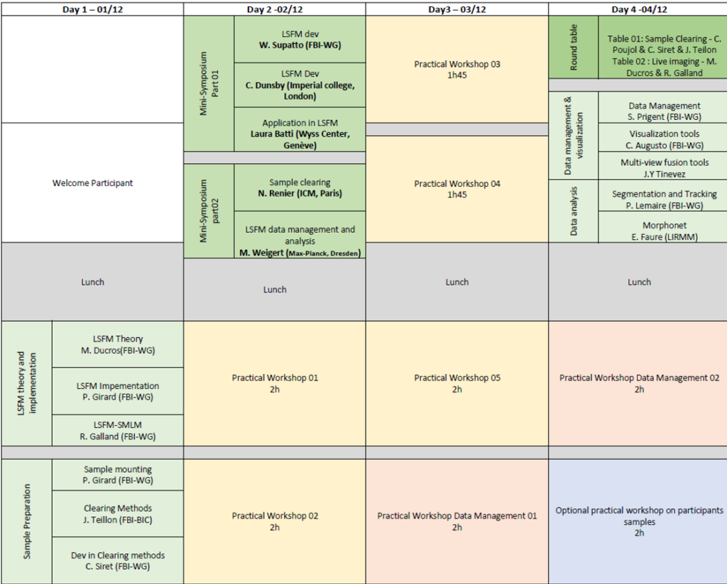
Program:

1.      Theoretical aspects of LSFM (15th to 26th of February 2021)

Here are the three main questions concerning the imaging with a light-sheet microscope: (1) what LSFM type should I use for my experiment, (2) How do I prepare and mount my sample, and (3) how to visualize and analyze my data sets. The first part of this training will address these three questions through three theoretical courses:

  • Course 1: Theoretical principles and numerous implementations overview of LSFM
    • P. Girard (Institut Jacques Monod, Paris-Centre)
  • Course 2: Sample preparation and mounting principles – highlight on clearing approaches
    • Carol Siret (CIML, Marseille)
  • Course 3: Reconstruction, Visualization and Analysis software overview.
    • Cesar Augusto Valades (Institut Curie, Paris-Centre),

2.      Practical demonstrations of several LSFM implementation and experiments (15th of February to 5th of March 2021)

In the second part of the training we will propose several videos on various systems available in the FBI laboratories and imaging platforms covering diverse types of LSFM design and applications.

Each video will feature a specific set-up and experts will present how to run an experiment on them focusing on three main aspects: (1) sample preparation and mounting methods, (2) image acquisition processes, and (3) visualization of the data-sets.

  • Lattice Light Sheet Microscope(Home-made  and 3i versions)
    • Mathieu Ducros (BIC, Bordeaux)
    • Ludovic Lecomte, Jean Salamero and Cesar Valaldes-Cruz (Institut Curie, Paris-Centre)
  • Single-objective Single Plane Illumination Microscope (soSPIM)Home-made
    • Rémi Galland (IINS, Bordeaux)
  • Dual inverted Single Plane Illumination Microscope (diSPIM)3i (Marianas)
    • Elric Esposito et Julien Fernandes (Institut Pasteur, Paris-Centre)
  • MuviSPIM – Luxendo
    • Sylvain De Rossi (MRI, Montpellier)
  • Ultramicroscope – LaVision Biotech
    • Carol Siret/Mathieu Fallet (CIML, Marseille)

3.      Questions & Answer interactive session (March 5th, 2021)

An online video session will conclude the training where FBI experts will answer all participants’ questions. You can ask questions either in advance in the comment box of the Youtube video, or during the Q&A session in a chat box. The Q&A session will be divided in sections, each related to a specific video.

To register:

In order to register to the Light-Sheet Fluorescence Microscopy remote training, please fill out the registration form available here.

Registration is free but mandatory in order to receive the links to the training videos.

Extended deadline: February 19th, 2021

We look forward to your participation !

Registration for France BioImaging Annual Meeting is now open!

France BioImaging is pleased to invite you to participate to France BioImaging 6th Annual Meeting.  For this edition, the meeting will be organized as a two-half days virtual meeting (from 9:00 AM to 1:00 PM) on February 4th & 5th, 2021.

This event, open to all members of the bioimaging community, aims to provide a platform to discuss pivotal subject matters in our field.

The 2021 program of the France BioImaging Annual meeting is built around two pillars:

  • February 4th: “Building and operating an integrated and open infrastructure for bioimaging
  • February 5th: “Latest and future developments in biological imaging

Registration is free but mandatory in order to receive the Zoom link: https://univ-nantes-fr.zoom.us/meeting/register/tJAof-itqjgjHNCQOGpsw4_2ERWm__3zUU0R

Program 

ZOOM Etiquette

We look forward to your participation!

The France BioImaging Image Contest is back for its 3rd edition!

This image contest is open to all within the imaging community: core facility staff and users, R&D labs teams and co-workers, students… Submit your best microscopy images for a chance to showcase your skills, research and creativity to the French bioimaging community and beyond, allowing people to see the visual appeal of the life sciences. Images from the contest will be featured on France BioImaging communication tools, online and in print.

France BioImaging and all the French community aims to develop and promote innovative imaging technologies and methods. But microscopy images can also take an artistic, creative look and make the invisible world beautiful.

We are all eager to see your work !

Prizes

1 to 3 images will be awarded depending on the quantity and quality of the entries submitted. France BioImaging will cover the registration fees for one 2022 microscopy related event of the winners’ choice (FOM, ELMI, EMC, COMULIS conference, etc.).

Important: Only French or foreign participants affiliated to a French institution can enter the contest. Foreign participants non-affiliated to a French institution can submit images and will be featured in the gallery, but will not be evaluated as part of the contest.

Submission deadline: Friday, October 15th, 2021, 23h59 UTC+2. 

Click here to consult the terms and conditions of the contest. When you are ready, submit your entry by filling the form below. You can check out last editions’s entries for inspiration. One participant can submit several entries (up to 3).


This form is currently closed for submissions.

Due to the current sanitary measures and uncertainty about future COVID-19 developments, the LSFM workshop to be held in Bordeaux in December has been cancelled. The organizers are currently working on a virtual format of the workshop. More details to come soon.

This workshop will be held in Bordeaux from December 1st to December 4th 2020 and aims to give an overview of the Light Sheet Fluorescence Microscopy techniques and their application possibilities, taking advantage of the available technologies and expertise within the France BioImaging research infrastructure and the broad light-sheet microscopy community. This workshop is organized by the members of the FBI Multiscale Light-sheet Imaging Working Group.

About the workshop

  • The workshop will last 3 ½ days
  • The workshop is built on the basis of an alternation between theoretical and practical sessions. Both will be in English and provided by national expert trainers.
  • On day 2 a mini-symposium will be organized to present the last developments in different LSFM domains (instrumentation, sample preparation and data analysis) done by international experts. This mini-symposium will be open to a larger audience in order to promote LSFM approaches to the local research community. Registration is free but mandatory (this does not apply to workshop participants). Register now
  • The workshop will encompass various theoretical aspects of LSFM from sample preparation, to image acquisition and first post-processing steps (3D reconstruction, visualization).
  • The workshop will span various scales that can be imaged with LSFM based on at least 4 different LSFM set-ups
  • It will be included in the future “FBI training passport” and corresponds to a level III module (“à la carte”). Therefore, “trainees” must demonstrate knowledge of Level I (basic imaging) and Level II (optical principles) imaging.

Objectives:

  • Describe the general principle of light-sheet microscopy
  • Present the three main instrumentations that have been developed depending of the sample scale to be imaged (1 to 100 µm; 100 µm to mm; mm to cm)
  • Give an overview of the sample preparation techniques
  • Present the challenges raised by the data management and analysis of LSFM data
  • Present the future of light-sheet microscopy

Audience:

This workshop is ideal for researchers, engineers, technicians, PhD students, and post-docs from public institutes and private companies with a prior knowledge in fluorescence microscopy techniques, and willing to become familiar with advanced techniques to answer specific biological questions.

Number of participants is limited to 20.

Practical workshops:

The practical workshops main objectives are to:

  • Provide an overview by experts of the 3 main implementations of LSFM depending of the sample size to be imaged:
    • Ultramicroscope (LaVision Biotech) – BIC
    • Invi Lattice Pro (Luxendo)
    • Z1 (Zeiss) – to be confirmed
    • LLS – BIC;
    • soSPIM – IINS
  • Give an overview of existing solutions for 3D reconstruction, Multiview merging and 3D+t visualization.

Participation fee

Academia: 200€
Industry: 600€

Preliminary program:

The workshop will last 3 ½  days with theoretical courses the morning and practical workshops the afternoon on 4 different set-up spanning the different sample sizes that can be imaged with LSFM:

  • Zeiss – Z1 (to be confirmed)
  • Luxendo – Invi Lattice Pro
  • LaVision – Ultramicroscope II
  • Lattice Light-sheet microscope – BIC
  • soSPIM – IINS

3 theoretical sessions will encompass various domains of LSFM:

  • General principle
  • Sample preparation
  • Data management

A mini-symposium will be organized to highlight recent national and international developments in LSFM.

Confirmed speakers:

  • Willy Supatto – Laboratoire D’Optique et Biosciences, Palaiseau – France
  • Christopher Dunsby – Faculty of Medicine, Imperial College, London – UK
  • Laura Batti – Wyss Center, Genève – Switzerland
  • Nicolas Renier – Laboratory of Structural Plasticity, ICM, Paris – France
  • Martin Weigert – EPFL, Lausanne

On the last day two round tables will be proposed to discuss about two hot topics in light-sheet fluorescent microscopy:

  • Clearing protocols
  • Live imaging: challenges and solutions

Participants are invited to bring their own samples if possible to test during an optional practical workshop on the last day.

A diner in Bordeaux is planned on day 2.

Venue:

The workshop will be held on the premises of the Bordeaux Imaging Center and the Interdisciplinary Institute for Neuroscience, in the Centre Broca Nouvelle Aquitaine, in Bordeaux, France.

Accomodation

Hotel booking is left to the participant’s initiative.

Here is a list of other hotels located in the historic center of Bordeaux city (quartiers des “Grands hommes”, “Quinconces”, “Hôtel de ville” and “Mériadeck”). All are close to the tramway stops of line A (direction Le Haillan-Rostan / Pin Galant – Stops: Hôpital Pellegrin, Saint-Augustin, François Mitterand):

Hôtel de France **

Hôtel Gambetta **

Hôtel la Porte Dijeaux **

Hôtel des 4 Sœurs **

Hôtel de l’Opéra **

Hôtel Le Chantry **

Citadines Centre Mériadeck Bordeaux ***

Hôtel Ibis style Bordeaux Mériadeck ***

La Maison du Lierre ***

Hôtel Ibis Mériadeck ***

Best Western Bordeaux-Bayonne Etche Ona ***

Adagio Bordeaux Gambetta ****

Mama Shelter

Local Organisers

Dr. Mathieu Ducros, Bordeaux Imaging Center

Dr. Rémi Galland, Interdisciplinary Institute for Neuroscience

Questions about the LSFM workshop can be addressed directly to the local organisation team : lsfm_workshop@france-bioimaging.org

Sponsors

[:en]France BioImaging is organising an “BioImage Analysis OpenDesk” session on November the 29th from 9:00 to 12:30, in its different nodes and online.

What is an OpenDesk ?

During the event, users in search for answers to image processing-related questions come to a dedicated spot, within the Facility. Individual questions are processed by BioImage Analysts, on a “first come-first served basis”.

Will I really get my image processing questions answered ?

We will work on that ! Depending on the topic, local BioImage Analyst may choose from one of those three options:
1-The problem might be solved locally, quickly: you will leave the Facility with either a procedure/advices on how to analyse your data.
2-The problem might be solved locally, but requires additional time to be processed: your Facility staff will propose an appointment so that a proper solution is found.
3-The problem might not be solved locally: we will take benefit of the France BioImaging infrastructure, through its dedicated transversal node (FBI-IPDM). Your local Facility staff will introduce you to specialists in the field, using remote communication means.

I’m in ! Where and when is it taking place ?

November the 29th, from 9:00 to 12:30.
Bordeaux — Bordeaux Imaging Centre, Photonic Unit, 1st floor, Centre Broca Nouvelle-Aquitaine
Montpellier
Paris Centre
Marseille — IBDM ground Floor  Luminy
Paris Sud
Nantes — Room 2 , ground floor IRS UN 8 quai Moncousu

If you can not join physically, join on https://rendez-vous.renater.fr/ipdm

By the way, what is FBI-IPDM ?

The objective of the BioImage Informatics – Image Processing & Data Management transversal node is to create a general framework and a complete and integrated image analysis and IT (Information Technology) solution to address a number of challenges in biological imaging and microscopy, as well as setting up a high performance grid-computing infrastructure dedicated to massive computational and data storage demands. The FBI-IPDM node proposes different IT frameworks to deal with the data flow from the different imaging nodes. FBI-IPDM node is thus transverse, by definition.[:fr]France BioImaging is organizing a “BioImage Analysis OpenDesk” session on November the 29th from 9:00 to 12:30, in its different nodes and online.

What is an OpenDesk ?

During the event, users in search for answers to image processing-related questions come to a dedicated spot, within the Facility. Individual questions are processed by BioImage Analysts, on a “first come-first served basis”.

Will I really get my image processing questions answered ?

We will work on that! Depending on the topic, local BioImage Analyst may choose from one of those three options:
1-The problem might be solved locally, quickly: you will leave the Facility with either a procedure/ advice on how to analyze your data.
2-The problem might be solved locally, but requires additional time to be processed: your Facility staff will propose an appointment so that a proper solution is found.
3-The problem might not be solved locally: we will take benefit of the France BioImaging infrastructure, through its dedicated transversal node (FBI-IPDM). Your local Facility staff will introduce you to specialists in the field, using remote communication means.

I’m in ! Where and when is it taking place ?

November the 29th, from 9:00 to 12:30.
Bordeaux — Bordeaux Imaging Centre, Photonic Unit, 1st floor, Centre Broca Nouvelle-Aquitaine
Montpellier
Paris Centre
Marseille — IBDM ground Floor Luminy
Paris Sud
Nantes — Room 2, ground floor IRS UN 8 Quai Moncousu

If you can not join physically, join on https://rendez-vous.renater.fr/ipdm

By the way, what is FBI-IPDM ?

The objective of the BioImage Informatics – Image Processing & Data Management transversal node is to create a general framework and a complete and integrated image analysis and IT (Information Technology) solution to address a number of challenges in biological imaging and microscopy, as well as setting up a high performance grid-computing infrastructure dedicated to massive computational and data storage demands. The FBI-IPDM node proposes different IT frameworks to deal with the data flow from the different imaging nodes. FBI-IPDM node is thus transverse, by definition.[:]

Les 29 Mai au 31 Mai prochain, nous organisons à Bordeaux une formation en Super Résolution qui traitera de la microscopie STED et des techniques basées sur la détection des molécules uniques (STORM, PALM, U-PAINT..). Elle se déroule avec une alternance entre cours théoriques (de la base vers les nouvelles techniques en développement), analyse d’images et ateliers pratiques devant les machines commerciales ou développées maison. Cette formation pourra se faire en anglais.

Dear colleagues, dear FBI community,

The National Coordination and Industry Board are proud to announce that the winners of the FBI Image Contest 2017 are:

1. Marie Irondelle – PiCT Institut Curie with “Biology’s Little Venice”

La petite Venise de la biologie © Carine Rossé, Emilie Lagoutte & Marie Irondelle, Institut Curie


La petite Venise de la biologie © Carine Rossé, Emilie Lagoutte & Marie Irondelle – Institut Curie
Confocal microscopy

Transparisation par U DISCO d’une glande mammaire murine régénérée à partir d’un transplant d’organoides mammaire murin.

2. Orestis Faklaris – Institut Jacques Monod – ImagoSeine with “The Tree of Life”


The Tree of Life © Orestis Faklaris, Nicolas Chevalier – Institut Jacques Monod
Ultramicroscope – light sheet microscopy

3D z-stack projection of transparised chicken embryo stomach. Label betaIII-tubulin – Alexa488.

3. Clémence Simon – UMR 8576 CNRS/Université Lille 1 with “When Chemistry Transcends Lignin”


Quand la chimie transcende la lignine ! 1 © Clémence Simon, Unité de Glycobiologie Structurale et Fonctionnelle, UMR8576
Microscopie confocale, MIP, BLISS

Application de la stratégie de double réaction chimique BLISS aux unités p-hydroxyphényle et guaïaicyle de la lignine sur coupe de racine de lin. Observation du double marquage (+ autofluorescence) par microscopie confocale et représentation par projection maximale d’intensité. Taille de l’image : 510 x 510 microns.

FBI Industry Committee Special Prize: Nathanaël Prunet – California Institute of Technology with “Arabidopsis Inflorescence”


Arabidopsis inflorescence 1 © Nathanaël Prunet, Caltech, Meyerowitz lab
Confocal microscopy

This is a live Arabidopsis inflorescence with young flower buds developing at the periphery. Cell walls have been stained with propidium iodide (grey). Fluorescent reporters were used to monitor the expression of the APETALA3 (AP3, green) and SUPERMAN (SUP, red) genes. AP3 is required for the development of stamens (the male organs), while SUP establishes the boundary between the male and female part of the flower. This picture was acquired using live confocal imaging, which allows us to describe the expression of several genes in both space and time, in the same live biological samples, with a precise cellular resolution. It finally allows us to understand a question that has been elusive for 25 years: how the male/female boundary is established during the formation of the flower. My research aims at understanding how flower buds are patterned as they form.

Thank you to all the participants for their great contributions:

  • Dario Donnarumma, Laboratoire Charles Coulomb UMR 5221 CNRS-UM
  • Filippo Piccinini, IRST
  • Aude Nommick, IBDM – Marseille University
  • Sébastien Marais, Bordeaux Imaging Center
  • Marie Held, Biochemistry, University of Liverpool, Levy Lab
  • Patrice Mascalchi, Bordeaux Imaging Center and Frédéric Saltel, INSERM U-1053, University of Bordeaux
  • Corrado Viotti, Institut de Biologie Moléculaire des Plantes, CNRS, Strasbourg – P. Genschik Lab
  • Marcello Delfini & Mathieu Fallet, CIML CNRS-INSERM-AMU
  •  Jonathan Daniel, Institut des Sciences Moléculaires
  • Laurence Dubreil, APEX-UMR703 PAnTher INRA Oniris
  • Pierre-Olivier Strale, Interdisciplinary Institute for Neuroscience
  • Clémence Simon, Unité de Glycobiologie Structurale et Fonctionnelle, UMR8576
  • Jérémie Teillon, INSERM U1034
  • Morgane Rabineau, Inserm
  • Eve Gazave & Nicolas Rabet, Institut Jacques Monod-CNRS
  • Nathanaël Prunet, Caltech, Meyerowitz lab
  • Françoise Geffroy, CEA-DRF-NeuroSpin-UNIRS, Midas Team
  • Valeria Davi, ImagoSeine – Institut Jacques Monod – CNRS
  • Anna Smirnova, University of Strasbourg – GMGM
  • Debora Olivier, Institut Pasteur
  • Orestis Faklaris, Institut Jacques Monod
  • Xavier Baudin, Institut Jacques Monod
  • Mathieu P. Dailly, CMAS
  • Lucie Sengmanivong, UMR 144, Institut Curie, Paris
  • Marie Irondelle, Institut Curie

Thank you also to the core facilities staff and heads for having forwarded the contest to their users and for providing them state of the art bioimaging.

The National Coordination

Image Contest FBI 2017

The France BioImaging Image Contest is back for its 2nd edition!

This image contest is open to all within the imaging community: core facility staff and users, R&D labs teams and co-workers, students… Submit your best microscopy images for a chance to showcase your skills, research and creativity to the French bioimaging community and beyond, allowing people to see the visual appeal of the life sciences. Images from the contest will be featured on France BioImaging communication tools, online and in print.

France BioImaging and all the French community aims to develop and promote innovative imaging technologies and methods. But microscopy images can also take an artistic, creative look and make the invisible world beautiful.

The National Coordination is eager to see your work !

Prizes

1 to 3 images will be awarded depending on the quatity and quality of the entries submitted. Prize winners will get to choose one option, between the following:

  • Registration and travel costs (flight from France to Hungary) to NEUBIAS Symposium (31 January-2 February 2018, Szeged, Hungary)*
  • Registration fees for Focus on Microscopy 2018 (25-28 March 2018, Singapore)
  • Registration fees for ELMI 2018 (5-8 June 2018, Dublin, Ireland)

* Prize winners already accepted to the NEUBIAS Training Schools may request that France BioImaging cover their fees as a prize. However, keep in mind that may you not win a prize, the registration fee for the event will still be due.


Submission deadline: Sunday 26 November 2017, 23h59 UTC+2. THE CONTEST IS NOW CLOSED.

Click here to consult the terms and conditions of the contest. When you are ready, submit your entry by filling the form below. You can check out last year’s entries for inspiration. One participant can submit several entries.


We are happy to announce the 4th Mini-symposium on Bioimage Informatics, which will take place the 27th of June 2017 in Rennes. This event is organized by the GDR ImaBio and France Bioimaging.

The idea of this annual workshop on Bioimage Informatics is to bring together experts from the fields of image analysis and biology, to promote exchanges between the scientists in these fields and to contribute to the creation and maintenance of a French community in the field of Bioimage informatics, which is continuing in gaining importance in the field of Bioimaging. This year, the workshop is organized as a one-day satellite of the “Imaging the cell” conference, held in Rennes the 28th-30th of June 2017 (http://www.atoutcom.com/imagingthecell2017/).

Details & registration: http://gdr-miv.fr/en/events/bioimageinformatics2017/

Correlative Microscopy (CLEM) takes an increasing value in scientific research.

The second France BioImaging course on correlative microscopies is jointly organized by Institut Jacques Monod, Institut Pasteur and Institut Curie.

The first day (free access, registration required) will be composed of a conference series from national and international speakers about the latest CLEM technologies as well as by participants who will follow the practical session in the following 4 days.

Practical training will be organized in 2 sessions of 2 days for 3 groups between the 3 organizing institutes (12 positions available).

The 2 sessions will be chosen among the 3 available:
– EMPACT2 CLEM / Tokuyasu (Institut Jacques Monod)
– Live Cell to HPF, Quick Freeze Substitution/fluorescence on sections and eC-CLEM alignment (Institut Curie)
– Live Cell to FIB-SEM / Cryo-CLEM (Institut Pasteur)

Registration on the FBI website, dead-line 31 March 2017

First day: free entrance
Practical sessions: 250.00 euros in total, selection on application form.

FBI Grand

On Friday April 14 2017, we will have the pleasure to welcome you to our Annual Meeting, to be held at the Curie Institute (Paris, France). This event, open to all members of the bioimaging community, aims to provide a platform to discuss pivotal subject matters in our field. This year, the France BioImaging Annual meeting will focus on the topic of “Future challenges in BioImaging”.

This main topic will be divided into six “challenges”, as described below:

CHALLENGES


Challenge 1: “QUANTIFICATION OF THE MOLECULAR DYNAMICS AND COORDINATION IN CELLS AND SMALL ORGANISMS, INCLUDING AT THE NANOMETER SCALE”
This session will focus on emerging in super-resolution, single-molecule and fluctuation microscopies. The session will bring together scientists working on new technological developments, on new probes, and new acquisition schemes including three-dimensional and in depth imaging. Specifically, we will aim to cover the following topics:

  • Quantification of dynamics in Super-Resolution, single-molecule, and fluctuation based microscopies.
  • Use of adaptive optics for in depth imaging in tissues or small organisms
  • New technologies for 3D imaging, including single-molecule localization and STED.
  • New probes for super-resolution and fluctuations techniques.
  • Micro-scale dynamics and molecular interactions by fluctuation techniques.
The main purpose will be to discuss and brainstorm on the key methodological challenges in this field in the near future (5 years).


Challenge 2: “IMAGING ARCHITECTURES AND PROCESSES OF LIFE, FROM MOLECULAR COMPLEXES TO MULTI CELLULAR SYSTEMS”
Imaging Microscopy to explore biological systems in space and time and across scales, including the observation of development, growth and aging of organisms, is a major challenge. This session is dedicated to future challenges in microscopy imaging to integrate molecular architecture, spatial and temporal control of gene expression, sub cellular dynamics and cell behaviors in morphogenesis and organogenesis.
Keywords: Multimodal an multiscale imaging; developmental biology; in toto 3D+time imaging; super resolution in correlative approaches; CLEM.


Challenge 3: “NEW FRONTIERS FOR IMAGING, SENSING, AND CONTROLLING BIOMOLECULES”
Methods for remote control of cellular processes by means of a trigger (light, magnetic field,…) have seen explosive growth. Recently, efforts have led to the engineering of powerful approaches in which fusing biomolecules with triggerable modules enables to turn their function on and off in response to the trigger action. Associated with intrumental tools (e.g. optical methods of active illumination or excitation beam shaping), these approaches have yielded an outstanding alternative  to standard genetic or pharmacological methods with much improved spatiotemporal resolution to analyze cell signaling and other molecular regulation in cells and multicellular complexes. On the other hand, new  probes and methods allow for visualization and sensing of molecules in live cell and during the development of multicellular organisms. This challenge should reflect the latest progresses on how the integration of chemistry and biology will frame our ways to investigate Life Sciences.

Challenge 4: “UNCONVENTIONAL IMAGING”

The combination of  imaging techniques with spatially and temporally tailored beams and reconstruction algorithms provide new approaches to cell imaging. These approaches exploit the properties of electromagnetic fields, such as polarization, spectrum or wavefront diversity, and new measurement schemes. We will discuss the implementation of such unconventional imaging techniques and the specific challenges that they face in terms of sensing, collection, data processing, and interpretation.

Unconventional imaging techniques include imaging from polarization/phase/aperture diversity, imaging using ultrafast pulses or wavefront control, imaging through turbid media


Challenge 5: BIOIMAGE INFORMATICS, IMAGE PROCESSING AND MICROSCOPY DATA MANAGEMENT”

This session will focus on emerging technologies in life science regarding microscopy, informatics & applied mathematics. The purpose is to bring together scientists working in the area of informatics and scientists working in life sciences who uses microscopy as a main tool in their research. Specifically, this session will cover some of the following themes:
– Visualization, manipulation and analysis of large multidimensional images
– Image correlation and fusion of multimodal and multiscale images
– Data mining and machine learning in biological studies from microscopy images
– Spatiotemporal Modeling and simulation of biological mechanisms
– Information Technologies for distributed computing, smart data storage and interactive image data bases.

Challenge 6: NEW BIOLOGICAL MODELS AND APPROACHES: HOW WILL THEY FRAME THE NEXT CHALLENGES IN BIOIMAGING?”

In recent years, the Life Sciences have experienced an impressive number of methodological revolutions, and many new fields of investigation have opened up. This is reflected in a diversification of our models of studies (new model organisms, plant and marine; stem cells hiPCs, IPCs…), new approaches of genetic engineering (genome editing, CRISPR/Cas), new large-scale programs (Brain imaging initiatives…) and new biological concepts (epigenetics…).
These discoveries and research directly impact or will necessarily impact current or future biological imaging technologies. The purpose of this session will be to reflect on and discuss the challenges ahead. It will allow, through the multi-disciplinarity represented within FBI and consistent examples, to define the major issues that our community will have to confront in the next 5 to 10 years.

Of the six challenges, four were selected to be part of the final program : Challenges 1, 2, 3 & 5. Challenges 4 & 6 will be represented during the poster session.

SCHEDULE & PROGRAM

Oral Sessions Schedule

Poster Session Program

CALL FOR ABSTRACTS

The call for abstracts is now closed.


However, you can still find specifications for your poster in the following document:

Abstract Submission & Guidelines

REGISTRATION

Registration is now closed. Thank you to all who submitted abstracts.

FREQUENTLY ASKED QUESTIONS

Who can participate?

The meeting is open to all members of the bioimaging community (within and outside the France BioImaging community) who are eager to share their thoughts, ideas and research on the challenges described above.

How can I participate?

Anyone can participate by submitting an abstract for an oral presentation as part of the sessions, or for a poster presentation.

How can I submit an abstract?

Abstracts are to be submitted at the time of registration.
Abstract Submission Guidelines
Abstract Template

Can I choose the kind of presentation I want to give?

During the registration process, one can choose whether to give an oral or a poster presentation. However, due to the limited amount of slots available for oral presentations, submissions that have not been selected to be part of a session can still be presented during the poster session. Candidates also have the option to apply for an oral presentation only, or a poster presentation only.

How much does the meeting cost?

Registration is free, and lunch and coffee breaks will be provided on site. However, travel and accommodation expenses are to be covered by participants.

LOCATION

Institut Curie, 12 rue Lhomond, 75005 Paris, France

CNRSLogoCurie