
Learn more about our 2021 France-BioImaging Image Contest winners!
As the 2022 edition of the France-BioImaging Image Contest admissions is coming to an end, we wanted to highlight our previous winners and their projects. Here is a quick throwback to our 2021 winners.
Before getting to the heart of the matter, we want to remind you that you still have time (before November 11th) to submit your best images and try to win your registration fees for one 2023 microscopy-related event! Please make sure you upload your images on the following link:
Last year, we enjoyed the winning images submitted for their artistic take and their quality. Thanks to Léna Meneux, Eunice HoYee Chan, Camille Boutin et Nicolas Brouilly for their beautiful images!
1st place: Léna Meneux, Eye Team, Institut des Neurosciences de Montpellier
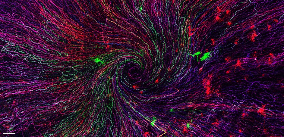
"The eye of the storm"
Sensory fibers of a mouse cornea imaged with a confocal microscope. The corneal nerves converge toward the centre forming a vortex. This particular transgenic mouse model allows stochastic expression of fluorescent proteins, unravelling the heterogeneity of the fiber origines inside the corneal epithelium.
Acknowledgements to Karine Loulier for the mouse model and Laetitia Hudececk for her help during the acquisition.
In the Institut des Neurosciences de Montpellier since 2020, Léna is a PhD student working in the team Eye lead by Dr. Frédéric Michon. This team is investigating the mechanisms related to the preservation and the integrity of the anterior part of the eye, including the lacrimal gland, the tears and the cornea. Léna’s project focuses on the cellular and molecular effects of the corneal innervation on the corneal homeostasis. The project goes further as they aim at highlighting new targets able to prevent and/or repair corneal damage.
The image she submitted for the 2021 France-BioImaging Image Contest (The eye of the storm) represents the sensory fibers of a mouse cornea. This innervation follows a typical pattern where all the nerves converge toward the centre forming a vortex. This particular transgenic mouse model allows random expression of fluorescent proteins, unravelling the heterogeneity of the fibers’ origin inside the corneal epithelium. As cornea is the most innervated tissue in the whole body, this model shows the differences between fibers. In pathological context, for example wound injury, it is thus possible to follow a specific fiber during the healing process.
France-Bioimaging sponsored her participation to the FOM (Focus on Microscopy) 2022 congress where she presented her project through a poster. Even though the congress was online, it gave her the opportunity to share her results with experts and as a consequence, to gather advice on her ongoing experiments.
2nd place: Eunice HoYee Chan, Muscle Dynamics Team, Developmental Biology Institute of Marseille (IBDM)
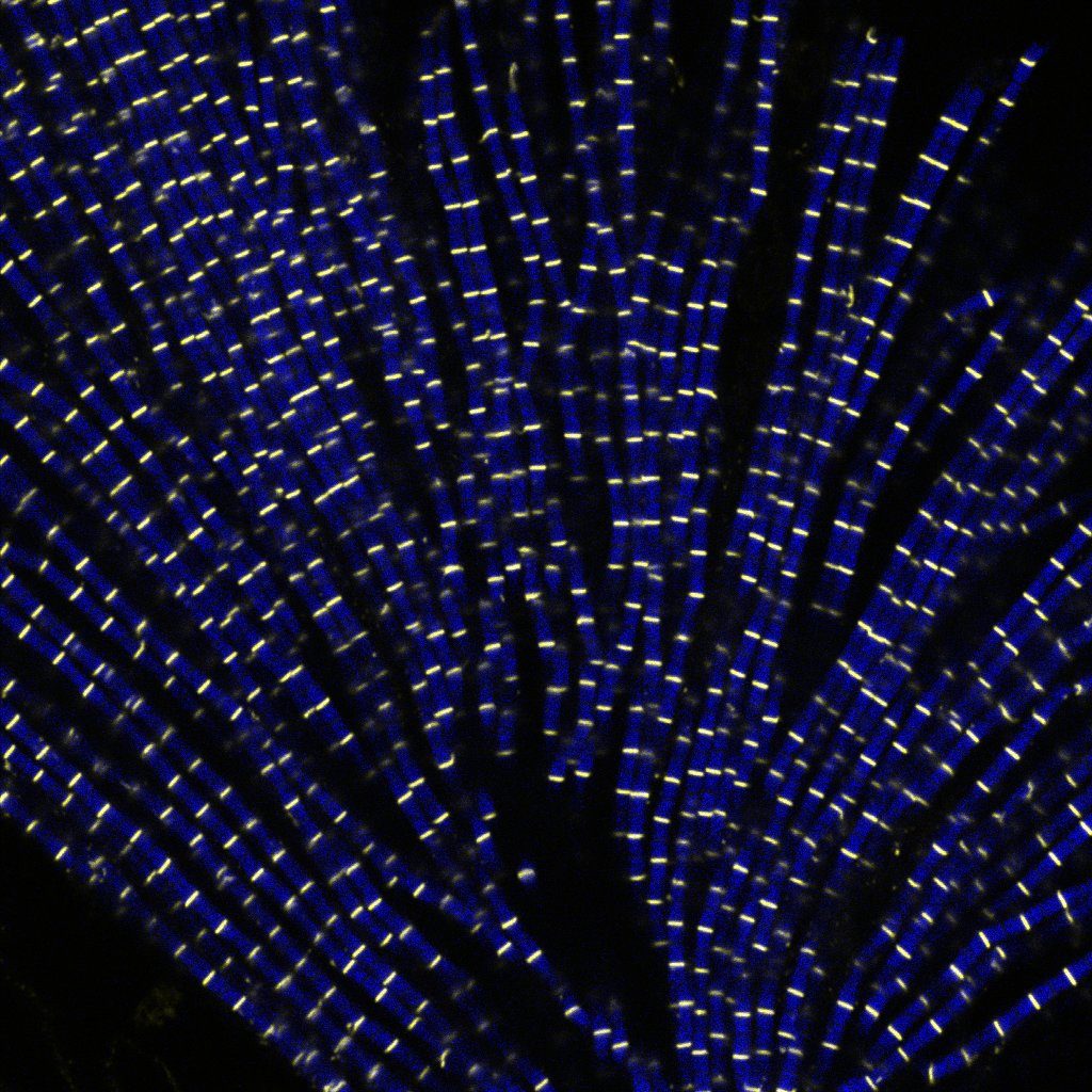
"Sarcomeric bouquet"
Myofibrils isolated from Drosophila indirect flight muscle labelled with titin (yellow) and actin (blue). Image captured from confocal microscope. We are studying the role of titin protein in muscle mechanics and organisation during development.
Research engineer in Frank Schnorrer's team at Institut de Biologie du Développement de Marseille (IBDM), Eunice focuses her research on Drosophila muscle dynamic and organisation during development using advanced biophysical and imaging techniques.
The image she submitted named “Sarcomeric bouquet" was from one of her very first muscle myofibrils isolation experiment. She dissected flight muscles from flies and labelled the individualised myofibrils with Llama nanobodies conjugated with different epitopes. Those labelled myofibrils were then subjected to various imaging methods including standard confocal microscopy, super resolution microscopy and cryo electron-tomogram. Using these novel labelling tools and imaging techniques, her team could study the dynamic and organisation of muscles during development in details.
France-BioImaging sponsored her registration to the 49th European Muscle Conference in Prague (22-26 September 2022). As she is new to the muscle field, this conference offered a great opportunity to have a broad view on different kind of state-of-the-art imaging techniques. Besides, she gave a presentation during the conference, highlighting her work and initiating discussion.
3rd Place: Camille Boutin, Biology of multiciliated cells Team, Developmental Biology Institute of Marseille (IBDM) & Nicolas Brouilly, PICsL Imaging facility, Electron Microscopy department
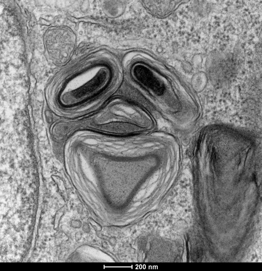
"Clown"
Lamellar structure in a differentiating multiciliated cell observed by transmission electron microscopy with a Tecnai G2 200kV FEI.
Camille is a researcher in Laurent Kodjabachian’s group at the Institut de Biologie du Développement de Marseille (IBDM). She develops projects as a principal investigator on the compartmentalization and sizing of multiciliated cells. With this in mind, she routinely uses confocal and super-resolution microscopy but also scanning and transmission electron microscopy and tomography.
Nicolas is in charge of the Electron Microscopy Unit of the Plateforme d’imagerie commune du site de Luminy (PICsL). In addition to the routine sample preparation and 2D TEM imaging, this imaging facility offers, to internal and external users, advanced sample preparation (cryo-methods, immunolabelling...) and advanced imaging (tomography, CLEM, serial blockface…).
To understand the production of multiple centrioles in multiciliate cells, they focused on the deuterosome, a membrane-less organelle that has been described 50 years ago but whose composition, organisation and function remain unknown to this day. In this context they have developed an inducible multiciliated cells line. This image was taken during the initial characterisation of this cell line by transmission electron microscopy.
Thanks to the France-Bioimaging Image Contest, Nicolas participated to the COST COMULIS Conference that was held by the Cyprus Institute in Nikosia. It was a great opportunity to exchange with the people at the cutting edge of the multi-modal imaging field. The program covered subjects such as the sample preparation for multi-modal imaging, image analysis and integrated industrial partners.