How to enhance brain intravital imaging? We asked Pierre Bourdoncle!

Recently, Pierre Bourdoncle, head of the IMAG’IC platform at the Cochin Institute (Paris Centre Node), and his team published a new protocol for intravital imaging of calvarial bone marrow. Today, he tells us more about their research and how it can enhance the study of diseases like leukemia.
Could you tell us a little about yourself and the project?
As the head of the IMAG’IC platform at the Cochin Institute, we have consistently advanced intravital imaging through multiphoton microscopy. For the past 25 years, we have been dedicated to enhancing intravital imaging at the Cochin Institute, with a focus on improving synchronization, laser technology, and OPO (Optical Parametric Oscillator, ed.) systems.
Why is the calvarial bone marrow such an interesting model to study hematopoiesis and vascular dynamics?
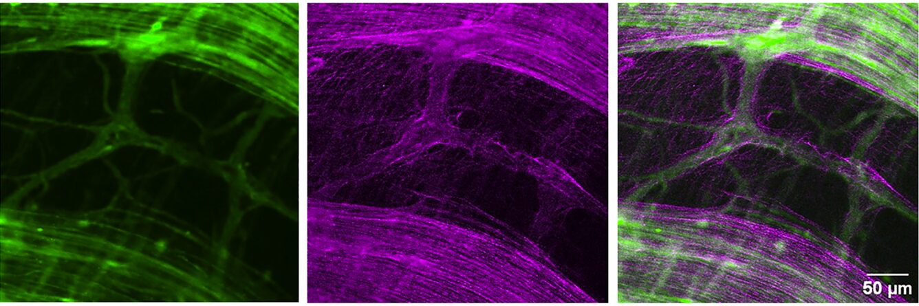
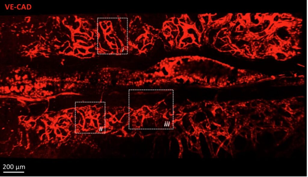
The calvarial bone marrow is an interesting model for studying hematopoiesis and vascular dynamics due to its unique anatomical features. Its thin structure allows for high-resolution imaging, facilitating the observation of cellular interactions and vascular networks. Additionally, it is easily accessible, making it ideal for experimental manipulations and real-time monitoring. This model provides valuable insights into the complex processes of blood cell formation and vascular development.
Your team has developed a custom-made titanium cranial implant. What advantages does it offer compared to existing methods?
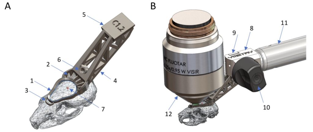
The stability of the imaging area has always been a major challenge in intravital microscopy. Indeed, the animal’s breathing and temperature variations complicate long-term acquisitions. Moreover, precise repositioning of the acquisition area over several days is essential for observing the evolution of the cellular environment. The development of titanium implants, as opposed to traditional resin 3D printing, allows for more robust fixation of the system to the microscope stage and, most importantly, limits the deformation of the implant.
What perspectives does this method open for the understanding of hematological diseases, such as leukemia?
This method opens significant perspectives for understanding hematological diseases like leukemia by enabling detailed visualization of disease progression and cellular interactions. It allows researchers to study the impact of treatments in real-time, enhancing the development of targeted therapies. Additionally, it facilitates the exploration of the bone marrow microenvironment’s role in disease pathogenesis.
What are your upcoming projects?
Following the same principle, we are collaborating with the company Ymetry to develop similar appendages adapted for soft organs. Our goal remains to maintain the acquisition area for as long as possible without any drift.
Click here to read the full scientific article!