Cell Dive: New technology available @H2P2
For several weeks now, the H2P2 histopathology platform located in Rennes (France BioImaging Bretagne-Loire Node) has become the European reference for the integral solution of Cell DIVE (https://www.leica-microsystems.com/products/light-microscopes/p/cell-dive/).
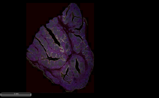
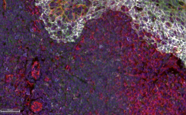
This technology brings great expectations for the research teams and the private companies with which we work. Leica’s Cell DIVE technology provides an in-depth solution for characterizing the tissue microenvironment using multiplexed imaging technology. Up to 60 biomarkers can be revealed in one tissue sample. An extensive list of antibodies is already validated and users can customize their own panel! The multiplexed Cell DIVE technology is based on successive immunolabeling of 4 antibodies conjugated with 4 fluorochromes (Cy2, Cy3, Cy5 and Cy7). The slides are digitized (x20 objective) as things progress and a final compiled image is obtained and can be analysed with the Halo Image Analysis Platform. This software allows users to do segmentation to highlight clusters, to define specific cell phenotypes, to analyse neighbourhood, heatmap…
For example, in cancer treatment research, researchers need a better understanding of the cellular architecture of normal and diseased tissues to develop better treatments and more accurately predict disease progression.
This technology has been developed by scientists for scientists over the last decade and several publications are available to date (https://www.leica-microsystems.com/products/light-microscopes/p/cell-dive/publications/).
For more information about the Cell Dive technology or to discuss your project, you can contact Nicolas Mouchet nicolas.mouchet@univ-rennes1.fr