Direct and simultaneous observation of transcription and chromosome architecture in single cells with Hi-M, Nature Protocols 2020
▽ Scroll down
Category: News from Nodes
Direct and simultaneous observation of transcription and chromosome architecture in single cells with Hi-M
Andrés M. Cardozo Gizzi, Sergio M. Espinola, Julian Gurgo, Christophe Houbron, Jean-Bernard Fiche, Diego I. Cattoni, Marcelo Nollmann
Simultaneous observation of 3D chromatin organization and transcription at the single cell level and with high spatial resolution may hold the key to unveil the mechanisms regulating embryonic development, cell differentiation and even disease. We have recently developed Hi-M, a technology that allows for the sequential labelling, 3D imaging and localization of multiple genomic DNA loci together with RNA expression in single cells within whole, intact Drosophila embryos. Importantly, Hi-M enables simultaneous detection of RNA expression and chromosome organization without requiring sample unmounting and primary probe re-hybridization. Here, we provide a step-by-step protocol describing the design of probes, the preparation of samples, the stable immobilization of embryos into microfluidics chambers, and the complete procedure for image acquisition. The combined RNA/DNA fluorescence in situ hybridization procedure takes 4-5 days including embryo collection. In addition, we describe image analysis software to segment nuclei, detect genomic spots, correct for drift and produce Hi-M matrices. A typical Hi-M experiment takes 1-2 days to complete all rounds of labelling and imaging and 4 additional days for image analysis. This technology can be easily expanded to investigate cell differentiation in cultured cells, or organization of chromatin within complex tissues.
ATP-driven separation of liquid phase condensates in bacteria
B. Guilhas, J.C. Walter, J. Rech, G. David, N.-O. Walliser, J. Palmeri, C., Mathieu-Demaziere, A. Parmeggiani, J.Y. Bouet, A. Le Gall1, M. Nollmann
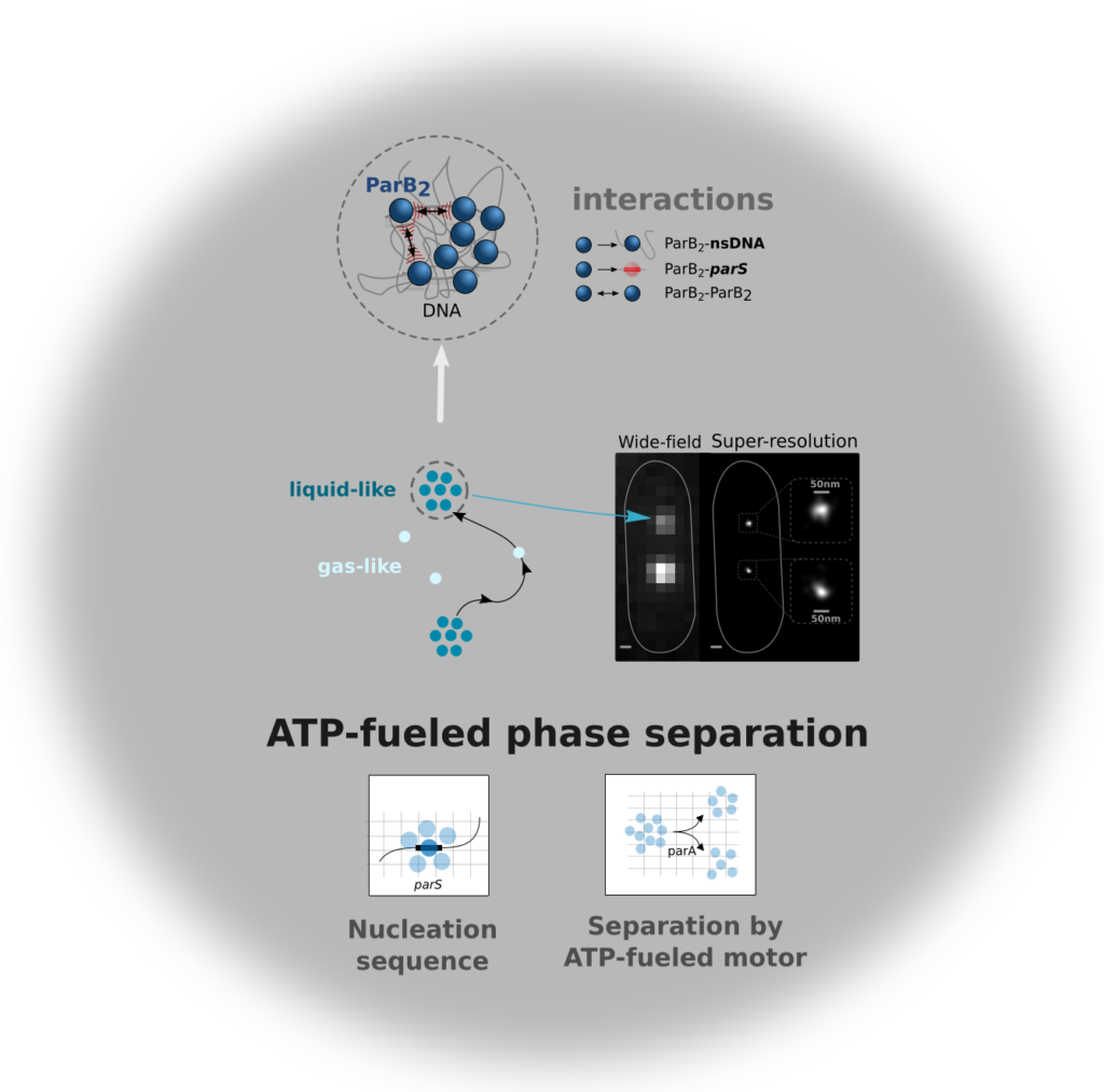
Liquid-liquid phase separated (LLPS) states are key to compartmentalise components in the absence of membranes, however it is unclear whether LLPS condensates are actively and specifically organized in the sub-cellular space and by which mechanisms. Here, we address this question by focusing on the ParABS DNA segregation system, composed of a centromeric-like sequence (parS), a DNA-binding protein (ParB) and a motor (ParA). We show that parS-ParB associate to form nanometer-sized, round condensates. ParB molecules diffuse rapidly within the nucleoid volume, but display confined motions when trapped inside ParB condensates. Single ParB molecules are able to rapidly diffuse between different condensates, and nucleation is strongly favoured by parS. Notably, the ParA motor is required to prevent the fusion of ParB condensates. These results describe a novel active mechanism that splits, segregates and localises non-canonical LLPS condensates in the sub-cellular space.
Guilhas et al. revealed that the bacterial DNA segregation apparatus behaves as a non-canonical phase separation system. This apparatus employs an ATP-powered motor that splits nanometer-sized condensates and localizes them robustly within the nucleoid to ensure faithful transmission of genetic material.
The ability to communicate effectively with each other is one of the strongest predictors for our chances to get ahead in life. In their latest publication in Science Advances, scientists and engineers from IGF-Montpellier (CNRS, INSERM, Univ. Montpellier), IPAM platform (BioCampus Montpellier, France-Bioimaging Montpellier Node) and ARO-Israel demonstrated that this also holds true for GnRH neurons.
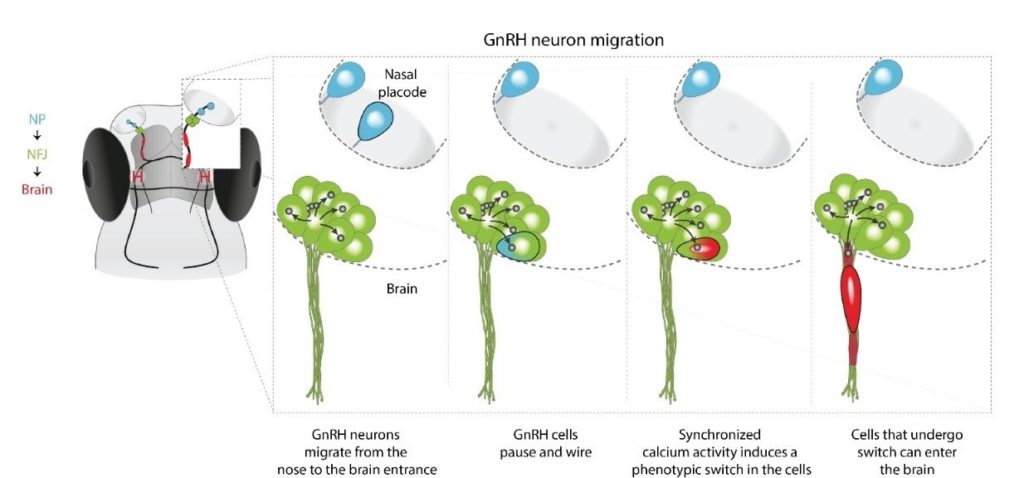
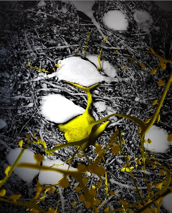
In humans and all vertebrates, species survival depends on a critical step during embryonic development: the migration of a small subset of GnRH neurons (about 2,000 in humans and less than 100 in fish) from the nose to the brain where they join the hypothalamus to control reproduction. Their latest results unveiled that GnRH neurons make a pause at the nose-brain frontier where they function as an inter-hemispheric network that is isolated from the rest of the brain. Only neurons that integrate into the network and are able to communicate with their neighbors will finally cross the barrier and make their way into the brain, towards their hypothalamic destination.
In other words, these GnRH neurons, that are critical for species persistence, face the same challenges like other immigrants: they must learn to communicate effectively if they are to integrate into their new world.
In this study, in vivo 2-photon microscopy was a key tool for:
Long term imaging with minimal bleaching and phototoxicity
Upright configuration enabling dorsal imaging of the fish in its natural position
Long-distance water-immersion objectives allowing imaging of deep tissue structures without sacrificing image quality
Fast calcium imaging
Imaging of red GECI using the higher wavelengths
Precise cell ablation
Photoactivation of ChR2 while monitoring Ca in the red channel
A graphical model illustrating the migration of a single GnRH neuron (marked by black border) from the nasal placode into the zebrafish brain.
Congratulations to Emmanuel Beaurepaire (CNRS Research Director from the Laboratory for Optics and Biosciences CNRS-INSERM-Polytechnique),PI of the ERC Synergy Grant project “HOPE”, and to Laurent Groc (CNRS Research Director ; Interdisciplinary Institute for Neuroscience), coordinator of theERC Synergy Grant project “ENSEMBLE“, Laurent Cognet (CNRS Research Director ; Laboratoire photonique numérique et nanosciences) and U. Valentin Nägerl (Professor at University of Bordeaux ; CNRS Research Director ; Interdisciplinary Institute for Neuroscience), both PIs of theERC Synergy Grant project “ENSEMBLE“.
These grants, each worth around 10 million euro over six years, are designed to enable groups of 2 to 4 scientists to tackle some of the world’s most challenging research problems, spanning several scientific disciplines.
The ERC Synergy Grant scheme is part of the EU’s research and innovation programme, Horizon 2020.
ERC Synergy Grant project “HOPE”
“Reverse engineering the assembly of the hippocampal scaffold with novel optical and transgenic strategies”
Emmanuel Beaurepaire, Directeur de recherche CNRS au Laboratoire d’optique et biosciences – LOB (CNRS/École polytechnique/INSERM),
Rosa Cossart, Directrice de recherche CNRS (Unité INSERM, Aix-Marseille Univ.)
Jean Livet, chercheur INSERM à l’Institut de la vision (CNRS, INSERM, Sorbonne Univ.)
At the heart of our brain, a structure plays a key role in memory, and more particularly in the acquisition and maintenance of our memories: the hippocampus. Classically considered as a “cognitive GPS” for space and time, it is also the seat of our episodic memory.
Over the last decade, the neural circuits of the hippocampus have been better described, in particular by the team of Rosa Cossart, director of the Institut de neurobiologie de la méditerranée (Inmed), but the nature, origin and remodeling of these circuits during development and pathologies remain to be understood.
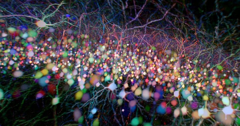
On the other hand, genetic engineering techniques for staining neurons, developed by Jean Livet, Inserm research director at the Institut de la vision, coupled with multi-photon microscopy developed by the team of Emmanuel Beaurepaire, CNRS research director at the Laboratoire d’optique et biosciences – LOB (illustration below / read the 2019 press release in French), have demonstrated their ability to accurately map the complex architecture of neuronal circuits and their evolution during development.
By combining these exceptional multidisciplinary advances, HOPE aims to answer three interdependent questions:
Is the architecture of the adult seahorse carried by specific circuits?
Are the circuits of the hippocampus pre-wired or shaped by experience?
How does this structure reorganize itself in pathological conditions?
HOPE aims to shed new light on the function of the hippocampus and the role of its neuronal circuits through the design of a new, non-invasive and universal method to monitor the growth and construction of brain circuits located deep in the brain, from their neurogenesis to adulthood, under normal and pathological conditions.
“Structure and functions of the brain extracellular space“
Laurent Groc (Research Director CNRS ; Interdisciplinary Institute for Neuroscience),
Erwan Bézard (Research Director INSERM; Institute of Neurodegenerative Disorders),
Laurent Cognet (Research Director CNRS ; Laboratoire photonique numérique et nanosciences)
U. Valentin Nägerl (Professor at University of Bordeaux ; Research Director CNRS ; Interdisciplinary Institute for Neuroscience)
The ENSEMBLE project aims at underpinning the molecular mechanisms of physiological and pathological brain function. This ambitious and innovative endeavor is based on our ability to develop new approaches in high-resolution microscopy at the service of a new conceptual framework in brain cell communication.
Credit: U.V. Nägerl
This project has roots in the international leadership of the Bordeaux communityin the fields of microscopy, nanophotonics, fundamental and translational neuroscience. The opportunity that is offered to these 4 investigators to break a frontier knowledge was permitted by the continuous support of local institutional actors. The installation of Prof. Valentin Nägerl’s laboratory in 2009 with a “Chaire Accueil” from the Regional Council of Aquitaine, the support of LabEx BRAIN, the Laphia Cluster and the IdEx of the University of Bordeaux provided the ground to build elementary blocks necessary for the challenging adventure of the ERC Synergy project (10 million euros, 6 years).
We are very pleased to announce that FBI Bretagne-Loire Node application to become a Euro BioImaging facility has been evaluated as highly recommended by the EuBI Scientific Advisory Board (SAB) and ratified by the EuBI Board on November 30th, 2020.
The Bretagne Loire Node became a node of the national infrastructure France BioImaging in November 2019 and applied to become a EuBI facility during the last EuBI Call for Nodes (June 2020).
The Bretagne Loire Node brings together four cellular imaging and histology facilities, two in Rennes (MRic and H2P2) and two in Nantes (MicroPIcell and APEX). These facilities have complementary expertise for live imaging and pathological anatomy. The added value of the Bretagne Loire Node is to be able to offer a continuum between biological imaging and medical imaging, through the development of a new line of services as well as methodological and technological transfer to users of microscopy technologies for preclinical studies.
As part of the Euro-BioImaging research infrastructure, the services provided by these facilities will be open to all scientists, regardless of their discipline or affiliation.
The PICsL-FBI microscopy core facility is located on two sites: Centre d’Immunologie de Marseille Luminy (CIML) and Institut de Biologie du Développement de Marseille (IBDM).The PICsL-FBI facility of the CIML called ImagImm (Imaging Immunity) via its microscopy resources – from the molecule to whole organisms – is dedicated to help its users deciphering cellular mechanisms in the fields of immunology.
Major research implications of the ImagImm facility:
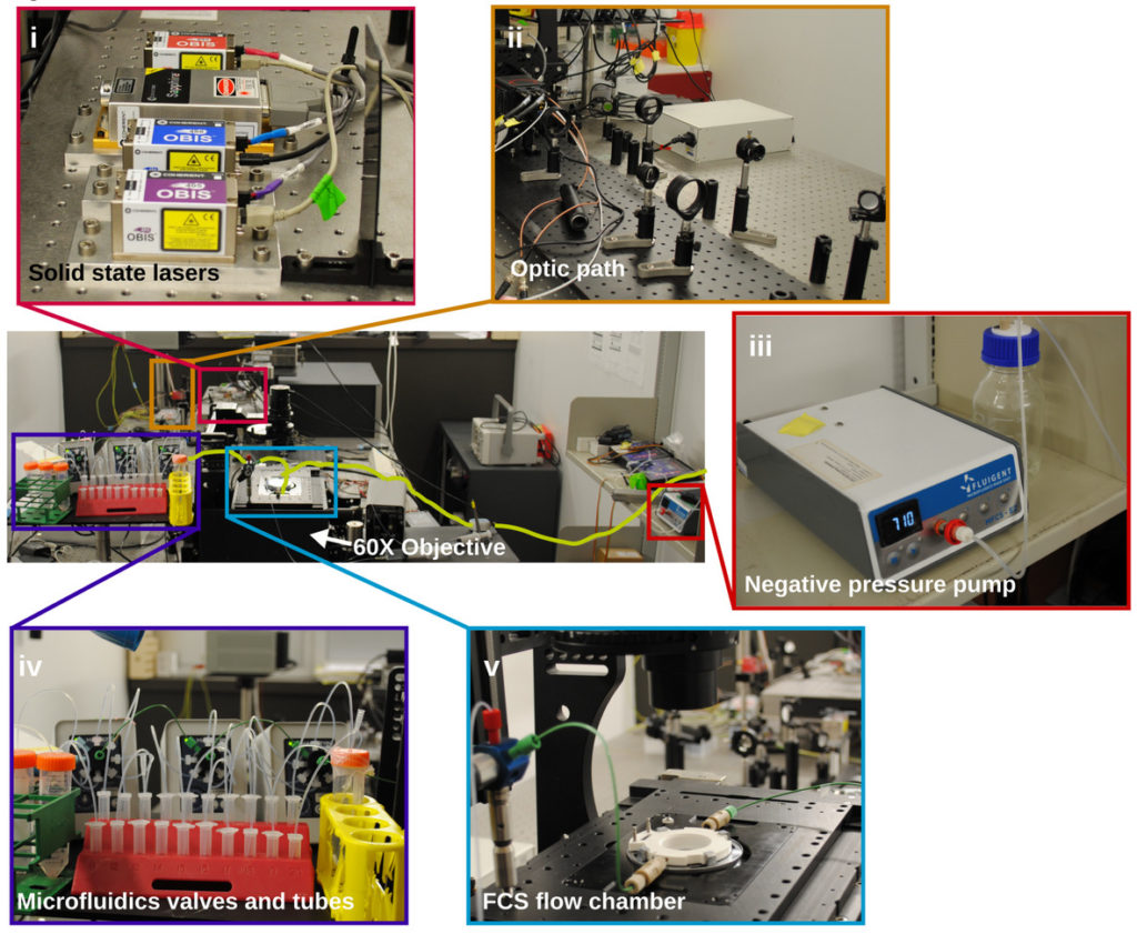
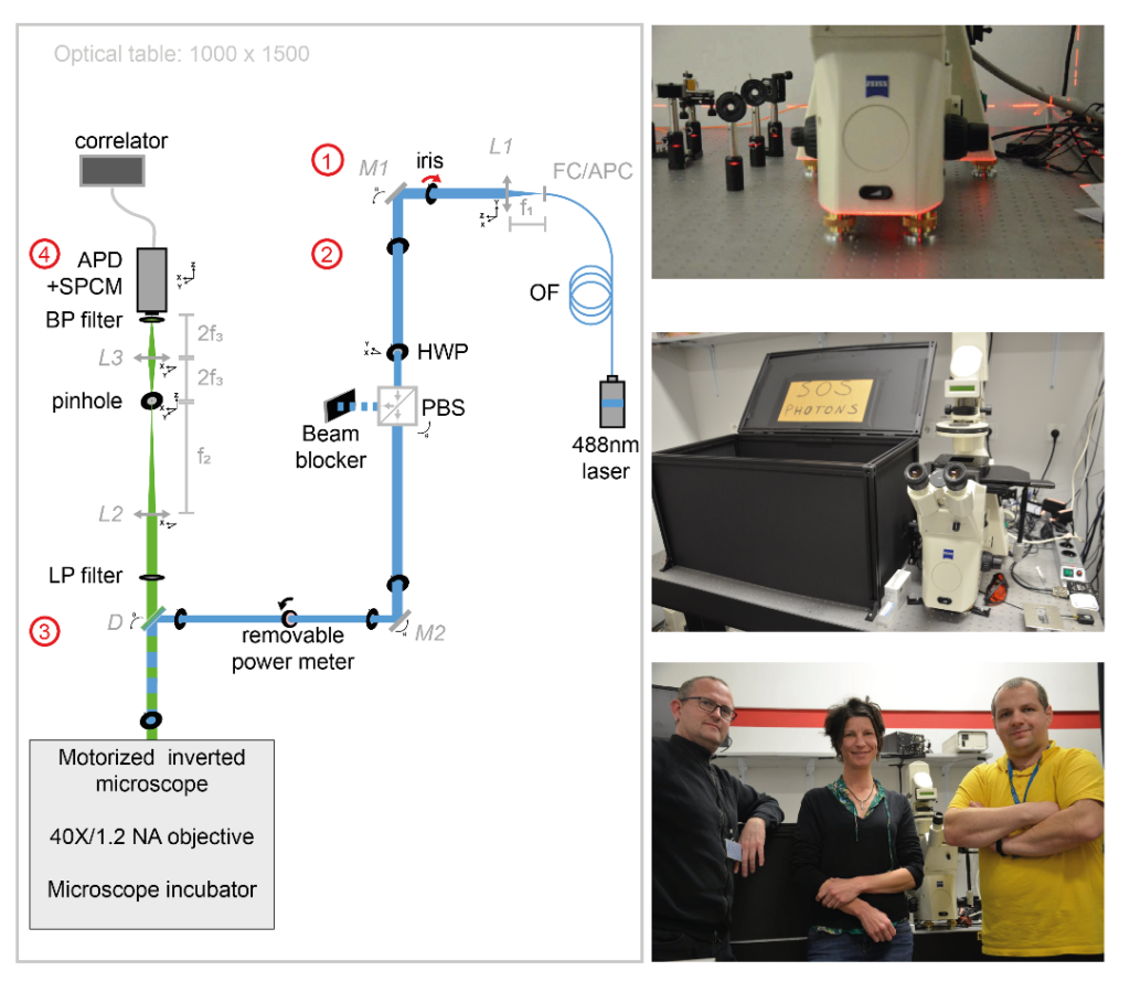
In collaboration with Tomasz Trombik (Faculty of Biotechnology, University of Wroclaw – Wroclaw, Poland), Sophie Brustlein (Institut de Convergences Centuri, AMU,CNRS – Marseille, France) and Nicolas Bertaux (Institut Fresnel, AMU, Centrale Marseille, CNRS – Marseille, France), Sébastien Mailfert and Didier Marguet published the procedure for implementing spot variation Fluorescence Correlation Spectroscopy (svFCS) measurements using a classical fluorescence microscope that has been customized1. This publication is following the technology transfer made in 2018: the svFCS developed by Didier Marguet’s lab was duplicated by Sébastien Mailfert and Sophie Brustlein and built from scratch in 7 days on site, in Poland.
Dynamic biological processes in living cells, including those associated with plasma membrane organization, occur on various spatial and temporal scales, ranging from nanometers to micrometers and microseconds to minutes, respectively. Such a broad range of biological processes challenges conventional microscopy approaches. The published protocol includes a specific performance check of the svFCS setup and the guidelines for molecular diffusion measurements by svFCS on the plasma membrane of living cells under physiological conditions. Additionally, a procedure for disrupting plasma membrane raft nanodomains by cholesterol oxidase treatment is provided and how these changes in the lateral organization of the plasma membrane might be revealed by svFCS analysis. This fluorescence-based method can provide unprecedented details on the lateral organization of the plasma membrane with the appropriate spatial and temporal resolution.
Figure 1:Schematic view of excitation and emission optical paths of the svFCS setup and pictures of the setup. The svFCS setup contains four modules: (1) the output of a fibered 488 nm laser is collimated, (2) a combination of a half-wave plate and polarizing beamsplitter sets the optical power, (3) the laser beam focused on the sample after traveling through a tube-lens free motorized microscope, and (4) the fluorescence is detected through a confocal-like detection path onto an avalanche photodiode coupled to a single photon counting module, which delivers a signal to a hardware correlator. Simplicity gives the system its sensitivity, robustness, and ease of use.
SAPHIR : a Shiny application to analyze tissue section images
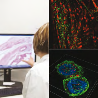
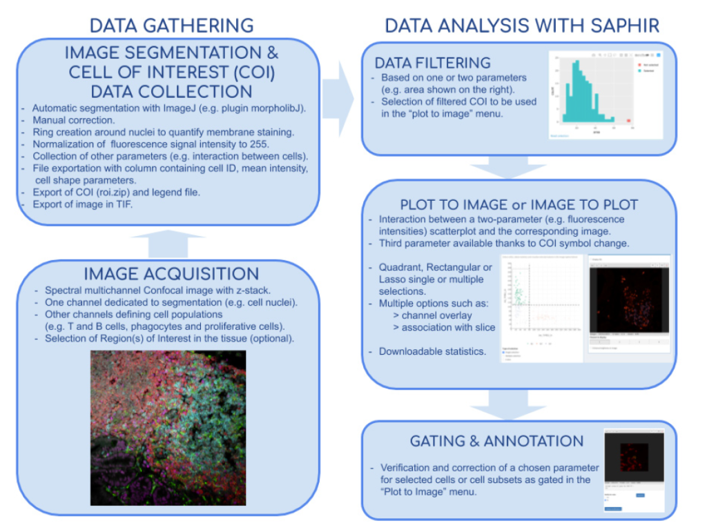
In collaboration with Hugues Lelouard (CIML, Inserm, CNRS, AMU) and Elodie Germani, Mathieu Fallet published a powerful method for both basic and medical research to study cell populations in tissues using immunofluorescence. Image acquisitions performed by confocal microscopy notably allow excellent lateral resolution and more than 10 parameter measurement when using spectral or multiplexed imaging. Analysis of such complex images can be very challenging and easily lead to bias and misinterpretation. They developed the Shiny Analytical Plot of Histological Images Results (SAPHIR), an R shiny application for histo-cytometry using scatterplot representation of data extracted by segmentation. It offers many features, such as filtering of spurious data points, selection of cell subsets on scatterplot, visualization of scatterplot selections back into the image, statistics of selected data and data annotation. This application allows to quickly characterize labeled cells, from their phenotype to their number and location in the tissue, as well as their interaction with other cells.
Figure 2: Flow chart of tissue image analysis from image acquisition and segmentation (left side) to extract data analysis with SAPHIR (right side)
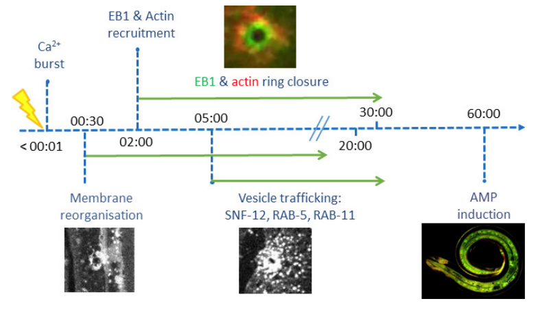
Wound healing in C. elegans
In collaboration with Nathalie Pujol and Jonathan Ewbank (CIML, Inserm, CNRS, AMU), Mathieu Fallet and Sébastien Mailfert participated in the project on the immune response by showing that wounding provokes a reorganization of plasma membrane subdomains3. The skin protects animals from infection and physical damage. In Caenorhabditis elegans, wounding the epidermis triggers an immune reaction and a repair response, but it is not clear how these are coordinated. Previous work implicated the microtubule cytoskeleton in the maintenance of epidermal integrity (Chuang et al., 2016). Taffoni et al. show the reorganization of the plasma membrane subdomains by a simple wounding system. This is followed by recruitment of the microtubule plus end-binding protein EB1/EBP-2 around the wound and actin ring formation, dependent on ARP2/3 branched actin polymerization. They show that microtubule dynamics are required for the recruitment and closure of the actin ring, and for the trafficking of the key signaling protein SLC6/SNF-12 toward the injury site. Without SNF-12 recruitment, there is an abrogation of the immune response. These results suggest that microtubule dynamics coordinate the cytoskeletal changes required for wound repair and the concomitant activation of innate immunity.
Figure 3: Time line of events
References:
Mailfert, S., Wojtowicz, K., Brustlein, S., Blaszczak, E., Bertaux, N., Łukaszewicz, M., Marguet, D., Trombik, T. Spot Variation Fluorescence Correlation Spectroscopy for Analysis of Molecular Diffusion at the Plasma Membrane of Living Cells, JoVE, 165, 1-19 (2020).
Germani, E., Lelouard, H., Fallet, M. SAPHIR: a Shiny application to analyze tissue section images, F1000Research, Faculty of 1000, 9, 1276-1285 (2020).
Taffoni, C., Omi, S., Huber, C., Mailfert, S., Fallet, M., Rupprecht, J-F,. Ewbank, J., Pujol., N. Microtubule plus-end dynamics link wound repair to the innate immune response, eLIFE, 9, e45047 (2020)
With his team members, Patrick Lemaire is studying the embryonic development of a small marine invertebrate, the sea squirt Phallusia mammillata, chosen for the simplicity and transparency of its embryos. His latest work has combined microscopy, image analysis and mathematical modeling approaches to describe, cell by cell, the embryogenesis of this animal and to analyze the role of communication between cells.
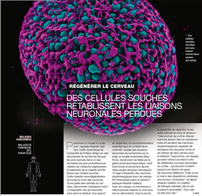
The image depicts a spheroid of human stem cells (green) and its actin cytoskeleton (purple), produced by Philippe Cohen during its PhD at Treefrog. This nice picture serves as an illustration for an article covering the use of stem cells for regenerative medicine. Acquisition was made by Philippe Cohen on a scanning confocal microscope and 3D rendering was done by Jérémie Teillon using Agave software. Agave is a free 3D visualization software, using light path-trace light rendering.
The Bordeaux Imaging Center team offers training and support on 3D commercial softwares such as Imaris and Arivis as well as other freeware such as Agave. Don’t hesitate to contact them (bic@u-bordeaux.fr) if you are interested in 3D rendering and visualization of your microscopy data!
During embryonic development, cells take on increasingly precise roles in the body as they divide. Be they skin cells, muscle cells or neurons, the different cell types that make up the embryo emerge gradually from a very fine orchestration of their positions and identities, coordinated by the signals they exchange with each other. Like us, the cells need to “talk” to each other to make decisions.
Screaming or whispering: the embryonic cell dilemma
In vertebrate embryos, cells have a very dynamic behaviour. They move around, exchange their neighbours or migrate over long distances. The signals they exchange therefore need to have a long range, which could be characterized as “shouting”. The study of the embryonic development of a sea squirt, a small marine animal with optically transparent embryos, has enabled scientists from several teams at CNRS and INRIA in France, in collaboration with a team from the European Molecular Biology Laboratory (EMBL, Germany), to capture and describe in detail a more discreet mode of cell communication.
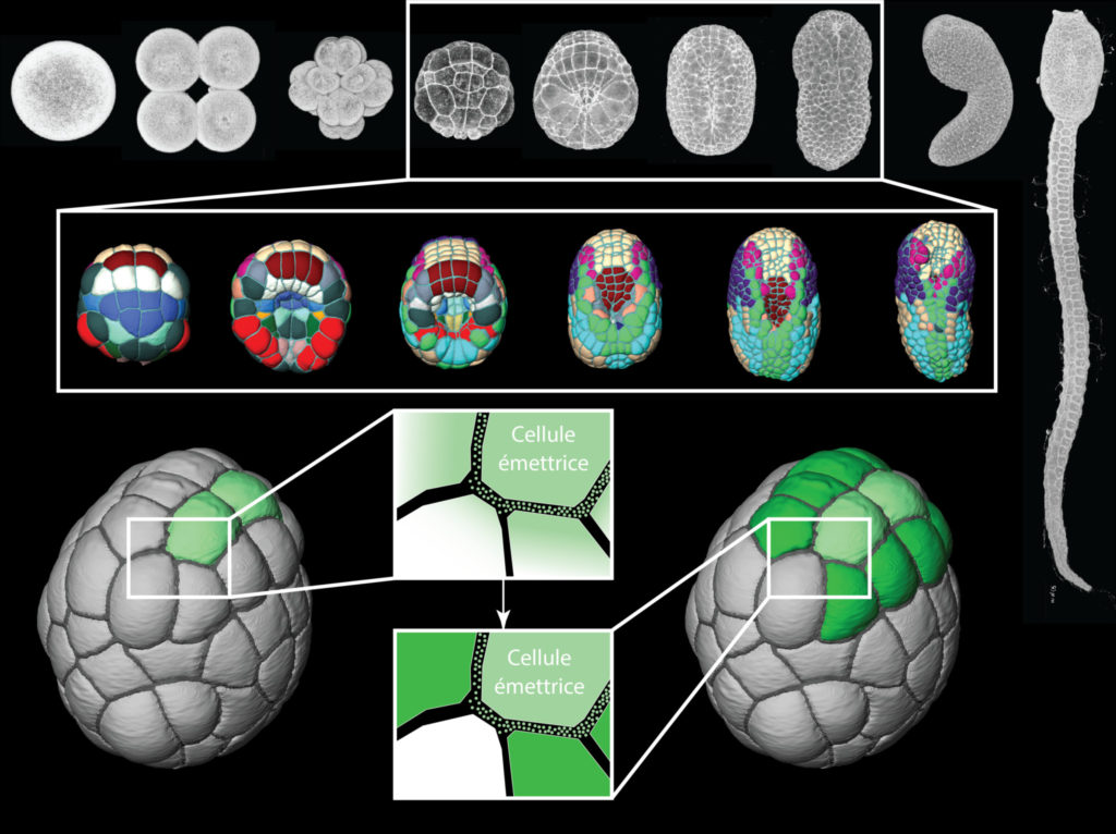
The scientists recorded the development of live embryos every two minutes with a new-generation « light-sheet » microscope. They then created software to automatically detect each cell and analyze its position, shape and neighbours up to an advanced stage of development. This work revealed an unusually reproducible mode of development, in which the same cell can be found in the same position across all embryos and where cells move very little in relation to each other. The authors of the study then annotated the films thus made with information on the cell type and the molecular signals emitted by each cell. Using mathematical modelling to integrate the quantitative description of the embryonic geometry with these annotations, their work suggest that cells communicate with very short-range signals. Moreover, the interpretation of these signals is modulated by the area of the contacts between cells. Unlike vertebrates, the cells of ascidian embryos thus have a static and fixed behaviour and the range of their “whispered” signals is very small.
Top: embryonic development of an ascidian from egg to tadpole. The part framed in white is the part of embryogenesis that we have imaged and then segmented (below, segmented cells coloured according to their cell fate). The lower part of the figure illustrates that the light green cells “whisper” instructions to their immediate neighbours by short-range signals.
This study indicates that the dynamics of cell movement varies greatly between animals and that these different modalities could be strongly related to the range of signals that the cells exchange with each other. By extending the repertoire of cellular communication mechanisms, this work opens new perspectives on the understanding of self-organization strategies of living forms.
Article: L. Guignard*, U.-M. Fiuza*, B. Leggio, J. Laussu, E. Faure, G. Michelin, K. Biasuz, L. Hufnagel, G. Malandain, C. Godin#, P. Lemaire# (2020) Contact-area dependent cell communications and the morphological invariance of ascidian embryogenesis (Science, July 10 2020 issue, https://science.sciencemag.org/content/369/6500/eaar5663)
During this year 2020, the MicroPICell facility from the Bretagne Loire Node acquired several imaging systems, some of which offer access to new technologies on the Nantes health research site:
a complete Zeiss Lighsheet 7 light sheet microscope associated to an X-Clarity clearing system and an Arivis Vision 4D Offline station,
a motorized Nanolive holotomographic microscope,
a high-end Nikon confocal microscope (resonant, spectral, FLIM, large field of view),
an Akoya CODEX system of multiplex fluorescent tissue labeling.
Holography offers a unique means to measure cells in their native environment: label-free, non-invasive, manipulation-free, and interference-free.
Moreover, the MicroPICell facility, in collaboration with the training organization of the CNRS, is organizing in March 2021 a training on histology: from sample preparation to markers validation by image analysis. This training (lectures, workshops) will take place over 4 days between 03/22/2021 and 04/24/2021.
We use cookies on our website to give you the most relevant experience by remembering your preferences and repeat visits. By clicking “Accept All”, you consent to the use of ALL the cookies. However, you may visit "Cookie Settings" to provide a controlled consent.
This website uses cookies to improve your experience while you navigate through the website. Out of these, the cookies that are categorized as necessary are stored on your browser as they are essential for the working of basic functionalities of the website. We also use third-party cookies that help us analyze and understand how you use this website. These cookies will be stored in your browser only with your consent. You also have the option to opt-out of these cookies. But opting out of some of these cookies may affect your browsing experience.
Necessary cookies are absolutely essential for the website to function properly. These cookies ensure basic functionalities and security features of the website, anonymously.
Cookie
Duration
Description
cookielawinfo-checkbox-analytics
11 months
This cookie is set by GDPR Cookie Consent plugin. The cookie is used to store the user consent for the cookies in the category "Analytics".
cookielawinfo-checkbox-functional
11 months
The cookie is set by GDPR cookie consent to record the user consent for the cookies in the category "Functional".
cookielawinfo-checkbox-necessary
11 months
This cookie is set by GDPR Cookie Consent plugin. The cookies is used to store the user consent for the cookies in the category "Necessary".
cookielawinfo-checkbox-others
11 months
This cookie is set by GDPR Cookie Consent plugin. The cookie is used to store the user consent for the cookies in the category "Other.
cookielawinfo-checkbox-performance
11 months
This cookie is set by GDPR Cookie Consent plugin. The cookie is used to store the user consent for the cookies in the category "Performance".
viewed_cookie_policy
11 months
The cookie is set by the GDPR Cookie Consent plugin and is used to store whether or not user has consented to the use of cookies. It does not store any personal data.
Functional cookies help to perform certain functionalities like sharing the content of the website on social media platforms, collect feedbacks, and other third-party features.
Performance cookies are used to understand and analyze the key performance indexes of the website which helps in delivering a better user experience for the visitors.
Analytical cookies are used to understand how visitors interact with the website. These cookies help provide information on metrics the number of visitors, bounce rate, traffic source, etc.
Advertisement cookies are used to provide visitors with relevant ads and marketing campaigns. These cookies track visitors across websites and collect information to provide customized ads.