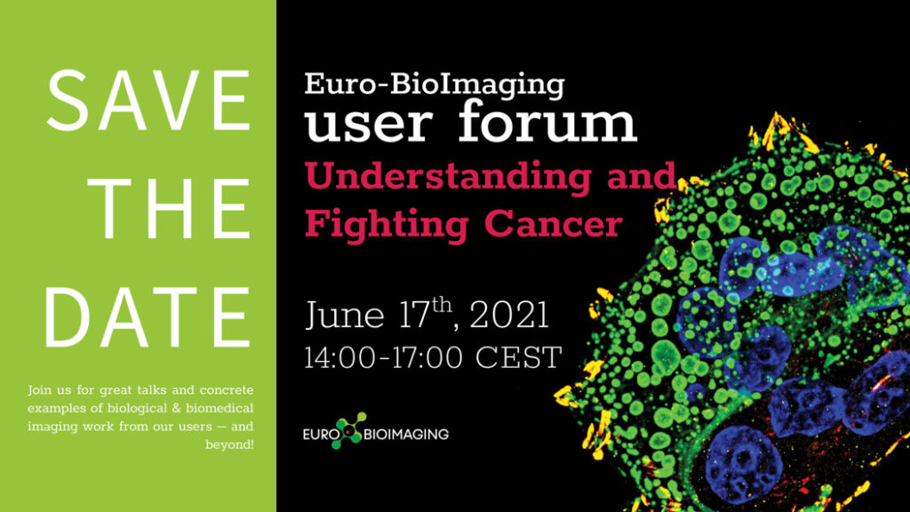
EuBI User Forum: “Understanding and Fighting Cancer” – 17/06
▽ Scroll down
Category: Announcement
Save the date! The European Research Infrastructure Euro BioImaging (EuBI) is organizing an online User Forum on “Understanding and Fighting Cancer”. The event takes place on June 17, 2021 from 14:00-17:00 CEST and will highlight the importance of cutting-edge imaging technologies in support of cancer research and showcase the specific expertise available at the EuBI Nodes.
In addition, keynote presentations from Kevin Brindle, University of Cambridge, and Frank Winkler, DKFZ, will further reveal the potential of biological and biomedical imaging technologies to boost cancer research.
The full program is coming soon! In the meantime, you can register here.
Quantifying translation in space and time during development
During development, precise control of gene expression allows the reproducible establishment of patterns, leading to the formation of organs at the right time and place.
The establishment of developmental patterns has been primarily studied at the transcriptional level. In comparison, the fate of these transcripts received little attention.
Dufourt*, Bellec* et al deployed the SunTag labeling method to image the dynamics of translation of individual mRNA molecules in living Drosophila embryos. This led to the discovery of translation factories and unmasked important heterogeneities in the efficiency of translation between identical mRNAs, demonstrating a novel layer of fine-tuning of gene expression.
Institut de Génétique Moléculaire de Montpellier (Univ.Montpellier/CNRS) 1919 route de Mende, 34090 Montpellier
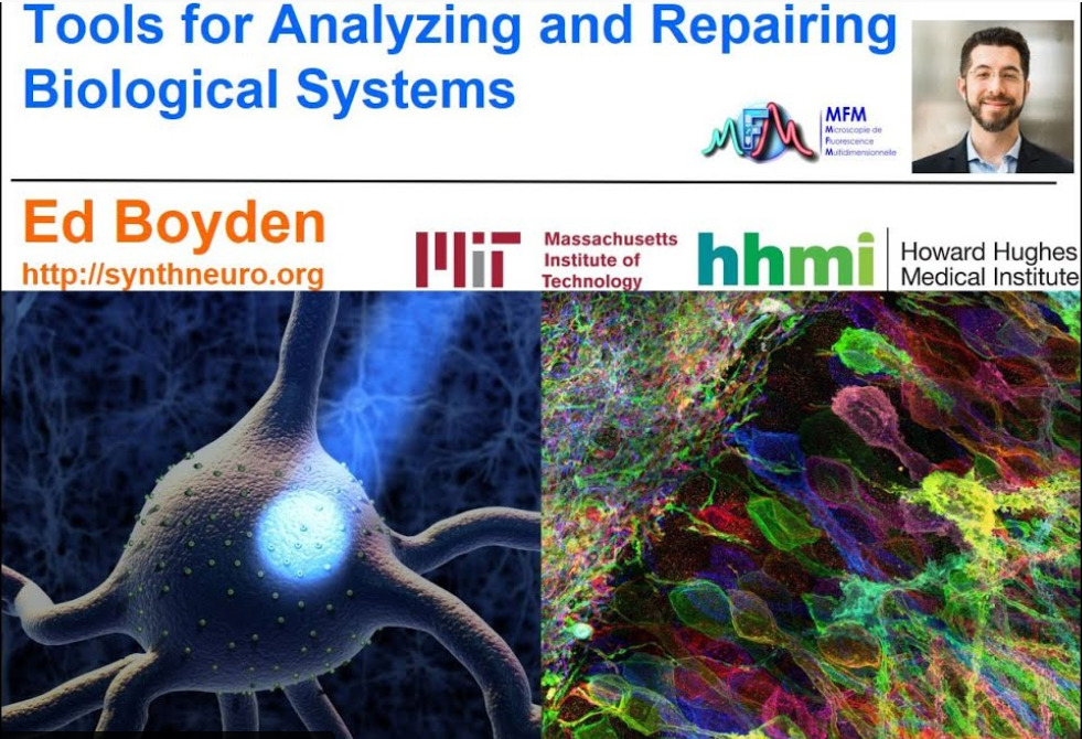
On May 25th, 2021, 15:00 CSET, our partner the French Network for Multidimensional Optical Fluorescence Microscopy will receive Edward S. Boyden* from the MIT, USA for a webinar on Expansion Microscopy: “Tools for analyzing and repairing biological systems”.
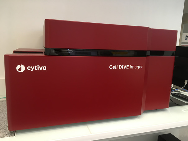
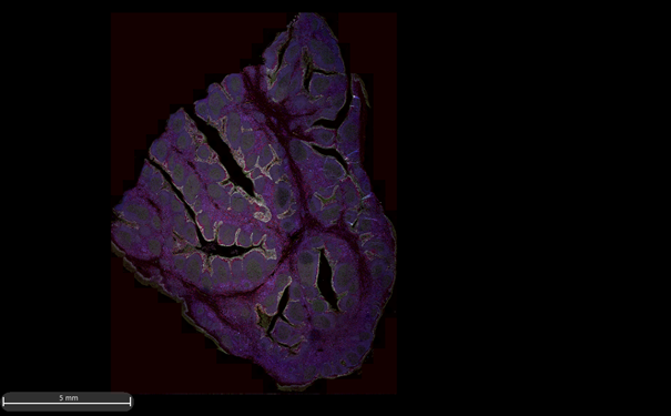
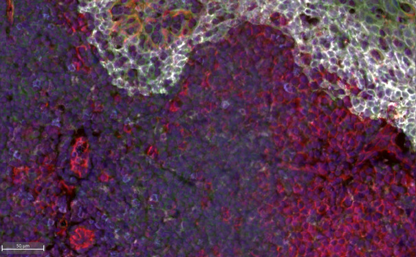
This technology brings great expectations for the research teams and the private companies with which we work. Leica’s Cell DIVE technology provides an in-depth solution for characterizing the tissue microenvironment using multiplexed imaging technology. Up to 60 biomarkers can be revealed in one tissue sample. An extensive list of antibodies is already validated and users can customize their own panel! The multiplexed Cell DIVE technology is based on successive immunolabeling of 4 antibodies conjugated with 4 fluorochromes (Cy2, Cy3, Cy5 and Cy7). The slides are digitized (x20 objective) as things progress and a final compiled image is obtained and can be analysed with the Halo Image Analysis Platform. This software allows users to do segmentation to highlight clusters, to define specific cell phenotypes, to analyse neighbourhood, heatmap…
For example, in cancer treatment research, researchers need a better understanding of the cellular architecture of normal and diseased tissues to develop better treatments and more accurately predict disease progression.
For more information about the Cell Dive technology or to discuss your project, you can contact Nicolas Mouchet nicolas.mouchet@univ-rennes1.fr
Grant Applications for organizing Virtual/Hybrid Training Schools are open!
COMULIS is now launching a call to financially support virtual and hybrid training schools fulfilling the following conditions:
the training school has to take place between the 1st of June and 30th of October 2021;
it has to be virtual (or hybrid);
it has to cover topics of the COMULIS network;
led by a COMULIS member or someone who is willing to become one;
COMULIS and COST support will have to be diplayed on the program, website, or any related document to the training school;
fees that can be covered by this grant include the technical setup of these training schools and training material:
If engaging a conference organiser, technician hourly rate if required on specific openings days before and during the event to assist with technical support, attendee management and monitoring, configuration and setup, communication.
customer support for attendees, live-stream tech support via email and/or chat.
pre-recording of keynotes/teaching sessions for training schools.
post-event process management: video editing, recording management.
Rental of rooms and audio-visual material
Consumables purchased for Training Schools
Photocopying and the printing of programmes, handouts, event materials, book of abstracts, book of proceedings, flyers etc
Maximum amount of a grant is 10000 euros, that will be reimbursed on presentation of invoices strictly related to eligible fees above.
Procedure
Filling an online form https://forms.gle/tJqaLmauZDA5V58o6 with check of the above conditions, (pedagogical) program, list of organizers, speakers and trainers, dates, provisional budget including usage of the COMULIS financial support in regards of one or several of the above categories of eligible expenses.
Deadline 1st of June. Notification of acceptance: 15th of June. (if needed earlier please do tell us, and we will do our best to meet you own deadline in case of co-funding).
Criteria among eligible proposals (fulfilling the above checklist) will be based on matching COMULIS objectives (www.comulis.eu) and scientific excellence. Proposal will be ranked by grade following this criteria, and funded until the available budget is used up. Three to ten possible grants, according to budget.
Direct and simultaneous observation of transcription and chromosome architecture in single cells with Hi-M
Andrés M. Cardozo Gizzi, Sergio M. Espinola, Julian Gurgo, Christophe Houbron, Jean-Bernard Fiche, Diego I. Cattoni, Marcelo Nollmann
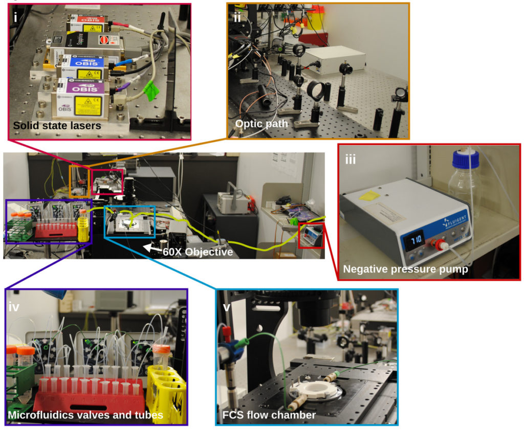
Simultaneous observation of 3D chromatin organization and transcription at the single cell level and with high spatial resolution may hold the key to unveil the mechanisms regulating embryonic development, cell differentiation and even disease. We have recently developed Hi-M, a technology that allows for the sequential labelling, 3D imaging and localization of multiple genomic DNA loci together with RNA expression in single cells within whole, intact Drosophila embryos. Importantly, Hi-M enables simultaneous detection of RNA expression and chromosome organization without requiring sample unmounting and primary probe re-hybridization. Here, we provide a step-by-step protocol describing the design of probes, the preparation of samples, the stable immobilization of embryos into microfluidics chambers, and the complete procedure for image acquisition. The combined RNA/DNA fluorescence in situ hybridization procedure takes 4-5 days including embryo collection. In addition, we describe image analysis software to segment nuclei, detect genomic spots, correct for drift and produce Hi-M matrices. A typical Hi-M experiment takes 1-2 days to complete all rounds of labelling and imaging and 4 additional days for image analysis. This technology can be easily expanded to investigate cell differentiation in cultured cells, or organization of chromatin within complex tissues.
ATP-driven separation of liquid phase condensates in bacteria
B. Guilhas, J.C. Walter, J. Rech, G. David, N.-O. Walliser, J. Palmeri, C., Mathieu-Demaziere, A. Parmeggiani, J.Y. Bouet, A. Le Gall1, M. Nollmann
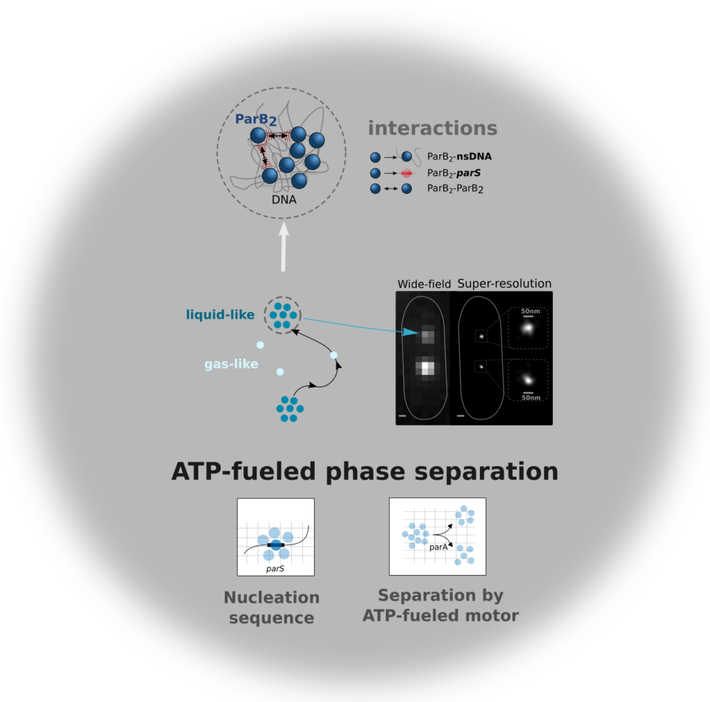
Liquid-liquid phase separated (LLPS) states are key to compartmentalise components in the absence of membranes, however it is unclear whether LLPS condensates are actively and specifically organized in the sub-cellular space and by which mechanisms. Here, we address this question by focusing on the ParABS DNA segregation system, composed of a centromeric-like sequence (parS), a DNA-binding protein (ParB) and a motor (ParA). We show that parS-ParB associate to form nanometer-sized, round condensates. ParB molecules diffuse rapidly within the nucleoid volume, but display confined motions when trapped inside ParB condensates. Single ParB molecules are able to rapidly diffuse between different condensates, and nucleation is strongly favoured by parS. Notably, the ParA motor is required to prevent the fusion of ParB condensates. These results describe a novel active mechanism that splits, segregates and localises non-canonical LLPS condensates in the sub-cellular space.
Guilhas et al. revealed that the bacterial DNA segregation apparatus behaves as a non-canonical phase separation system. This apparatus employs an ATP-powered motor that splits nanometer-sized condensates and localizes them robustly within the nucleoid to ensure faithful transmission of genetic material.
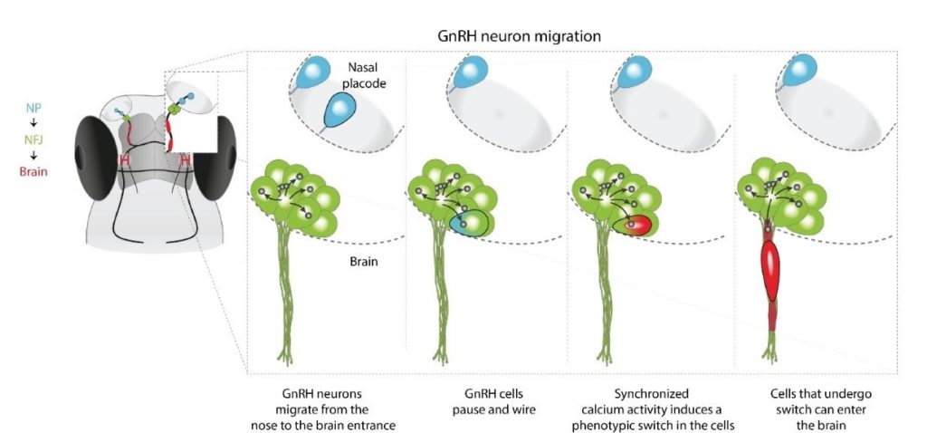
The ability to communicate effectively with each other is one of the strongest predictors for our chances to get ahead in life. In their latest publication in Science Advances, scientists and engineers from IGF-Montpellier (CNRS, INSERM, Univ. Montpellier), IPAM platform (BioCampus Montpellier, France-Bioimaging Montpellier Node) and ARO-Israel demonstrated that this also holds true for GnRH neurons.
In humans and all vertebrates, species survival depends on a critical step during embryonic development: the migration of a small subset of GnRH neurons (about 2,000 in humans and less than 100 in fish) from the nose to the brain where they join the hypothalamus to control reproduction. Their latest results unveiled that GnRH neurons make a pause at the nose-brain frontier where they function as an inter-hemispheric network that is isolated from the rest of the brain. Only neurons that integrate into the network and are able to communicate with their neighbors will finally cross the barrier and make their way into the brain, towards their hypothalamic destination.
In other words, these GnRH neurons, that are critical for species persistence, face the same challenges like other immigrants: they must learn to communicate effectively if they are to integrate into their new world.
In this study, in vivo 2-photon microscopy was a key tool for:
Long term imaging with minimal bleaching and phototoxicity
Upright configuration enabling dorsal imaging of the fish in its natural position
Long-distance water-immersion objectives allowing imaging of deep tissue structures without sacrificing image quality
Fast calcium imaging
Imaging of red GECI using the higher wavelengths
Precise cell ablation
Photoactivation of ChR2 while monitoring Ca in the red channel
A graphical model illustrating the migration of a single GnRH neuron (marked by black border) from the nasal placode into the zebrafish brain.
L’appel à candidature pour le prix « Pierre Favard » 2021 de la Société Française des Microscopiesest ouvert.
Ce prix récompense depuis 1989 des travaux de thèse réalisés dans une université française dans le domaine de la microscopie électronique, photonique, en champ proche, ou de la sonde atomique. Un prix est attribué en Sciences du Vivant et un autre en Sciences de la Matière. Ils sont décernés tous les deux ans. Pour cette édition, ils couronneront des travaux de thèse soutenus entre le 1er mars 2019 et le 28 février 2021. Chaque lauréat(e) est invité(e) à donner une conférence à l’issue de la remise du prix, lors de la réunion biennale de la Société qui est prévue à Reims du 5 au 9 Juillet 2021. Les prix (1000 € chacun) sont sponsorisés par des compagnies.
La date limite de dépôt des candidatures est fixée au 21 mars 2021.
1. Les candidats doivent fournir un exemplaire papier et une version « pdf » de leurs travaux de thèse, un curriculum vitae d’une page maximum et le fichier modèle ci-dessous dûment complété. Le dossier (papier et électronique) devra être envoyé au président de jury de leur discipline avec copie électronique au secrétariat de la SFμ (sfmu@sfmu.fr) et à la présidente (catherine.venien-bryan@upmc.fr).
2. Les candidats doivent être membre de la Société à la date de dépôt de leur dossier. 3. La Société Française des Microscopies prend en charge l’inscription au congrès de Reims, l’hébergement des lauréats (à hauteur de 90€/nuit) et rembourse le billet aller-retour en 2e classe (train) ou classe économique (pour la voie aérienne), sur présentation des factures originales et d’un ordre de mission sans frais.
Les deux présidents de jury nommés par le Conseil d’administration de la SFμ sont mandatés pour composer un jury de plusieurs personnes. Chaque jury est chargé de sélectionner et de classer les trois meilleurs candidats. Le Conseil de la SFμ, sur proposition des jurys et sur rapport des présidents, désigne les deux lauréats.
Registration for France BioImaging Annual Meeting is now open!
France BioImaging is pleased to invite you to participate to France BioImaging6th Annual Meeting. For this edition, the meeting will be organized as atwo-half days virtual meeting (from 9:00 AM to 1:00 PM) on February 4th & 5th, 2021.
This event, open to all members of the bioimaging community, aims to provide a platform to discuss pivotal subject matters in our field.
The 2021 program of the France BioImaging Annual meeting is built around two pillars:
February 4th: “Building and operating an integrated and open infrastructure for bioimaging“
February 5th: “Latest and future developments in biological imaging“
A l’occasion de la MorningTech Photonique & Santé organisée par Photonics Bretagne et Biotech Santé Bretagne le 2 février 2021 de 9h30 à 12h00, Marc Tramier, coordinateur du Nœud Bretagne-Loire de France BioImaging présentera “lestechnologies photoniques des plateformes de Biogenouest au service de l’imagerie pré-clinique“.
Imaging of proteins, cells, and tissues is critical to understanding health and disease. On December 2nd, 2020, the Chan Zuckerberg Initiative (CZI) announced nearly $32 million in funding to support biomedical imaging researchers, technology development, and the BioImaging North America international network of bioimaging facilities and communities. CZI also opened a new Request for Applications (RFA) aimed at supporting technology development that will allow researchers to see the inner workings of cells, including proteins, at near-atomic resolution to better understand what causes disease and how to develop treatments.
Frontiers of Imaging: Visual Proteomics
The Frontiers of Imaging initiative, part of CZI’s broader Imaging program, supports the development of disruptive imaging technologies that connect biological scales across organs, cells, and proteins, allowing researchers to directly visualize biological processes at the necessary resolution and context to obtain a mechanistic understanding of health and disease. As part of the Frontiers initiative, the new Visual Proteomics Imaging RFA supports technology development that will allow researchers to see the inner workings of cells, including proteins, at near-atomic resolution. CZI invites scientists to apply for this 2 1/2-year grant opportunity to support the development of hardware, software, and methods. Examples of research themes could include hardware and software development to enhance contrast and resolution for electron tomography, high-resolution correlated light and electron microscopy (CLEM), and FIB-SEM; sample preparation improvements for electron tomography; and development of software or new computational techniques and algorithms for identifying protein molecules inside cells and segmenting sub-cellular structures.
The Visual Proteomics Imaging RFA accept applications until February 17, 2021 at 5 p.m. Pacific Time.
For administrative and programmatic inquiries, technical assistance, or other questions pertaining to this RFA, please contact: sciencegrants@chanzuckerberg.com.
We use cookies on our website to give you the most relevant experience by remembering your preferences and repeat visits. By clicking “Accept All”, you consent to the use of ALL the cookies. However, you may visit "Cookie Settings" to provide a controlled consent.
This website uses cookies to improve your experience while you navigate through the website. Out of these, the cookies that are categorized as necessary are stored on your browser as they are essential for the working of basic functionalities of the website. We also use third-party cookies that help us analyze and understand how you use this website. These cookies will be stored in your browser only with your consent. You also have the option to opt-out of these cookies. But opting out of some of these cookies may affect your browsing experience.
Necessary cookies are absolutely essential for the website to function properly. These cookies ensure basic functionalities and security features of the website, anonymously.
Cookie
Duration
Description
cookielawinfo-checkbox-analytics
11 months
This cookie is set by GDPR Cookie Consent plugin. The cookie is used to store the user consent for the cookies in the category "Analytics".
cookielawinfo-checkbox-functional
11 months
The cookie is set by GDPR cookie consent to record the user consent for the cookies in the category "Functional".
cookielawinfo-checkbox-necessary
11 months
This cookie is set by GDPR Cookie Consent plugin. The cookies is used to store the user consent for the cookies in the category "Necessary".
cookielawinfo-checkbox-others
11 months
This cookie is set by GDPR Cookie Consent plugin. The cookie is used to store the user consent for the cookies in the category "Other.
cookielawinfo-checkbox-performance
11 months
This cookie is set by GDPR Cookie Consent plugin. The cookie is used to store the user consent for the cookies in the category "Performance".
viewed_cookie_policy
11 months
The cookie is set by the GDPR Cookie Consent plugin and is used to store whether or not user has consented to the use of cookies. It does not store any personal data.
Functional cookies help to perform certain functionalities like sharing the content of the website on social media platforms, collect feedbacks, and other third-party features.
Performance cookies are used to understand and analyze the key performance indexes of the website which helps in delivering a better user experience for the visitors.
Analytical cookies are used to understand how visitors interact with the website. These cookies help provide information on metrics the number of visitors, bounce rate, traffic source, etc.
Advertisement cookies are used to provide visitors with relevant ads and marketing campaigns. These cookies track visitors across websites and collect information to provide customized ads.