The brain: a network of neurons… and supporting cells
Our brain is made up of neurons…but not only neurons! Neurons are specialized cells that transmit information to other cells, whether nerve or non-nerve. Information travels as electrical impulses along the axon, a sort of “highway” that carries the nerve signal.
At the end of each axon are synapses, small communication zones composed of:
- the axon terminal of the presynaptic neuron (which sends the signal),
- the membrane of the postsynaptic neuron (which receives it),
- and the synaptic cleft, the narrow gap where neurotransmitters are released to pass the message.
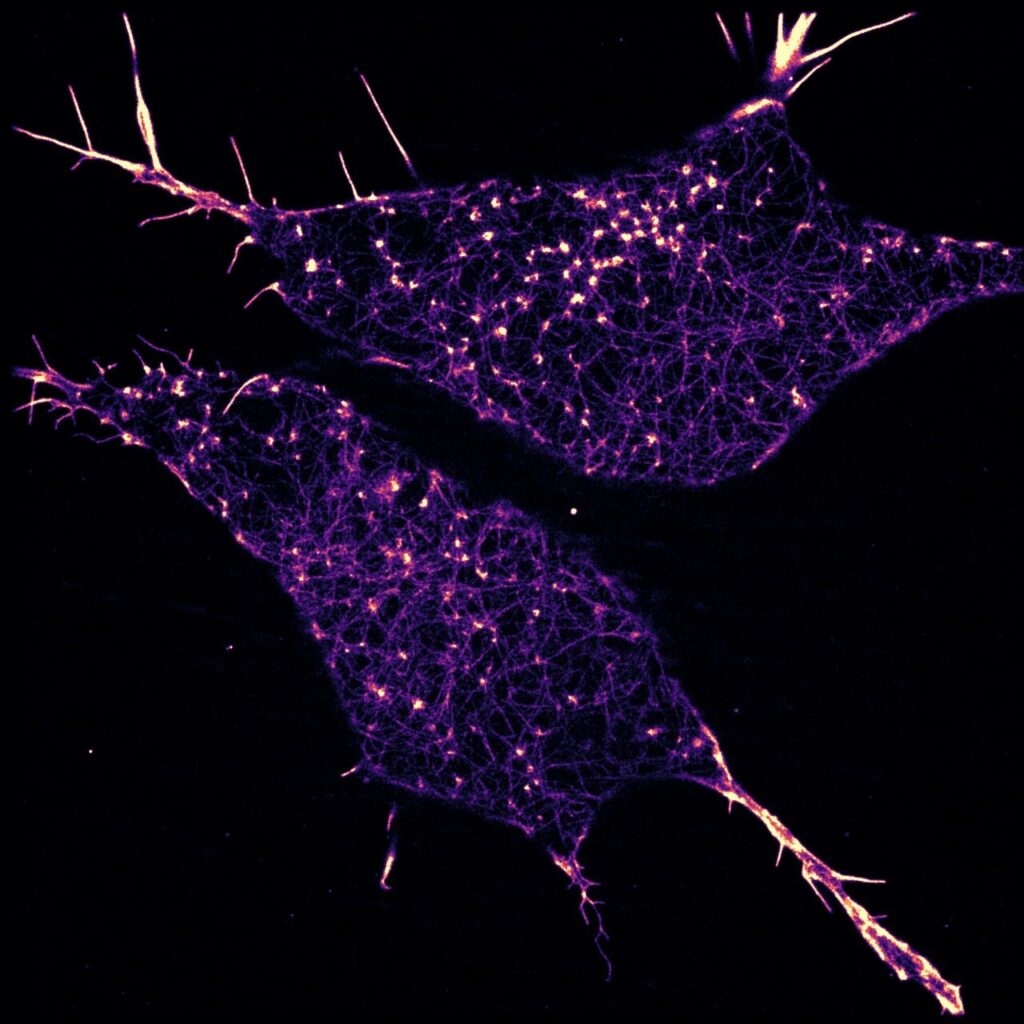
To keep neurons functioning properly, the brain also contains glial cells, which provide structural and metabolic support and regulate many physiological processes. Among them are astrocytes, star-shaped cells located close to synapses.
Since the 1990s and the discovery of the “tripartite synapse” concept, scientists have revealed the crucial role astrocytes play in information transmission.
However, astrocytes still hold some secrets, particularly concerning their finest extensions, called “leaflets”!
Cutting-edge imaging strategies for unprecedented results
To explore the structure and function of astrocytes in greater detail, the authors developed an innovative multimodal imaging workflow, combining two-photon and electron microscopy.
The electron microscopy experiments were led by several France-BioImaging units: CEA-Grenoble, Grenoble Institute of Neurosciences and the TIMC laboratory. The 2-photon imaging was performed at Lausanne University.
- FIB-SEM microscopy to reveal the 3D ultrastructure of astrocytes and, in particular, their leaflets. This technique was combined with 3D immunolabeling to detect the presence of specific proteins:
- IP₃R1, a marker of the endoplasmic reticulum involved in calcium signaling,
- Connexin-43, a protein found at gap junctions connecting different structures.
- 2-photon calcium imaging to monitor real-time calcium variations in live leaflets in 3D, thereby assessing their role in cell-to-cell communication.
Architecture of the leaflets
Leaflets are extremely thin extensions (<250 nm), sometimes smaller than the wavelength of visible light, which explains why they long escaped direct observation.
They contain tiny segments of endoplasmic reticulum (i-ER) but no mitochondria, indicating a specialization for rapid and localized calcium signaling.
Several leaflets are interconnected via gap junctions, forming micro-networks within the astrocyte.

Figure 1. Leaflets are complex subdomains of the astrocyte interconnected by gap junctions. (B) Top: segmentation of a complete astrocyte fragment, including: bottom left: shafts with inner diameter >250 nm (gray); bottom right: leaflets, ≤250 nm (green). Bars: 3 μm.

Figure 3. i-ER: Minuscule organelles observed specifically in the leaflet domains of astrocytes. (C) Examples of i-ER saccules (magenta overlaid to EM images). Top: i-ERs surrounded by associated ribosomes (magenta arrowheads). Bottom: i-ERs contacting the astrocyte plasma membrane (yellow arrowheads). Bar: 500 nm.
Interaction with synapses
3D reconstructions revealed that the majority of synapses (around 90%) are shared by several leaflets of a single astrocyte.
Thus, one astrocyte can link together multiple synapses within the same neuronal network.
This finding challenges the “one synapse ↔ one astrocyte” model: in reality, astrocytes connect neurons to one another through their leaflets.

Figure 2. Leaflets are the astrocyte’s specialized subdomains that interact with synapses and preferentially embed multiple vs. individual synapses. (B) Arrangement of synaptic elements with leaflets and other astrocyte structures; 2D FIB-SEM images overlaid with segmentation map (leaflet: green; axonal bouton: yellow; spine: orange; synaptic cleft: red). Left: five synapses encompassed in a leaflet domain, contacted by leaflet units. Bar: 0.5 μm. Top right: synapse in direct contact with a shaft (S, gray); bar: 0.5 μm. Middle right: synapse in contact with both leaflets emerging from a shaft and the shaft itself; bar: 0.5 μm. Bottom right: synapse in contact with leaflets emerging from cell body (soma). Bar: 1 μm.
Calcium activity in leaflets
2-photon calcium imaging revealed very rapid, localized calcium micro-events, often triggered by neuronal activity.
These signals depend on IP₃R1 receptors in the i-ER and can propagate from one leaflet to another via gap junctions.
When they merge, these signals create broader calcium waves capable of synchronizing several neighboring synapses.

Figure 7. Leaflet domains host multiple synapses from independent circuits and display complex Ca2+ events that can integrate their ongoing activities in space and time. (B) Left: single-plane time-series capturing a representative multi-originated Ca2+ event in “Leaflets” (pseudocolor Ca2+ intensity scale, see “Two-photon 3D+t imaging data analysis” in STAR Methods). “Mitochondria” regions (azure pseudocolor) display an independent event. Right: oriented graph representation of the evolution of the event in “Leaflets,” visualizing spatial and time aspects, directionality, and intensity. (i), (ii), and (iii): timings of the images on the left. Arrowheads in (i) and (ii) identify distinct origination sites, and in (iii) their merging into a larger, long-lasting event. Bar: 1 μm. (C) Left: time-series as in (B), here capturing a Ca2+ event in “Leaflets” with three distinct originations (arrowheads). Two of them appear simultaneously in (i), the third, delayed, at another location in (ii), while in (iii) they all merge into a larger, long-lasting, high-intensity Ca2+ elevation. Noteworthy, the event expands around “Mitochondria” regions without touching them. Bar: 1 μm. Right: oriented graph representation of the Ca2+ event’s dynamics as in (B).
Conclusion
The results show that astrocytes are not mere support cells, but true biological computation units, where each leaflet acts as a functional “pixel” in a mosaic capable of interpreting and integrating multiple neuronal signals through the language of calcium.
This new astrocyte-oriented approach could pave the way for further studies exploring other brain regions to determine whether similar mechanisms exist elsewhere in the nervous system.
Read the scientific article here!
Benoit L, Hristovska I, Liaudet N, Jouneau PH, Fertin A, de Ceglia R, Litvin DG, Di Castro MA, Jevtic M, Zalachoras I, Kikuchi T, Telley L, Bergami M, Usson Y, Hisatsune C, Mikoshiba K, Pernet-Gallay K, Volterra A. Astrocytes functionally integrate multiple synapses via specialized leaflet domains. Cell. 2025 Sep 24:S0092-8674(25)01028-1. doi: 10.1016/j.cell.2025.08.036. Epub ahead of print. PMID: 40997814.