Learn more about our 2022 France-BioImaging Image Contest winners!
As the 2023 edition of the France-BioImaging Image Contest admissions is still running, we wanted to highlight our previous winners and their projects. Here is a quick throwback to our 2022 winners.
Before getting to the heart of the matter, we want to remind you that you still have time (before November 10th) to submit your best images and try to win your registration fees for one 2024 microscopy-related event! Please make sure you upload your images on the following link:
Last year, we enjoyed the winning images submitted for their artistic take and their quality. Thanks to Carole SIRET, Magalie BENARD and Frédéric FERCOQ for their beautiful images!
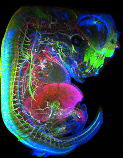
- 1st Place: Carole SIRET, Van de Pavert Team, Centre d'Immunologie de Marseille-Luminy

"Little Monster"
The embryonic formation of lymph nodes, small organs essential for the immune response, is now known. Using light sheet microscopy, scientists were able to determine the dynamics at work in this 13.5-day-old mouse embryo. In blue, the lymphoid cells (LTi), derived from the haematogenous endothelium, a specific tissue of the embryo. They pass into the liver where they proliferate before migrating through the body to give rise to lymph nodes. The 3D information obtained thus makes it possible to follow the interactions of lymph nodes with their environment, in particular with nerve cells, in green, and blood vessels, in white. The lymphatic endothelial cells and some macrophages are visible in red.
Lightsheet Microscopy
Carole Siret is a Research engineer, expert in Lightsheet microscopy, at the Centre d’Immunology Marseille Luminy (CIML) since 2018. She is working in Dr Serge van de Pavert team where they study immune system development. They are particularly interested in the lymph nodes (LN) formation during mouse embryogenesis.
The image she submitted is a projection from a lightsheet acquisition on the UMII (Miltenyi). This image illustrates an E13.5 mouse embryo stained for neurons, LTi (Tissue inducer cells which are the precursor cells for the lymph node), lymphatic and blood vessels. This acquisition was done in the context of the study of the role of Cxcl12 in embryonic LN formation. From previous work it is clear that Cxcl13 and Ccl21 are not expressed present near blood vessels, but it likely that some chemokines, possibly Cxcl12, could be expressed on the endothelial cells. We focus on Cxcl12 since this chemokine has shown to be important for the attraction of several hematopoietic cells. Although it was shown that the receptor for Cxcl12, Cxcr4, is expressed by the mature hematopoietic inducer cells, it is not clear whether it also expressed by the progenitor hematopoietic inducer cells. Next to the possible attraction of hematopoietic cells towards the lymph node anlagen, Cxcl12 is involved in the attraction of nerve fibers. Therefore, the possible role of Cxcl12 could be to both attract hematopoietic cells as well as nerve fibers to initiate a region which is permissive to form lymph nodes.
Thanks to the France-Bioimaging Image Contest, Carole participated to the SFI Congress, where, this year, it was a special joint conference both between the Société Française d’Immunologie (SFI) and the Deutsche Gesellschaft für Immunologie (DGfI). It was a great opportunity to exchange with people at the cutting edge of the immunology field.
- 2nd Place: Magalie BENARD, Plateforme de Recherche en IMAgerie CEllulaire de Normandie (PRIMACEN), Research infrastructure HeRacLeS, Inserm US 51, CNRS UAR 2026,
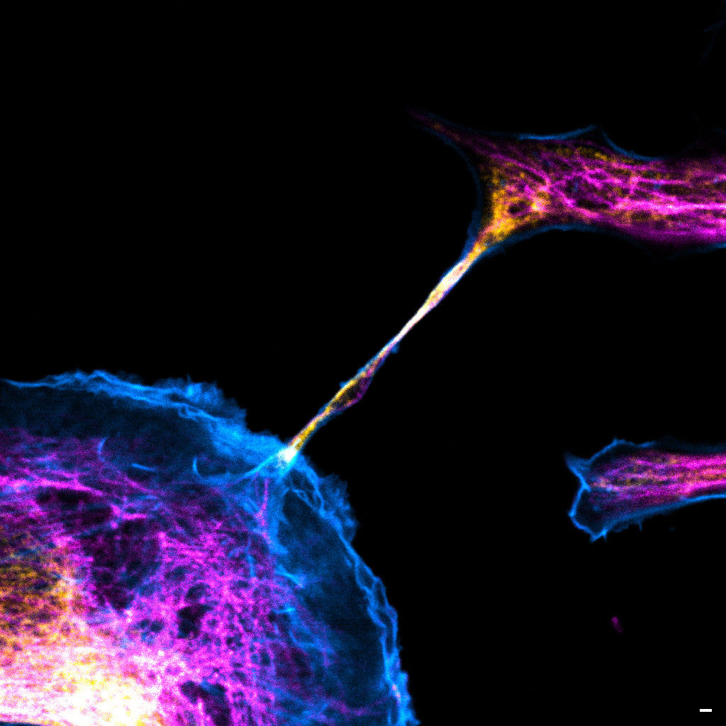
"The communication link with others"
Image of a cellular interconnection between two human tumor cells whose cytoskeleton has been labeled with anti-tubulin (ATTO-647N), anti-vimentin (AlexaFluor594) antibodies and with Phalloidin probe (AlexaFluor488). Scale bar 1µm.
Confocal microscopy
Magalie Bénard is a Research Engineer and the Technical Manager at the Cellular Imaging Facilty PRIMACEN (Plate-forme de Recherche en IMAgerie CEllulaire de Normandie).
The image she submitted is a confocal image representing a cellular interconnection tunneling nanotube (TNTs) between two human tumour cells. In a cancer case, some cells are able to express spontaneously TNTs with cytoskeleton protein composition corresponding to specific role of this communication mechanism. In the winning image, the TNT is composed of tubulin (magenta), actin (cyan) and vimentin (yellow) proteins. Called TNT1, this nanotube allows the transfer of intracellular elements such as RNA, proteins or organelles. Moreover, due to the thinness of TNTs, their photo-sensitivity and their fragility, live-cell imaging is technically challenging with regards to potentially damaging methods. Magalie and her team have developed an adapted method to observe TNTs in living cell with high resolution imaging (STED) enhanced by FLIM by using red and near infrared probes.
France-Bioimaging sponsored her participation to the ELMI (European Light Microscopy Initiative Meeting June 6-9, 2023) congress. During this event, she had the chance to present her project through a poster. This congress also offered a great opportunity to have an overview and the last updates on state-of-the-art imaging techniques.
- 3rd Place: Frédéric FERCOQ, Parasites et Protistes Libres (PPL), Museum National d'Histoire Naturelle
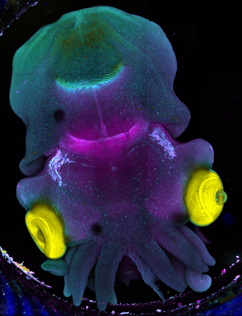
"Sepia"
Stage 25 cuttlefish embryo (Sepia officinalis) observed under a confocal microscope.
The cuttlefish was cleared and the tissue autofluorescence was captured.
This image was produced in collaboration with Laure BONNAUD-PONTICELLI and Luis MOLINA from the BOREA laboratory.
Confocal microscopy
Frédéric Fercoq is a postdoc scientist in the Parasitology laboratory of the Muséum National d’Histoire Naturelle (MNHN) in Paris. My main interest is on how myeloid cells participate to the control of parasitic infections, but sometimes at the price of collateral tissue damage. This project involves a lot of microscopy of immune cells, parasites and host tissues to analyse the complex interactions taking place at the site of infection.
The image he submitted has nothing to do with his main project! As he has the chance to work on very different topics and models, this image was acquired as a proof of concept for imaging full embryos of the cuttlefish Sepia officinalis for Frédéric's colleagues Laure BONNAUD-PONTICELLI and Luis MOLINA (BOREA laboratory, MNHN). They work on the nervous system of cephalopod and on the influence of environmental factors during its development. They are now optimizing fluorescent staining for neuronal markers to test the effect of light on the nervous system in situ.
France-Bioimaging sponsored his participation to the FOM (Focus on Microscopy) 2023 congress in Porto. He had the chance to be granted the opportunity to both present his current project through a poster and to give an oral presentation. He was also amazed by the new avenues opened up by the cutting-edge imaging techniques presented throughout the conference.
Want to be the next winner of our FBI Image Contest? Apply through the following link before November 10th, 2023: france-bioimaging.org/fbi-special-events/france-bioimaging-image-contest-2023