The BIC is setting up in a brand new space
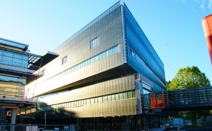
In the last weeks of October 2016, the BIC has settled in a brand new building, constructed by the Regional Council of Aquitaine as part of the Neurocampus project. This building, of around 13 000 m2, is shared with the Interdisciplinary Institute for Neuroscience (IINS) and the Institute for Neurodegenerative Disorders (IMN). This building, constructed in two years, cost 47 M€ and is part of a large project to develop Neuroscience and imaging in Aquitaine. The new building is conveniently located and connected by footbridges between the Magendie Neuroscience center and the Center for functional genomics (CGFB) that hosts several core facilities.
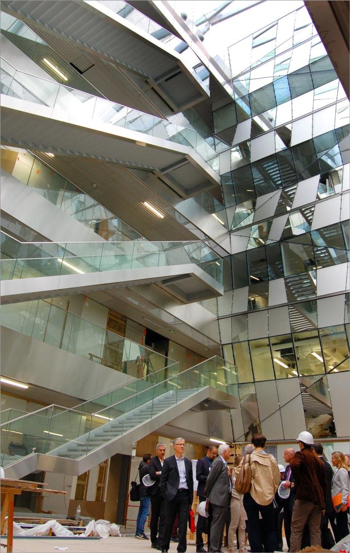
In total, the BIC will occupy 1000 m2, split between the CGFB and the new building. The major part in the new building is dedicated to photonic microscopy. Electron microscopy instruments, including two brand new ones coming in 2017, will be dispatched between the CGFB and Neurocampus building. In these new spaces, users have access to a culture room and also a room with analysis stations. Other rooms are dedicated to each kind of microscopy (one room for live cells imaging, one room for multiphoton, one room for confocal, one room for new scanning electron microscope etc…). Special rooms are dedicated to host R&D projects as well as confidential collaborations with industry.
Development of training capacities at the BIC – joint projects with the Cajal School of Neuroscience
The BIC has engaged for many years in active training programs for imaging at all levels (beginners to advanced training) for local, national and transnational users. The BIC personnel also participates extensively to various theoretical and hands on training/showcase activities in France and abroad (MifoBio, NeuBias, etc…). Within the strategy to develop the BIC-FBI training, we are engaging a partnership with the Cajal Advanced Neuroscience Training Program to develop special ima ging training for Neuroscience. The Cajal school is a European FENS and IBRO initiative in partnership with Bordeaux Neurocampus and the Champalimaud Foundation, which offers state-of-the-art hands-on training courses in neuroscience.
Construction of a light sheet microscope for super resolution imaging inside living samples
Fast and non-damaging imaging of single molecules inside live organisms is essential to study physiologically relevant biochemical mechanisms occurring at the subcellular level. For example, the dynamic organization of transmitter receptors at the membrane of excitatory neurons should, ideally, be studied in vivo in the brain of animal models. Unfortunately super resolution techniques such as PALM1, STORM23 and uPAINT4 are mostly restricted to the sample external surfaces and are unable to image inside live samples.
For these reasons the Bordeaux Imaging Center is developing a new light sheet microscope specially dedicated to image single molecules into live samples. Light sheet fluorescence microscopy (LSFM) is recognized as the method of choice to image thick live samples. Compared to other fluorescence imaging modalities such as wide field, confocal, structured illumination, two-photon or STED, LSFM strongly reduces out of focus fluorescence, decreases photobleaching and phototoxicity, and improves temporal resolution. Among the numerous technical implementations of LSFM 5, we decided to build a lattice light sheet microscope (LLS) because it has been specifically designed to perform super resolution imaging in thick live samples 6. Indeed In LLS the illumination beam is shaped by a spatial light modulator (SLM) to produce a < 1 µm thick excitation plane over a length of > 50 µm at the sample. A 1.1 NA detection objective ensures efficient light collection required for high localization precision. Illumination and detection objectives are both long working distance and water immersion, thus allowing observation of live samples up to 5 mm in diameter. (Fig 1 A)
Our LLS microscope is mostly based on the documentation freely and kindly shared by Eric Betzig’ group (HHMI Janelia Farms, USA).
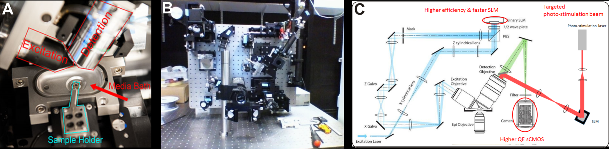
Fig 1. (A) The sample is placed at the intersection of the excitation and detection objective optic axes in a temperature controlled perfusion chamber. It is held at the tip of motorized arm on a 5 mm diameter cover slip (from 6). (B) The LLS microscope under construction in June 2016. (C) In blue and green the optical path of the excitation and detection beams respectively (from 6). A higher efficiency SLM, higher QE camera should improve the light budget compared to the original specifications. In addition, a targeted laser beam (red) will allow precise photo-conversion of light sensitive molecules.
We made a few modifications compared to the original specifications of the LLS as described in 6 : our microscope will be equipped with a laser combiner including 4 high power lasers at 405 nm (300mW), 488 nm (1 W), 560 nm (2 W), 642 nm (2W), a higher efficiency SLM (Fourth Dimension DD QXGA) and a sCMOS camera with improved quantum efficiency (Hamamatsu ORCA Flash V2). These improvements should mitigate the weak throughput of the LLS beam path, and, in turn, improve molecule localization precision and/or time resolution. In addition, a targeted photostimulation beam will be coupled through the detection objective to photo stimulate or photoconvert with a high spatial and temporal resolution photosensitive molecules.
STORM, PALM and PAINT imaging modalities will be fully compatible with the constructed LLS.
The microscope construction by Mathieu Ducros, INSERM research Engineer on the BIC, started in April (Fig 1B). First images are expected by the end of 2016. Once our LLS is fully operational and running, it will be accessible to all BIC users under the supervision of a local engineer.
For this project we are supported financially by the GIS IBiSA, LABEX brain and FBI.
References
- Betzig, E. et al. Imaging intracellular fluorescent proteins at nanometer resolution. Science 313, 1642–1645 (2006).
- Rust, M. J., Bates, M. & Zhuang, X. Sub-diffraction-limit imaging by stochastic optical reconstruction microscopy (STORM). Nat. Methods 3, 793–795 (2006).
- van de Linde, S. et al. Direct stochastic optical reconstruction microscopy with standard fluorescent probes. Nat. Protoc. 6, 991–1009 (2011).
- Giannone, G. et al. Dynamic superresolution imaging of endogenous proteins on living cells at ultra-high density. Biophys. J. 99, 1303–1310 (2010).
- Santi, P. a. Light sheet fluorescence microscopy: a review. J. Histochem. Cytochem. 59, 129–138 (2011).
- Chen, B.-C. et al. Lattice light-sheet microscopy: Imaging molecules to embryos at high spatiotemporal resolution. Science (80-. ). (2014). doi:10.1126/science.1257998
Bordeaux Imaging Center: http://www.bic.u-bordeaux.fr/
UMS 3420 CNRS-Université de Bordeaux, US4 INSERM
Contact: bic[at]u-bordeaux.fr
Photo Credits: www.bordeaux-neurocampus.fr