
Dans le cadre du programme “Imaging 4 All – Access Track”, le Dr Brice Tonfack, de l’Université de Yaoundé (Cameroun), a bénéficié d’un grant pour collaborer avec Jean-Luc Verdeil, le responsable scientifique de la plateforme d’imagerie MRI-PHiV du CIRAD à Montpellier. Ce séjour allant au-delà de l’aspect technologique, a permis de structurer une collaboration scientifique sérieuse et durable entre le laboratoire du Dr Tonfack et le MRI-PHiV.

Nous avons rencontré Jean-Luc et Brice, qui nous ont fait le plaisir de répondre à nos questions pour mieux comprendre les enjeux et bénéfices de cet échange.
English-speaking readers, the interview can be found in English at the end of the article.
Brice, pouvez-vous brièvement vous présenter ?
[Brice] Je suis Dr Libert Brice TONFACK, Maître de Conférences en Biotechnologies Végétales et Environnement à l’Université de Yaoundé I depuis 2011. Mon parcours s’est construit entre le Cameroun, la France et l’Afrique du Sud, avec un doctorat en biotechnologies végétales et une expérience postdoctorale à l’Université de Pretoria. Mes recherches portent sur la valorisation des plantes tropicales sous-exploitées, l’agriculture durable en conditions de stress et la génomique fonctionnelle, avec pour objectif de relier recherche fondamentale et applications concrètes au service du développement durable en Afrique.
Sur quel projet de recherche travaillez-vous actuellement ?
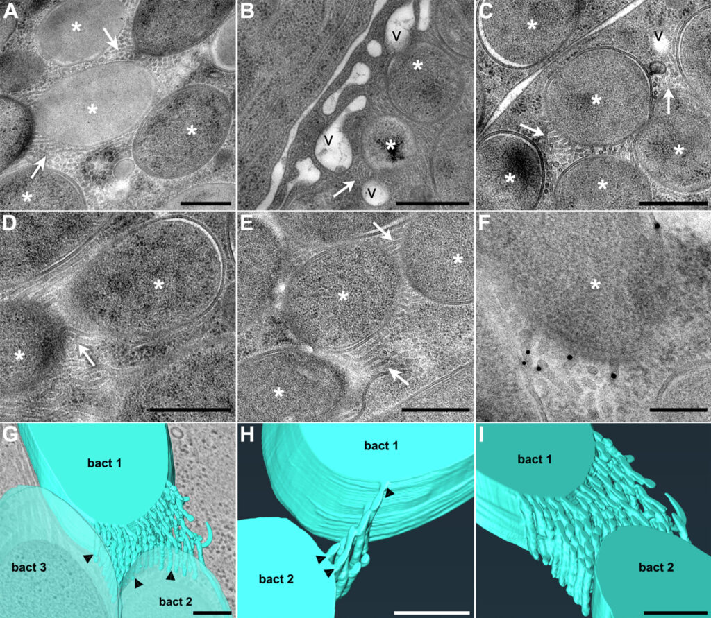
[Brice] Je travaille actuellement sur un projet consacré à la valorisation d’espèces tropicales sous-exploitées du genre Aframomum, en collaboration avec le CIRAD à Montpellier. À l’aide d’outils de bioimagerie et de microscopie, nous étudions la diversité et la structure des graines. L’enjeu est à la fois scientifique et sociétal : mieux comprendre ces espèces encore peu étudiées, révéler leur potentiel médicinal, alimentaire ou cosmétique, et contribuer à leur conservation ainsi qu’au développement des communautés qui en dépendent.
Brice, comment avez-vous vécu le programme Imaging 4 All – Access Track ?
[Brice] Mon expérience avec ce programme a été très enrichissante. Ce séjour a permis de poser les bases d’une collaboration scientifique ambitieuse et durable entre mon laboratoire et la plateforme MRI-PHiV du CIRAD. L’accueil, l’organisation et l’accompagnement scientifique ont été exemplaires, et les infrastructures de pointe ont permis de générer des données de très grande qualité. Le soutien financier du programme a été déterminant pour la réussite du projet et a eu un impact fort sur mes travaux et mes collaborations internationales.


Pourquoi avoir choisi une plateforme France-BioImaging ?
[Brice] J’ai choisi une plateforme France-BioImaging car l’imagerie végétale reste encore peu représentée, et la MRI-PHiV de Montpellier est l’une des rares infrastructures de haut niveau dédiées à la recherche sur les plantes. J’ai été mis en contact avec Jean-Luc Verdeil par Jean Salamero, puis une première visite de la plateforme en 2024 a permis d’initier les échanges scientifiques et de faire émerger un projet commun, ensuite construit collectivement à distance.
En quoi ce séjour a-t-il fait avancer votre projet ?
[Brice] Ce séjour a permis d’obtenir des données inédites que je n’aurais pas pu acquérir dans mon laboratoire. Nous avons réalisé une caractérisation complète des graines d’Aframomum par imagerie non invasive et analyses histologiques et histochimiques avancées. Ces approches ont généré des jeux de données riches et prometteurs, constituant une avancée méthodologique majeure et posant les bases de travaux collaboratifs approfondis.

Jean-Luc, comment la plateforme a-t-elle bénéficié de cette collaboration ?
[Jean-Luc] Cette collaboration a été très stimulante pour l’ensemble de l’équipe de la MRI-PHiV. Elle nous a permis de travailler sur un matériel biologique original et peu étudié, présentant une grande richesse morphologique et biochimique. Le projet nous a amenés à combiner plusieurs modalités d’imagerie dans une approche intégrative, renforçant notre expertise méthodologique, notamment sur des échantillons complexes riches en métabolites secondaires. Les échanges avec Brice ont été particulièrement riches et son regard de biologiste tropical a donné une nouvelle dimension aux images produites.
En quoi cette collaboration a-t-elle enrichi vos approches scientifiques ?
[Brice] Cette collaboration a renforcé ma conviction que l’imagerie est un outil central pour comprendre le fonctionnement du végétal: une bonne image valant mille mots! Elle m’a conduit à repenser l’ensemble de la chaîne expérimentale, de la préparation des échantillons à l’analyse des images. L’imagerie, en interaction avec la physiologie, la biochimie, la génomique et l’agronomie, ouvre un véritable changement de paradigme scientifique.
[Jean-Luc] Travailler avec Brice sur des espèces tropicales orphelines a profondément enrichi ma manière d’aborder l’imagerie végétale, en la replaçant au cœur de questions biologiques, écologiques et sociétales concrètes. L’imagerie ne doit pas être perçue comme une discipline isolée, mais comme un langage transversal reliant la physiologie, la biochimie, la génomique, l’agronomie et l’écologie. Cette collaboration nous a amenés à repenser la conception des protocoles et la finalité des données produites, et a également été très riche sur le plan humain, en instaurant une relation de confiance essentielle à des partenariats durables.
Brice, qu’avez-vous retiré de cette expérience sur le plan professionnel ?
[Brice] Au-delà de l’accès aux technologies, cette expérience a fortement renforcé ma visibilité internationale et celle de mon institution. J’ai pu me familiariser avec des outils de microscopie de haut niveau et générer un volume important de données, qui seront analysées en étroite collaboration avec l’équipe de la MRI-PHiV.
Comment Global BioImaging favorise-t-il des collaborations équilibrées ?
[Jean-Luc] Les initiatives portées par Global BioImaging dépassent une simple logique d’accès aux équipements. Elles encouragent la co-construction de projets, la reconnaissance des expertises et des priorités scientifiques des pays du Sud, et contribuent à réduire les inégalités d’accès aux technologies. Elles bénéficient autant aux chercheurs invités qu’aux plateformes hôtes et créent un cadre de confiance propice à des collaborations durables. J’ai par ailleurs énormément apprécié de travailler avec Brice, que je ne connaissais que très peu avant cette collaboration. Cette rencontre a été pour moi une expérience humainement très enrichissante, tant sur le plan personnel que culturel et scientifique, et elle a largement contribué à la qualité, à la confiance et à la profondeur de notre collaboration.
Comment envisagez-vous la suite de cette collaboration ?
[Brice] Oui, clairement. L’analyse des données nécessitera une collaboration étroite sur au moins un an, avec des publications, des communications scientifiques et de nouveaux séjours de recherche. À plus long terme, nous envisageons des échanges d’étudiants, des actions de formation et des projets communs, notamment en biotechnologie forestière et en imagerie appliquée aux écosystèmes marins.
[Jean-Luc] Cette collaboration constitue le point de départ d’un partenariat scientifique structurant et durable. À court terme, l’analyse conjointe des données ouvrira la voie à plusieurs publications. À plus long terme, nous souhaitons développer des échanges de jeunes chercheurs, des formations en imagerie et répondre ensemble à de futurs appels à projets autour de l’imagerie végétale et du phénotypage des plantes tropicales.


