On November 20th and 21th 2024, we have the pleasure to invite you to our Annual Meeting, to be hosted by our brand new FBI Alsace Node at the Bibliothèque Nationale et Universitaire (BNU) de Strasbourg (6 Pl. de la République, 67000 Strasbourg).
We will be happy to celebrate yet another year of achievements and developments in bioimaging with all the members of the community.
With a focus on “Live functional imaging: From chemical synthesis of the probes to instrumentation”, this edition aims to bring together chemists, microscope builders and biologists developping tools to probe life.
The scientific sessions will explore the development of new probes (organic, inorganic, nanoparticles, fluorescent proteins, hybrid materials), how to leverage their optical properties (spectra, quantum yield, lifetime, photostability, switching between dye states, etc…) and new instrumentations taking advantage of these new probes for life science.
France-BioImaging Mission Officers, Technology WGs, facilities or R&D teams are invited to present news, innovations or any achievements to the community with a poster. We strongly encourage you to submit an abstract for a poster presentation during your registration!
Registration is free but mandatory. Please note that the BNU capacity is limited and registrations will be accepted on a first come first served basis.
As part of the scientific activities that it wishes to carry out and in continuation of its 1st seminar (Dec. 2022), the FBI “Preclinical Microscopy” Working Group is organizing its 1st webinar on February 1 from 2:00 pm to 4:00 pm. The objective of these scientific meetings is to present research projects, technologies and regulatory frameworks related to light microscopy from the organ/organoid to the living animal.
Program
2:00 pm – Welcoming & Introduction
2:15 pm – “Visualization of the ticking of circadian clock cells in freely moving mice” by Xavier Bonnefont (Institute of Functional Genomics, Montpellier).
3:00 pm – “Regulatory aspects of the use of animals for scientific purposes” by Isabelle Bardou (Cyceron, Biomedical Imaging Platform, Caen).
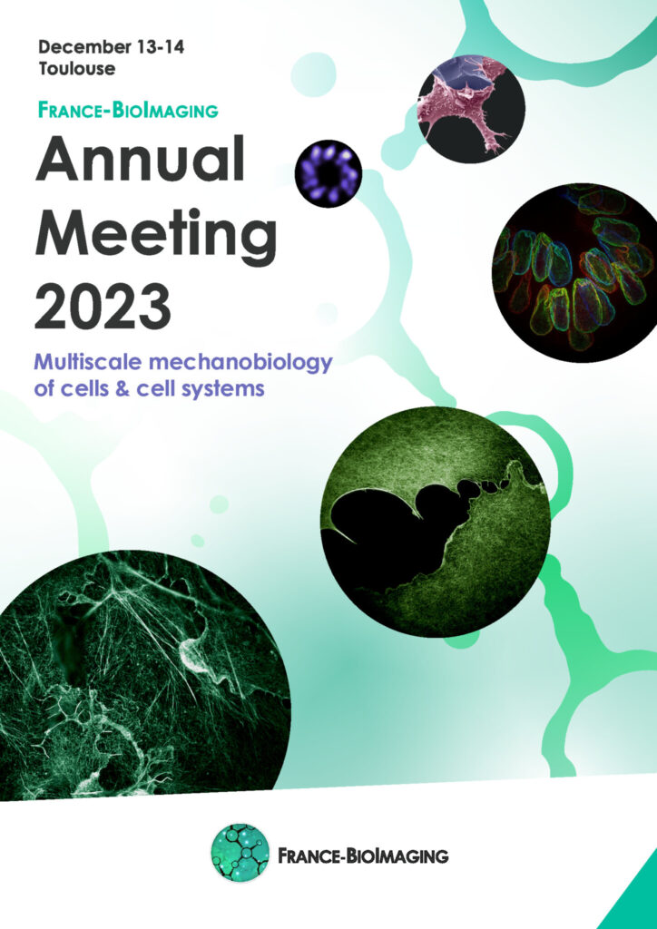
On December 13th and 14th 2023, we have the pleasure to invite you to our Annual Meeting, to be hosted by our brand new FBI Toulouse Node at the Centre de Biologie Intégrative of the Université Toulouse 3 – Paul Sabatier.
We will be happy to celebrate yet another year of achievements and developments in bioimaging with all the members of the community.
This year, the Annual Meeting will focus on “Multiscale mechanobiology of cells and cell systems“, a topic specially selected for being one of Toulouse node’s expertise.
Mechanobiology aims to apply biophysical approaches to measure and perturb forces in complex physiological systems, and to develop in vitro systems including different types of organoids, enabling the controlled manipulation of cells and tissues and the measurement of mechanical forces to study cell mechanics and morphogenesis.
This “mechanobiology” theme is including (i) how cells generate and transmit forces, (ii) the impact of forces on cell/tissue dynamics, (iii) the impact of extrinsic forces on cells and tissues, and (iv) the development of new tools for manipulating and measuring forces in cells and tissues in a controlled, non-invasive way.
We invite the France-BioImaging Community to present their mechanobiology-related projects during the second day of the Annual Meeting around a dedicated session with selected talks. We strongly encourage you to submit an abstract for a talk or a poster presentation during your registration!
The winner of the best talk and the best poster presentation will win their registration fees for one 2024 microscopy related event of their choice!
Core facilities will also have an opportunity to present a poster.
En venant de Bordeaux, contourner Toulouse par l’Est (direction Montpellier) ; avant le péage prendre la sortie no 23 direction Rangueil ou la no 19 direction Ramonville, puis suivre Université Paul Sabatier.
En venant de Montpellier, après le péage, prendre la sortie no 19 direction Ramonville, puis suivre Université Paul Sabatier.
Une navette et le tramway relient l’aéroport et le centre ville de Toulouse (Transports en communs)
De là, il est possible de prendre le métro (lignes A ou B) pour rejoindre la station « Ramonville St Agne » (ligne B, direction Ramonville) : environ 1h de trajet
Le CBI est à 500 mètres à l’Ouest de la station et accessible à pieds ou en bus.
Il est également possible de prendre un taxi depuis l’aéroport : compter au moins 1/2h de trajet hors heures de pointe
En train
Depuis la Gare SNCF Matabiau:
Prendre le métro LIGNE A direction Basso Cambo, changer à la station “Jean Jaurès” et prendre la Ligne B pour rejoindre la station « Ramonville St Agne » : comptez environ 40 minutes depuis la gare.
Le CBI est à 500 mètres à l’Ouest de la station et accessible à pieds ou en bus.
Hôtels conseillés :
Nous vous conseillons de privilégier des hôtels en centre-ville.
Prise en charge des missions:
Se rapprocher de votre noeud FBI (fonds mission), sauf pour les intervenants qui seront directement contactés pour la prise en charge de leur missions.
France-BioImaging and all the French community aims to develop and promote innovative imaging technologies and methods. But microscopy images can also take an artistic, creative look and make the invisible world beautiful, allowing people to see the visual appeal of the life sciences.
We enjoyed the diversity of the images submitted with many different microscopy techniques, models and applications represented. A big thank you to all the participants!
The National Coordination Team and the Executive Board are proud to announce the winners of the FBI Image Contest 2023:
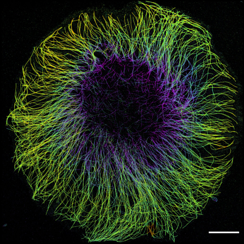
1st Place: Laurent LE, Lévêque-Fort Team, Institut des Sciences Moléculaires d’Orsay
“In the blink of an eye”
COS7 fixed cell. Alpha-tubulin labeled with DNA-PAINT and imaged with Atto 647N. Axial information is obtained by virtual-SAF measurement known as DONALD.
SMLM Fluorescence Microscopy with DNA-PAINT with DONALD detection
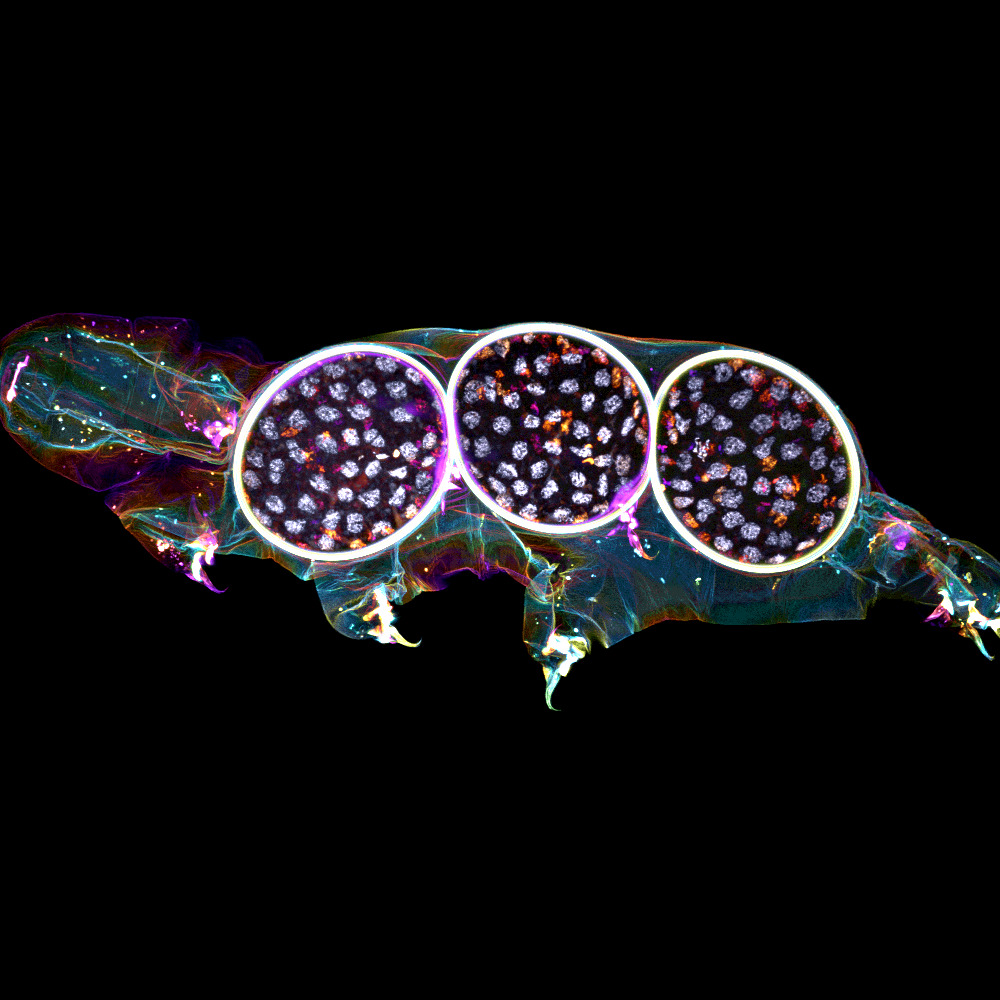
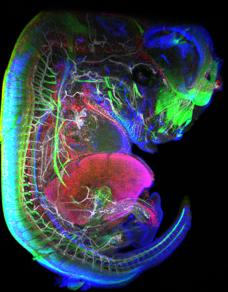
2nd Place:Gonzalo QUIROGA-ARTIGAS, Team Contrôle cytoplasmique de la stabilité du génome, Centre de recherche en Biologie Cellulaire de Montpellier
“Tardigrade embryos protected by mother’s molt”
Tardigrades commonly align the time of molting with egg laying. In this image we observe a tardigrade molt covering three developing embryos (DNA in white). The microscopy technology applied was confocal microscopy, and the research aimed to investigate the synchronization of embryo development in tardigrades.
Confocal microscopy
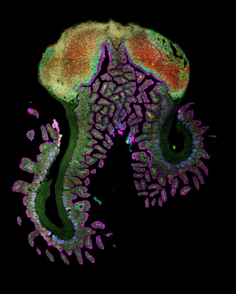
3rd Place:Hugues LELOUARD, Gorvel team, Centre d’Immunologie de Marseille Luminy
“Intestinal octopus”
Small intestine section from a LyzM-eGFP mouse containing one Peyer’s patch and stained for proliferative cells (Ki-67, yellow), Paneth cells (UEA-I, blue), epithelial cells (EpCAM, magenta), naive B cells (IgD, red), T cells (CD3, orange), helper T cells/macrophages (CD4, cyan), phagocytes (CD11c, turquoise), monocyte-derived phagocytes (GFP, green).
10-color spectral confocal microscopy
Congratulations to the winners!
Explore all the images submitted here:
As stated in the Terms & Conditions of the contest, foreign participants non-affiliated to a French institution are featured in the gallery, but were not evaluated as part of the contest.
The FBI Working Group Correlative Light-Electron Microscopy is organizing a workshop!
This workshop will take place at the Bordeaux Imaging Center (FBI Bordeaux node) from January 31st to February 2nd, 2024.
Correlative Light and Electron microscopy (CLEM) increases our capacity of biological investigation. By combining light microscopy and electron microscopy, this complementary approach takes advantages of both techniques. Light imaging provides valuable functional information thanks to its labeling power, whereas Electron microscopy excels at high resolution.
The Bordeaux Imaging Center has developed several workflows such as In-resin fluorescence and Array tomography. Both helps to determine 3D ultrastructure of a targeted area or a whole sample at different resolutions. In the framework of the FBI CLEM workshop, participants can choose one of these 2 different practicals, In-resin fluorescence and Array Tomography, in which a local specialist will show them the workflows.
Workshop 1: “In-Resin Fluorescence” – From HPF to tomography, by way of freeze-substitution and on-section fluorescence observation.
Workshop 2:“Array tomography” – From serial section to acquisition with confocal and SEM.
It is firstly intended to scientists with expertise in electron microscopy, who are expected to use the chosen technique.
2 workshops of 4 people each (you can apply only to one workshop)
A practical manual will be provided, covering every step.
You want to attend this workshop? Please fill the following form before December 22nd, 2023:
The candidates whose research projects suit the workshop the best will be selected. We will get in touch with you after the Christmas break to confirm your participation at this workshop.
This form is currently closed for submissions.
Nous vous invitons au premier webinaire sur l’Initiative Commune Afrique-France pour l’Imagerie Biologique qui se déroulera le 6 décembre 2023 à 11h00 CET!
En coordination avec l’African BioImaging Consortium et Africa Microscopy Initiative et dans le cadre du programme Horizon Europe, l’Initiative Commune Afrique-France pour l’Imagerie Biologique vise à étendre et renforcer les collaborations entre collègues africains et français intéressés par l’utilisation de microscopie avancées pour leurs propres projets de recherche. C’est dans cette optique que nous avons lancé deux appels à projet : l’un favorisant l’accès aux plateformes de bioimagerie de France-BioImaging, l’autre consistant à un programme de jumelage.
Ce webinaire sera notamment l’opportunité d’en apprendre plus sur les projets sélectionnés et sur les bénéfices de cette initiative pour les scientifiques africains et français.
Webinaire
The France-BioImaging Image Contest is back for its 5th edition!
This image contest is open to all within the imaging community: core facility staff and users, R&D labs teams and co-workers, students… Submit your best microscopy images for a chance to showcase your skills, research and creativity to the French bioimaging community and beyond, allowing people to see the visual appeal of the life sciences. Images from the contest will be featured on France-BioImaging communication tools, online and in print.
France-BioImaging and all the French community aims to develop and promote innovative imaging technologies and methods. But microscopy images can also take an artistic, creative look and make the invisible world beautiful.
We are all eager to see your work !
Prizes
1 to 3 images will be awarded depending on the quantity and quality of the entries submitted. France-BioImaging will cover the registration fees for one 2024 microscopy related event of the winners’ choice (FOM, ELMI, EMC, COMULIS conference, etc.).
Important: Only French or foreign participants affiliated to a French institution can enter the contest. Foreign participants non-affiliated to a French institution can submit images and will be featured in the gallery, but will not be evaluated as part of the contest.
Submission deadline: Friday, November 10th, 2023, 23h59 UTC+2.
We are happy to announce our 8th France-BioImaging Annual Meeting! Happening on December 13th and 14th 2023, this year’s edition will be hosted by our new Toulouse node at the Centre de Biologie Intégrative (Toulouse, France).
The Annual meeting will highlight France-BioImaging’s development and perspectives. Imaging scientists and users from the infrastructure’s nodes will present their key projects and demonstrate how they have benefited from France-BioImaging and its community.
More information about the program and the registration coming soon!
France BioImaging and all the French community aims to develop and promote innovative imaging technologies and methods. But microscopy images can also take an artistic, creative look and make the invisible world beautiful, allowing people to see the visual appeal of the life sciences.
We enjoyed the diversity of the images submitted with many different microscopy techniques, models and applications represented. A big thank you to all the participants!
The National Coordination Team and the Executive Board are proud to announce the winners of the FBI Image Contest 2022:
1st Place: Carole SIRET, Van de Pavert Team, Centre d’Immunologie de Marseille-Luminy
“Little Monster”
The embryonic formation of lymph nodes, small organs essential for the immune response, is now known. Using light sheet microscopy, scientists were able to determine the dynamics at work in this 13.5-day-old mouse embryo. In blue, the lymphoid cells (LTi), derived from the haematogenous endothelium, a specific tissue of the embryo. They pass into the liver where they proliferate before migrating through the body to give rise to lymph nodes. The 3D information obtained thus makes it possible to follow the interactions of lymph nodes with their environment, in particular with nerve cells, in green, and blood vessels, in white. The lymphatic endothelial cells and some macrophages are visible in red.
Lightsheet Microscopy
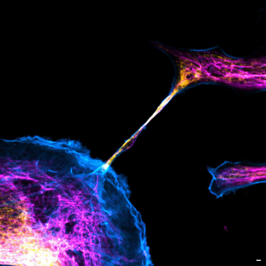
2nd Place:Magalie BENARD, Plateforme de Recherche en IMAgerie CEllulaire de Normandie (PRIMACEN), Research infrastructure HeRacLeS, Inserm US 51, CNRS UAR 2026,
“The communication link with others”
Image of a cellular interconnection between two human tumor cells whose cytoskeleton has been labeled with anti-tubulin (ATTO-647N), anti-vimentin (AlexaFluor594) antibodies and with Phalloidin probe (AlexaFluor488). Scale bar 1µm.
Confocal microscopy
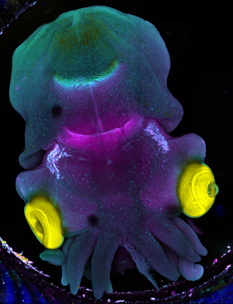
3rd Place:Frédéric FERCOQ, Parasites et Protistes Libres (PPL), Museum National d’Histoire Naturelle
“Sepia”
Stage 25 cuttlefish embryo (Sepia officinalis) observed under a confocal microscope. The cuttlefish was cleared and the tissue autofluorescence was captured.
This image was produced in collaboration with Laure BONNAUD-PONTICELLI and Luis MOLINA from the BOREA laboratory.
Confocal microscopy
Congratulations to the winners!
Explore all the images submitted here:
As stated in the Terms & Conditions of the contest, foreign participants non-affiliated to a French institution are featured in the gallery, but were not evaluated as part of the contest.
In the framework of FBI-AT 2022 on Multiscale Fluorescence Imaging, which will be held in Paris from November 21st to 25th, 2022 (program available here), we have decided to open more widely the plenary lectures given by the invited researchers as well as the introductory lectures to each of the workshops.
Remote access to these lectures will be free but registration is mandatory. You will receive the connection link upon registration.
Registration form
This form is currently closed for submissions.
The France-BioImaging Image Contest is back for its 4th edition!
This image contest is open to all within the imaging community: core facility staff and users, R&D labs teams and co-workers, students… Submit your best microscopy images for a chance to showcase your skills, research and creativity to the French bioimaging community and beyond, allowing people to see the visual appeal of the life sciences. Images from the contest will be featured on France-BioImaging communication tools, online and in print.
France-BioImaging and all the French community aims to develop and promote innovative imaging technologies and methods. But microscopy images can also take an artistic, creative look and make the invisible world beautiful.
We are all eager to see your work !
Prizes
1 to 3 images will be awarded depending on the quantity and quality of the entries submitted. France-BioImaging will cover the registration fees for one 2023 microscopy related event of the winners’ choice (FOM, ELMI, EMC, COMULIS conference, etc.).
Important: Only French or foreign participants affiliated to a French institution can enter the contest. Foreign participants non-affiliated to a French institution can submit images and will be featured in the gallery, but will not be evaluated as part of the contest.
Submission deadline: Friday, November 11th, 2022, 23h59 UTC+2.
France-BioImaging, with its partner the GDR IMABIO, organizes the 4th edition of the FBI-AT: an advanced microscopy workshop to be held in Paris from November 21st to 25th, 2022.
The aim of this France-BioImaging-Advanced Training is to train microscopy users on the most advanced imaging techniques that will allow them to perform molecular studies at the cellular level as well as in thick samples. In particular, recent developments on fluorescent probes will be highlighted. The workshop will benefit from state-of-the-art equipment available on several of the Parisian Node Imaging facilities.
This year’s edition will have plenary lectures given by experts in the microscopy development field. These seminars will be advertised as a series and will be broadcasted for a large audience. In addition, specific techniques will be introduced.
Hands-on practicals will train attendants on these techniques every afternoon in different sites in Paris including Institut Curie, Institut Pasteur, Institut Cochin, Institut Jacques Monod, Institut de Psychiatrie et Neurosciences de Paris and ENS Paris. Access to this part of training will be restricted to selected and registered trainees.
To guaranty access to set-ups and proper training, each practical session will host only 3-4 persons. The sessions will be run in parallel.
Apply now, attendance will be limited to 25 participants!
FBI-AT is ideal for researchers with a basic training in microscopy willing to become familiar with advanced techniques to answer their specific biological questions, or to be exposed to new developments that will allow them to tackle new questions in their project. We will consider applications from early career researchers (PhD students, post-docs), technical staff members and more senior scientists.
We use cookies on our website to give you the most relevant experience by remembering your preferences and repeat visits. By clicking “Accept All”, you consent to the use of ALL the cookies. However, you may visit "Cookie Settings" to provide a controlled consent.
This website uses cookies to improve your experience while you navigate through the website. Out of these, the cookies that are categorized as necessary are stored on your browser as they are essential for the working of basic functionalities of the website. We also use third-party cookies that help us analyze and understand how you use this website. These cookies will be stored in your browser only with your consent. You also have the option to opt-out of these cookies. But opting out of some of these cookies may affect your browsing experience.
Necessary cookies are absolutely essential for the website to function properly. These cookies ensure basic functionalities and security features of the website, anonymously.
Cookie
Duration
Description
cookielawinfo-checkbox-analytics
11 months
This cookie is set by GDPR Cookie Consent plugin. The cookie is used to store the user consent for the cookies in the category "Analytics".
cookielawinfo-checkbox-functional
11 months
The cookie is set by GDPR cookie consent to record the user consent for the cookies in the category "Functional".
cookielawinfo-checkbox-necessary
11 months
This cookie is set by GDPR Cookie Consent plugin. The cookies is used to store the user consent for the cookies in the category "Necessary".
cookielawinfo-checkbox-others
11 months
This cookie is set by GDPR Cookie Consent plugin. The cookie is used to store the user consent for the cookies in the category "Other.
cookielawinfo-checkbox-performance
11 months
This cookie is set by GDPR Cookie Consent plugin. The cookie is used to store the user consent for the cookies in the category "Performance".
viewed_cookie_policy
11 months
The cookie is set by the GDPR Cookie Consent plugin and is used to store whether or not user has consented to the use of cookies. It does not store any personal data.
Functional cookies help to perform certain functionalities like sharing the content of the website on social media platforms, collect feedbacks, and other third-party features.
Performance cookies are used to understand and analyze the key performance indexes of the website which helps in delivering a better user experience for the visitors.
Analytical cookies are used to understand how visitors interact with the website. These cookies help provide information on metrics the number of visitors, bounce rate, traffic source, etc.
Advertisement cookies are used to provide visitors with relevant ads and marketing campaigns. These cookies track visitors across websites and collect information to provide customized ads.